- ContentsContents
- Article InfoArticle Info
- CiteCite
- MetricsMetrics
- CommentComment
- RelatedRelated
- FigsFigs
- TabsTabs
- MapMap
- TaxaTaxa
- RefsRefs
- CitedCited
- NanopubsNanopubs
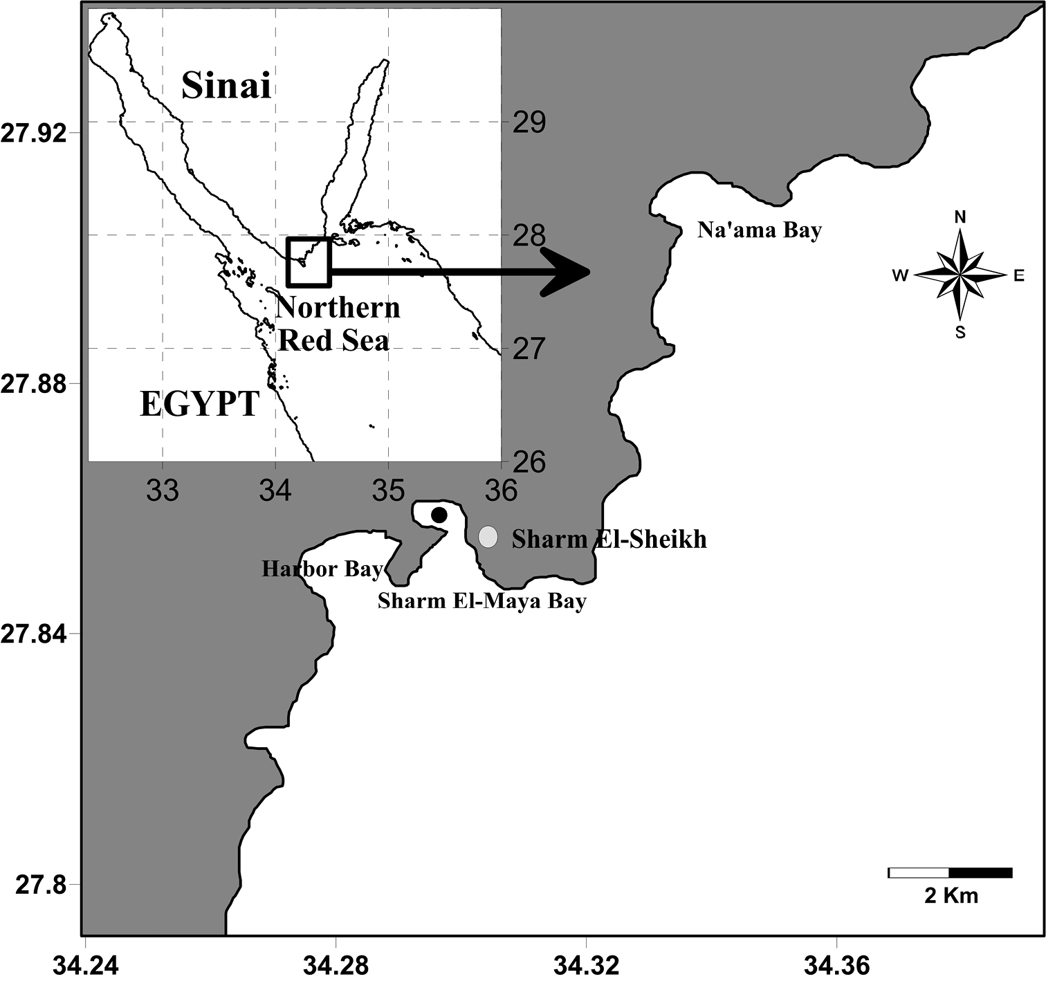
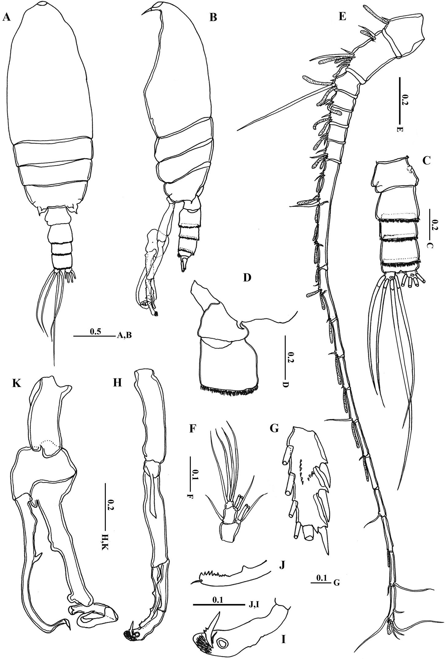
Location of the sampling site (Black circle).
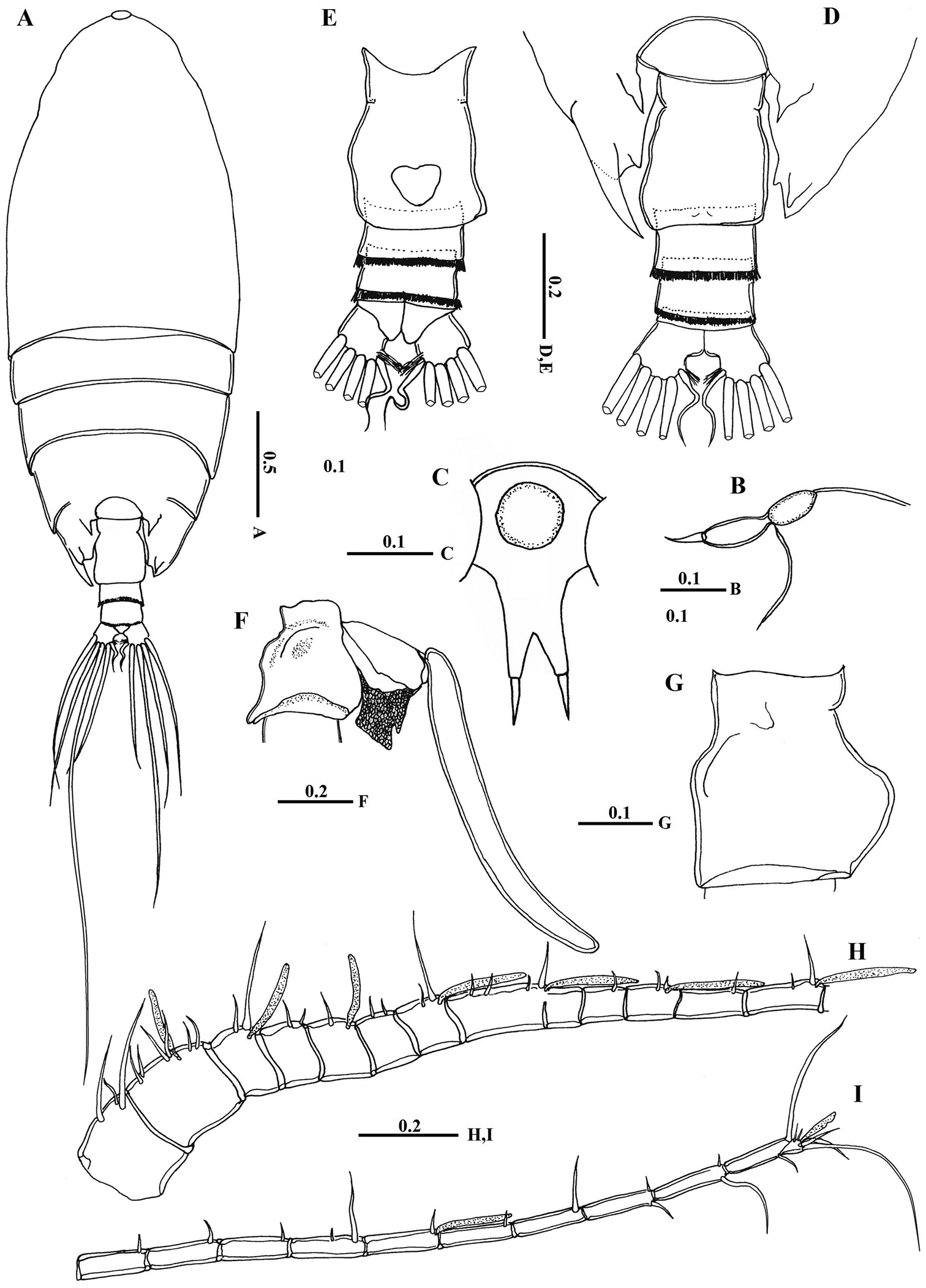
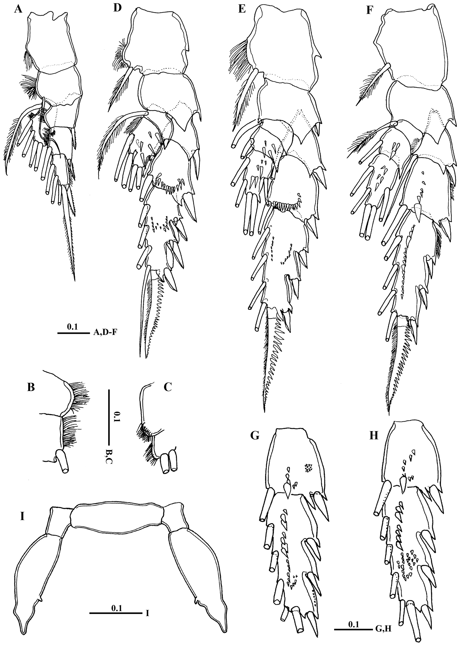
Macandrewella cochinensis female from the northern Red Sea. A habitus, dorsal view B rostrum, lateral view C rostrum, ventral view D posterior prosome and urosome, dorsal view E urosome, ventral view F genital double-somite with spermatophore, lateral view (right) G genital double-somite, lateral view (right) H–I antennules. All scale bars in mm.
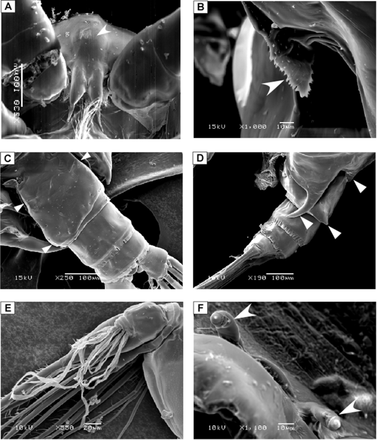
SEM micrographs of Macandrewella cochinensis female from the northern Red Sea. A rostrum and cuticular lens indicated by arrow, ventral view B serration of postero-dorsolateral process of prosomal end indicated by arrow, lateral view C urosome, anterodorsal protrusions and posterodorsal swelling on left side indicated by arrows, dorsal view D urosome, posterodorsal swelling on left side indicated by arrow, lateral view (left) E maxillary endopod F leg 5 indicated by arrow.
Macandrewella cochinensis female from the northern Red Sea. A antenna B mandibular gnathobase cutting edge C mandibular palp D maxillule E maxilla F maxilla endopod G maxilliped. All scale bars in mm.
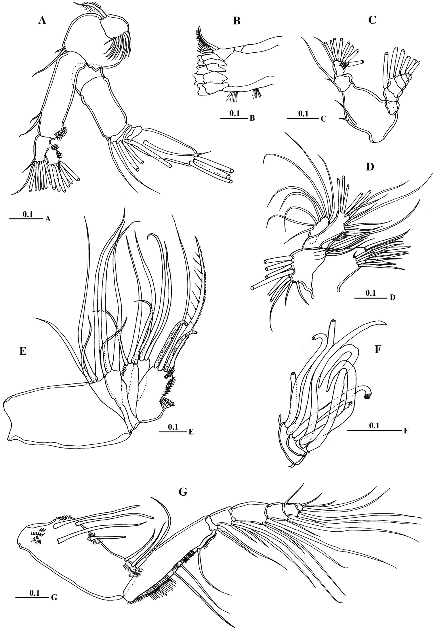
Macandrewella cochinensis female from the northern Red Sea. A Leg 1, anterior surface B medial margin of first and second exopodal segments of Leg 1 C lateral distal margin of leg 1 endopod D leg 2, posterior surface E Leg 3, posterior surface F leg 4, posterior surface G–H second and third exopodal segments of leg 4, anterior surface I leg 5, anterior surface. All scale bars in mm.
Macandrewella cochinensis male from the northern Red Sea. A habitus, dorsal view B habitus, lateral view C urosome, dorsal view D first and second urosomal segment, lateral view (right) E left antennule F maxilliped, terminal endopod segments G Exopod segment 3 of leg 2 H left leg 5 I terminal portion of left exopodal of leg 5 J terminal portion of left endopod of leg 5 K right leg 5. All scale bars in mm.

SEM micrographs of Macandrewella cochinensis male from the northern Red Sea. A genital somite, dorsal view B distal part of leg 5.