

- ContentsContents
- Article InfoArticle Info
- CiteCite
- MetricsMetrics
- Comment1Comment
- RelatedRelated
- FigsFigs
- TabsTabs
- MapMap
- TaxaTaxa
- DataData
- RefsRefs
- CitedCited
- NanopubsNanopubs
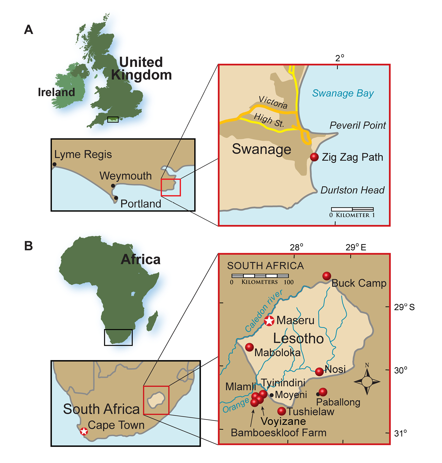
Heterodontosaurid localities. A Locality (red dot) for Echinodon becklesii on the southern coast of England B Heterodontosaurid localities in South Africa and Lesotho. Locality(ies)/taxon identification: Nosi/Abrictosaurus consors; Mlamli, Tushielaw, Tyinindini/Heterodontosaurus tucki; Bamboeskloof Farm, Buck Camp, Paballong/Lycorhinus angustidens; Maboloka/Heterodontosauridae incertae sedis; Voyizane/ Pegomastax africanus gen. n. sp. n.
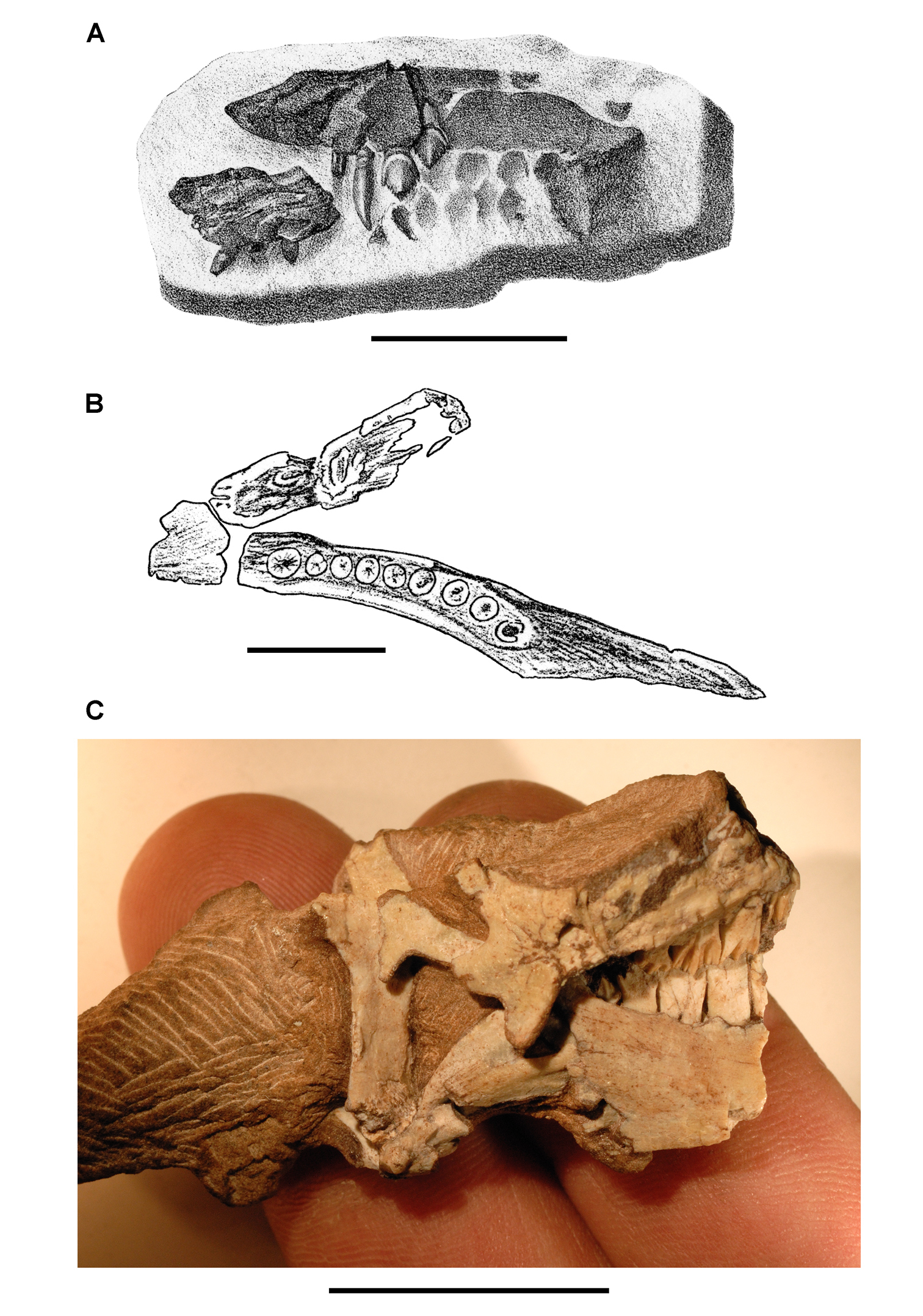
Early heterodontosaurid discoveries. A Lithographic drawing of the right and left premaxillae and the anterior portion of the left maxilla in lateral view of Echinodon becklesii (NHMUK 48209; from Owen 1861) B Drawing of lower jaws in dorsal view of Geranosaurus atavus (SAM-PK-K1871; from Broom 1911) C Photograph of the posterior portion of a subadult skull in right lateral view of Heterodontosaurus tucki (AMNH 24000). Scale bars equal 1 cm in A and 2 cm in B and C.
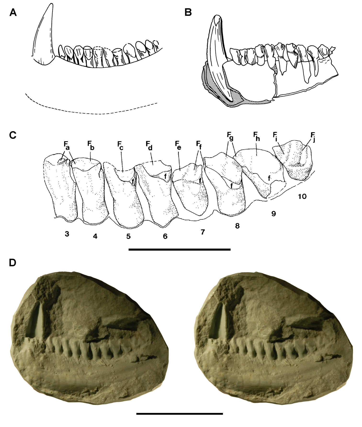
Early heterodontosaurid discoveries from southern Africa. A Drawing of the left dentary of the holotype of Lycorhinus angustidens (SAM-PK-K3606) in lateral view (from Haughton 1924) B Drawing of the left dentary of the holotype of Lycorhinus angustidens (SAM-PK-K3606) in medial view (reversed from Haughton 1924) C Left dentary teeth 3-10 in lateral view based on a natural cast (UCRC PVC10) of the holotype of Lycorhinus angustidens (from Hopson 1980) D Stereopair of a natural silicone cast of the holotype of Lycorhinus angustidens (UCRC PVC10). Abbreviations: 3-10 dentary tooth 3-10 Fa-j tooth-to-tooth wear facet a-j f accessory facet. Scale bars equal 1 cm in C and 3 cm in D.
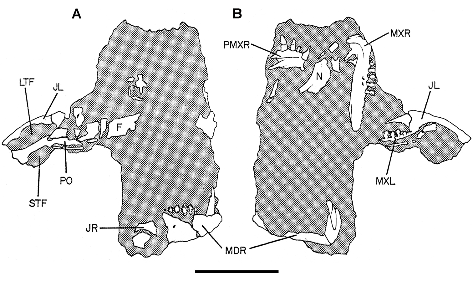
Controversial early heterodontosaurid specimen from southern Africa. Drawing of specimen NHMUK RU A100 as embedded in matrix in A top and B bottom views (from Thulborn 1970). Abbreviations: F frontal JL left jugal JR right jugal LTF left lateral temporal opening MDR right mandible MXL left maxilla MXR right maxilla N left nasal PMXR right premaxilla PO postorbital STF supratemporal fossa. Scale bar equals 5 cm.
More recent heterodontosaurid discoveries from southern Africa. A Partial skull of Abrictosaurus consors in left lateral view (NHMUK RU B54) B Lower jaws of Pegomastax africanus gen. n. sp. n. (SAM-PK-K10488) in right ventrolateral view. Scale bars equal 2 cm in A and 1 cm in B.
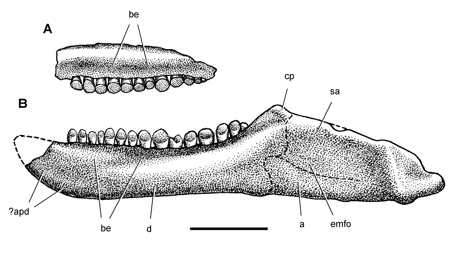
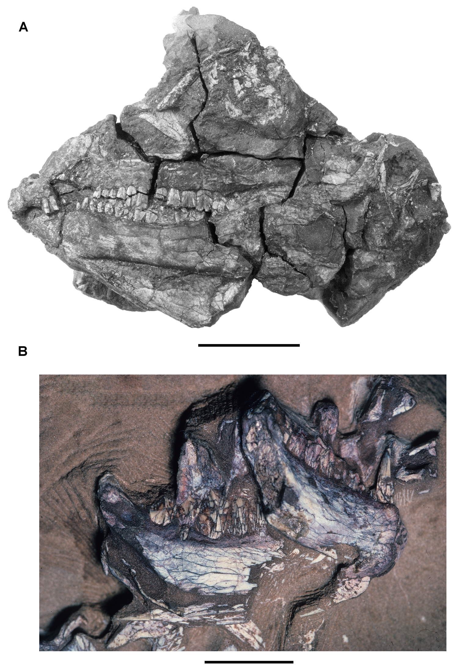
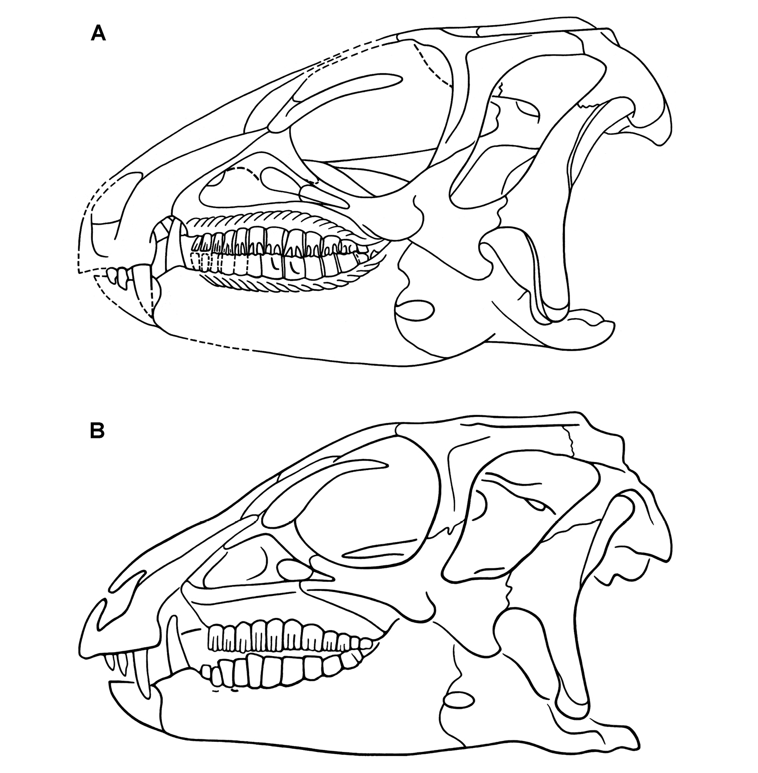
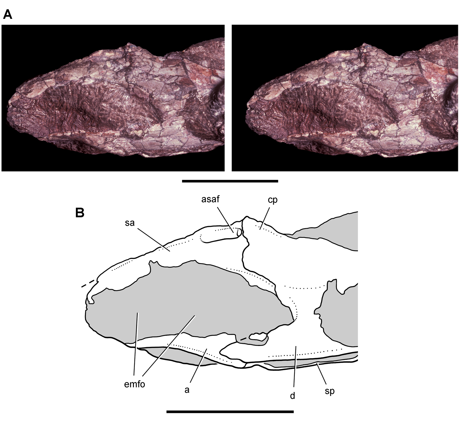
Cranial remains of Pisanosaurus mertii from the Upper Triassic Ischigualasto Formation of Argentina. Drawings of a partial right maxilla in medial view (A) and right lower jaw (reversed) in lateral view (B) (from Bonaparte 1976). Dashed lines indicate estimated sutures and edges. Scale bar equals 2 cm in B. Abbreviations: a angular apd articular surface for the predentary be buccal emargination cp coronoid process d dentary emfo external mandibular fossa sa surangular.
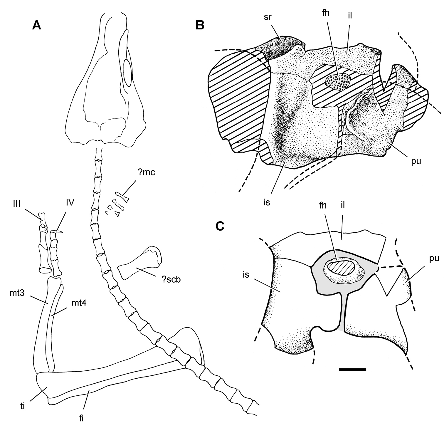
Postcranial remains of Pisanosaurus mertii from the Upper Triassic Ischigualasto Formation of Argentina. Drawings of the holotype (PVL 2577) in the field (from Bonaparte 1976) (A) and impression of central portion of the right pelvic girdle and head of the femur in medial view as previously interpreted (from Bonaparte 1976) (B) and in this study (C). Dashed lines indicate estimated edges; hatching in B indicates broken bone and matrix; hatching in C indicates broken bone; tone in C indicates matrix. Scale bar for B and C equals 1 cm. Abbreviations: III, IV pedal digit III, IV fh femur head fi fibula il ilium is ischium mc metacarpal mt3, 4 metatarsal 3, 4 pu pubis scb scapular blade sr sacral rib ti tibia.
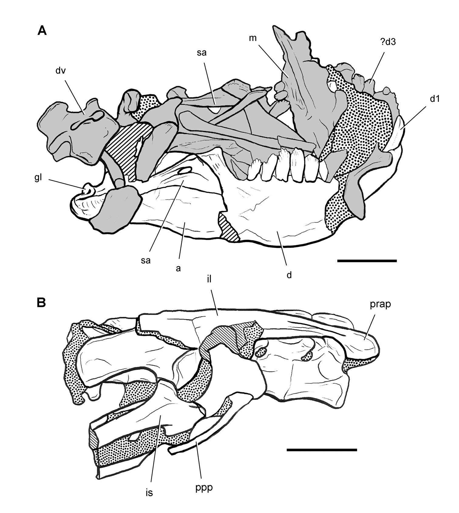
Skeletal remains of Manidens condorensis from the Middle Jurassic Cañadón Asfalto Formation of Chubut Province, Argentina. Drawings of major cranial (A) and postcranial (B) blocks (from Pol et al. 2011). Stipple indicates matrix; hatching indicates broken bone; grey tone in A shades the right maxilla, left lower jaw and other bones to highlight the right lower jaw. Scale bars equal 1 cm. Abbreviations: a angular d dentary d1, 3 dentary tooth 1, 3 dv dorsal vertebra gl glenoid il ilium is ischium m maxilla ppp postpubic process prap preacetabular process sa surangular.
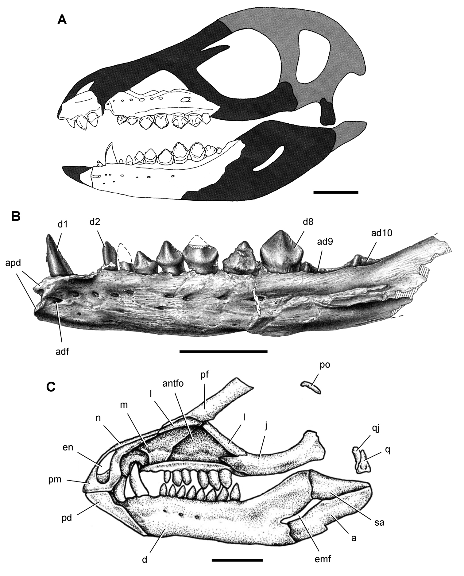
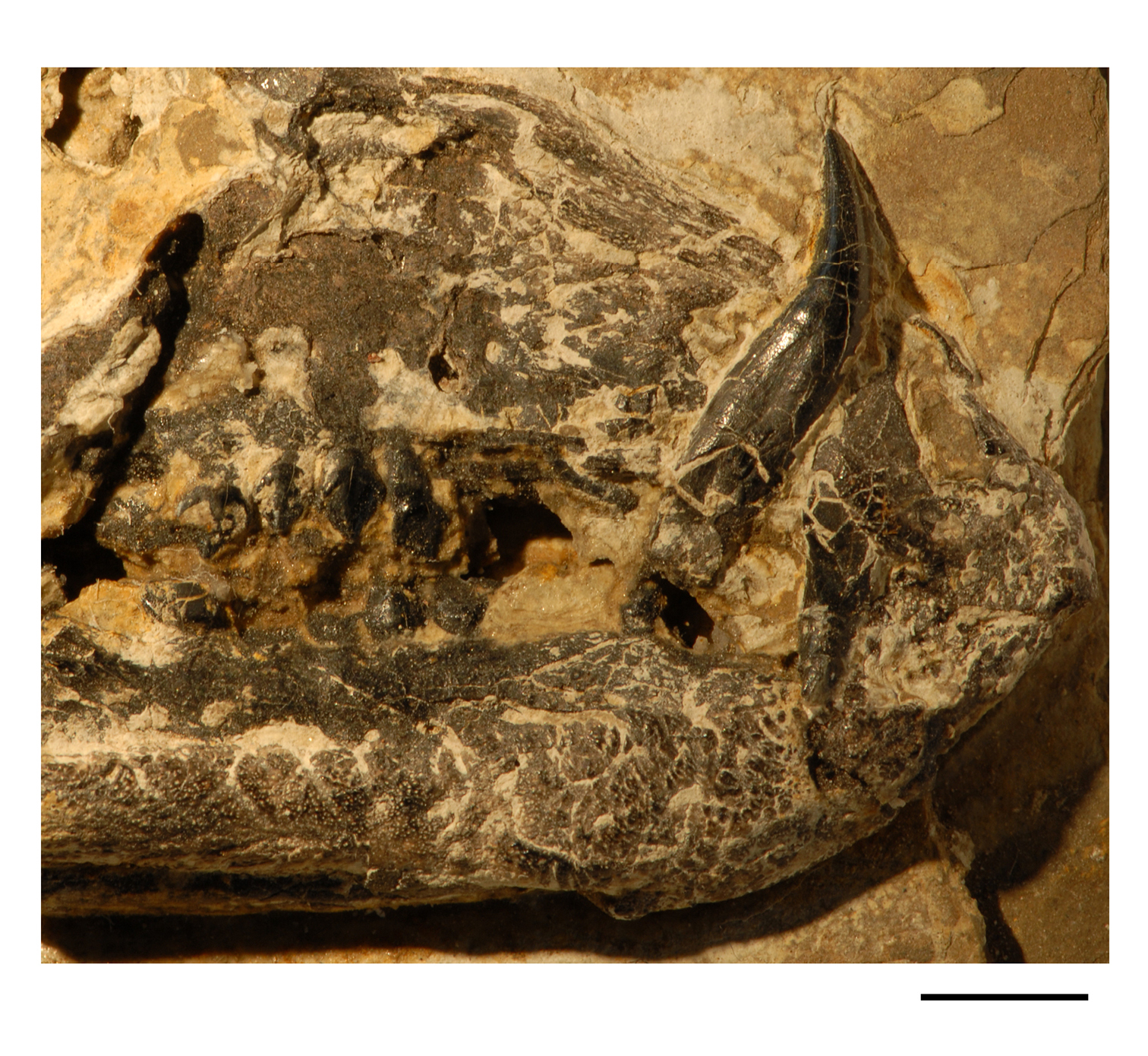
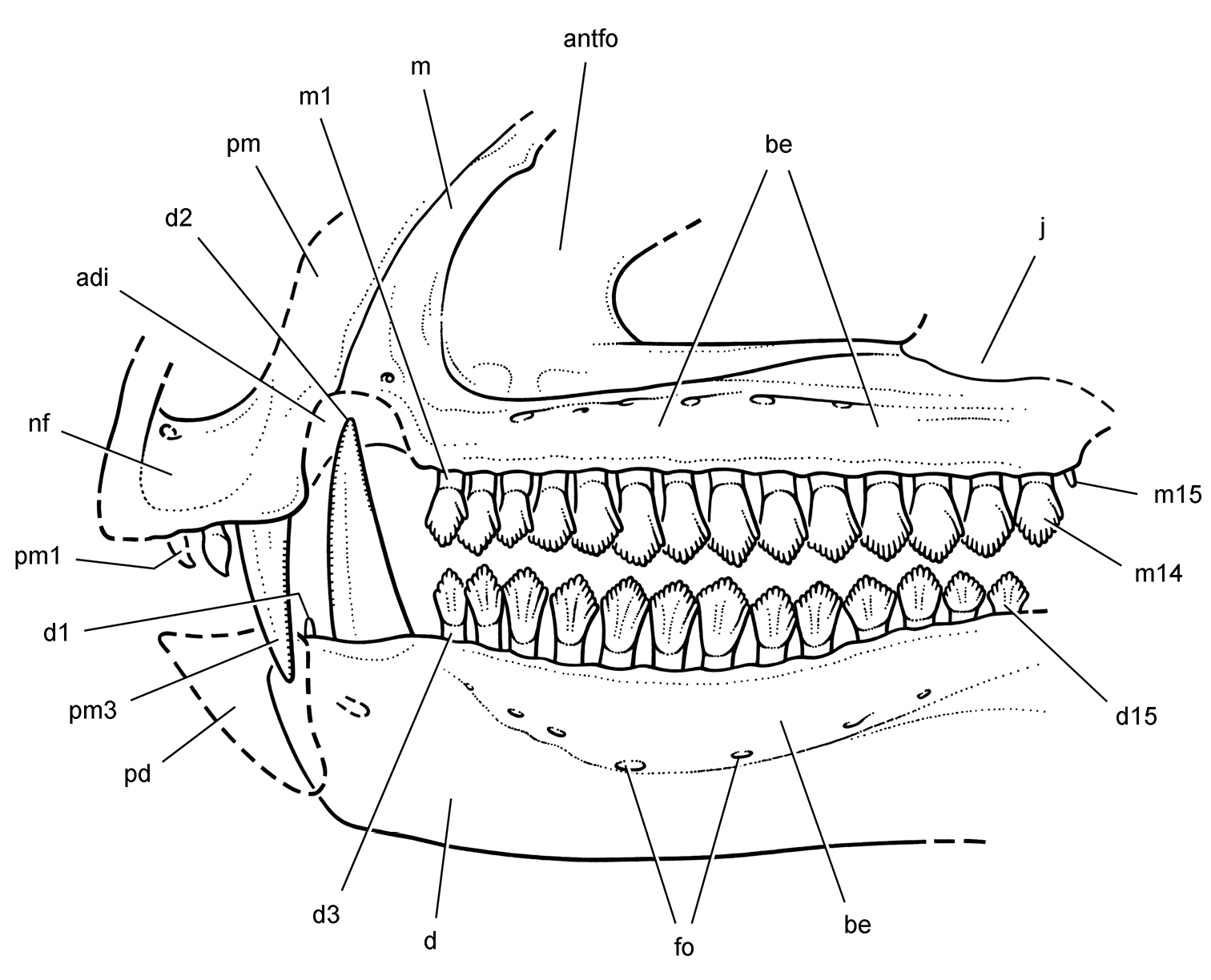
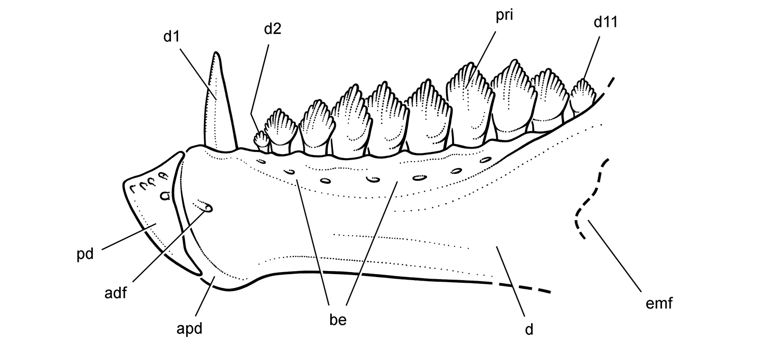
More recent heterodontosaurid discoveries from northern locales. A Jaws of Fruitadens haagarorum from the Upper Jurassic Morrison Formation in Colorado, USA (based on LACM 115747, 128258; reversed from Butler et al. 2010) B Left dentary in lateral view of an undescribed heterodontosaurid from the Lower Jurassic Kayenta Formation of Arizona (from Sereno et al. unpublished) C Partial skull of Tianyulong confuciusi from the Yixian Formation of Liaoning Province, PRC (STMN 26-3; reversed from Zheng et al. 2009). Abbreviations: a angular ad 9, 10 alveolus for dentary tooth 9, 10 adf anterior dentary foramen antfo antorbital fossa apd articular surface for the predentary d dentary d1, 2, 8 dentary tooth 1, 2, 8 emf external mandibular fenestra en external nares j jugal l lacrimal m maxilla n nasal pd predentary pf prefrontal pm premaxilla po postorbital q quadrate qj quadratojugal sa surangular. Scale bar equals 1 cm in A and C and 5 mm in B.
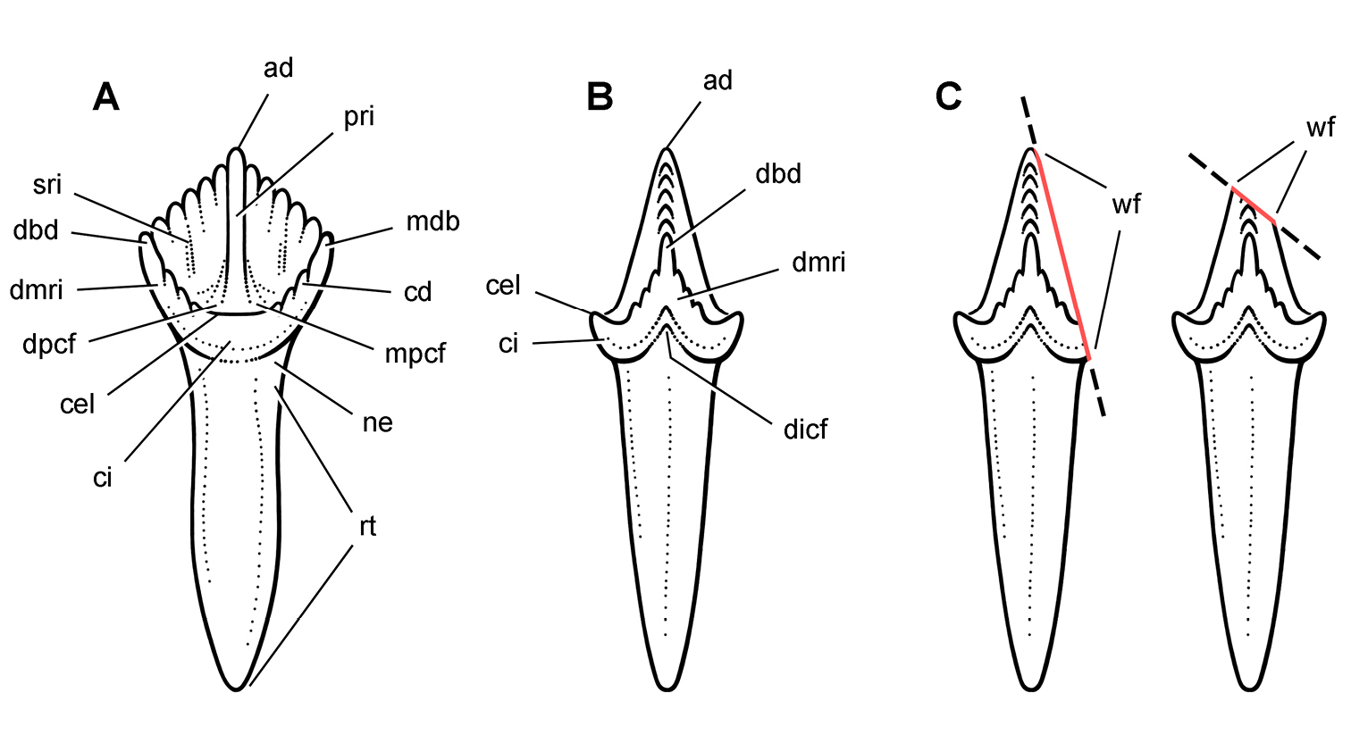
Cheek tooth terminology. A Postcaniniform maxillary or dentary tooth in labial or lingual view, respectively. B Postcaniniform maxillary or dentary tooth in distal view. C Pair of worn postcaniniform maxillary or dentary teeth in proximal or distal view showing low-angle (left) and high-angle (right) wear facets (red line). The angle of incidence (dashed line) of each wear facet (red line) is measured away from the vertical axis of the crown. Abbreviations: ad apical denticle cd cingular denticle cel cingular ectoloph ci cingulum dbd distal basal denticle dicf distal intercingular fossa dmri distal marginal ridge dpcf distal paracingular fossa mbd mesial basal denticle mpcf mesial paracingular fossa ne neck pri primary ridge rt root sri secondary ridge wf wear facet.
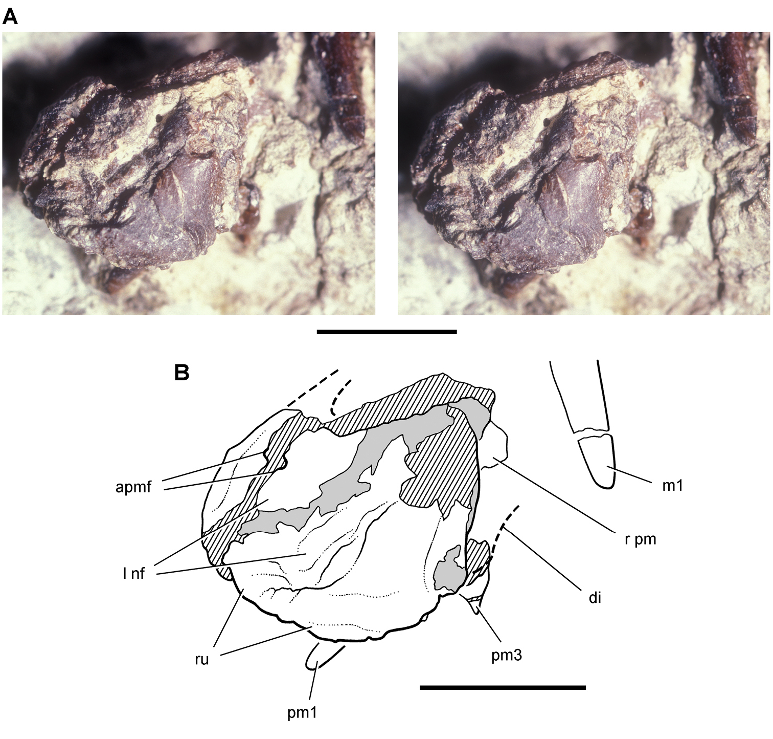
Premaxilla of Echinodon becklesii from the Lower Cretaceous Purbeck Formation of England. Left premaxilla in anterdorsolateral view (NHMUK 48209). Stereopair (A) and line drawing (B). Hatching indicates broken bone; dashed lines indicate estimated edges; tone indicates matrix. Scale bars equal 5 mm. Abbreviations: apmf anterior premaxillary foramen di diastema l left m1 maxillary tooth 1 nf narial fossa pm premaxilla pm1, 3 premaxillary tooth 1, 3 r right ru rugosity.
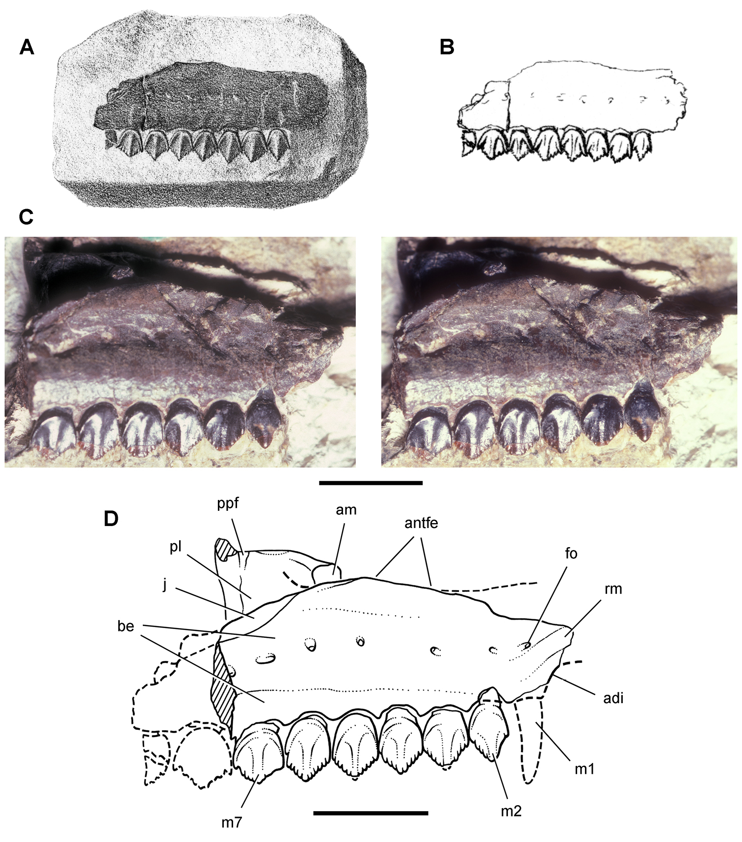
Maxilla of Echinodon becklesii from the Lower Cretaceous Purbeck Formation of England. Right maxilla in lateral view (NHMUK 48211). Lithograph (A) and line drawing (B) from Owen (1861). Stereopair (C) and line drawing (D). Hatching indicates broken bone; dashed lines indicate estimated edges. Scale bars equal 5 mm. Abbreviations: adi arched diastema am articular surface for the maxilla antfe antorbital fenestra be buccal emargination fo foramen j jugal m1, 2, 7 maxillary tooth 1, 2, 7 pl palatine ppf postpalatine foramen rm rim.
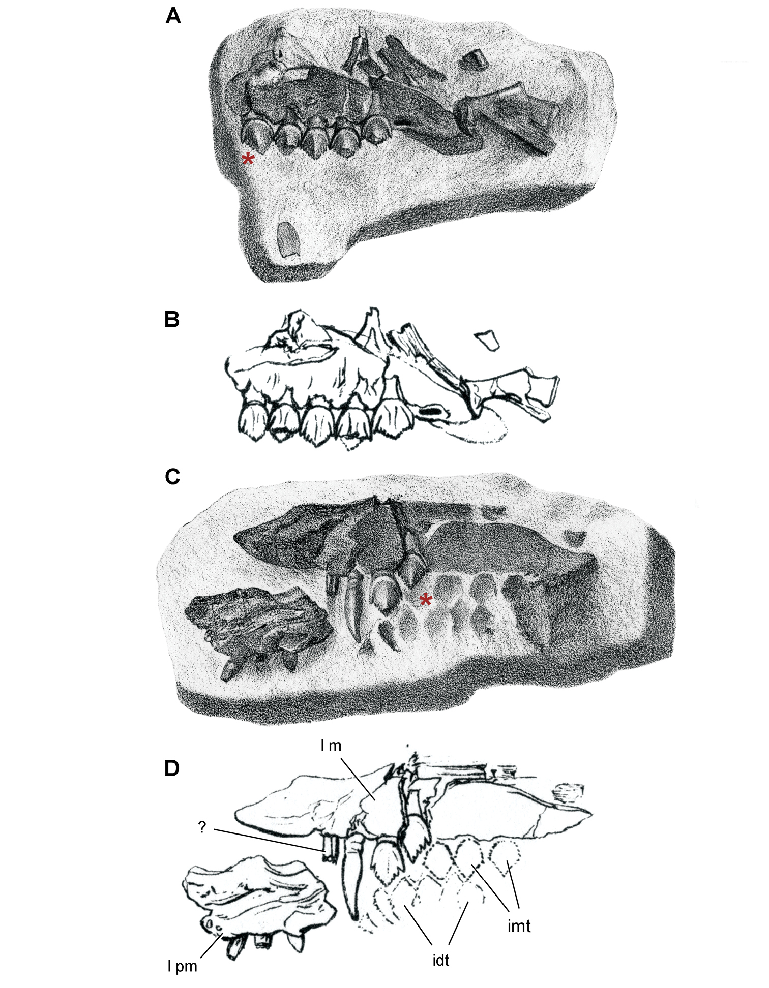
Maxilla of Echinodon becklesii from the Lower Cretaceous Purbeck Formation of England. Part (NHMUK 48210) and counterpart (NHMUK 48209) of a block preserving portions of the snout of a skull. A, B Part preserving the posterior portion of the left maxilla and portions of the left lacrimal, jugal and ectopterygoid in medial view (NHMUK 48210; reversed from Owen 1861). C, D Counterpart preserving portions of the right and left premaxillae and the anterior portion of the left maxilla in lateral view (NHMUK 48209; from Owen 1861). A red asterisk marks a crown on the part (A) and its impression on the counterpart (C). Abbreviations: idt impressions of dentary teeth imt impressions of maxillary teeth l left m maxilla pm premaxilla.
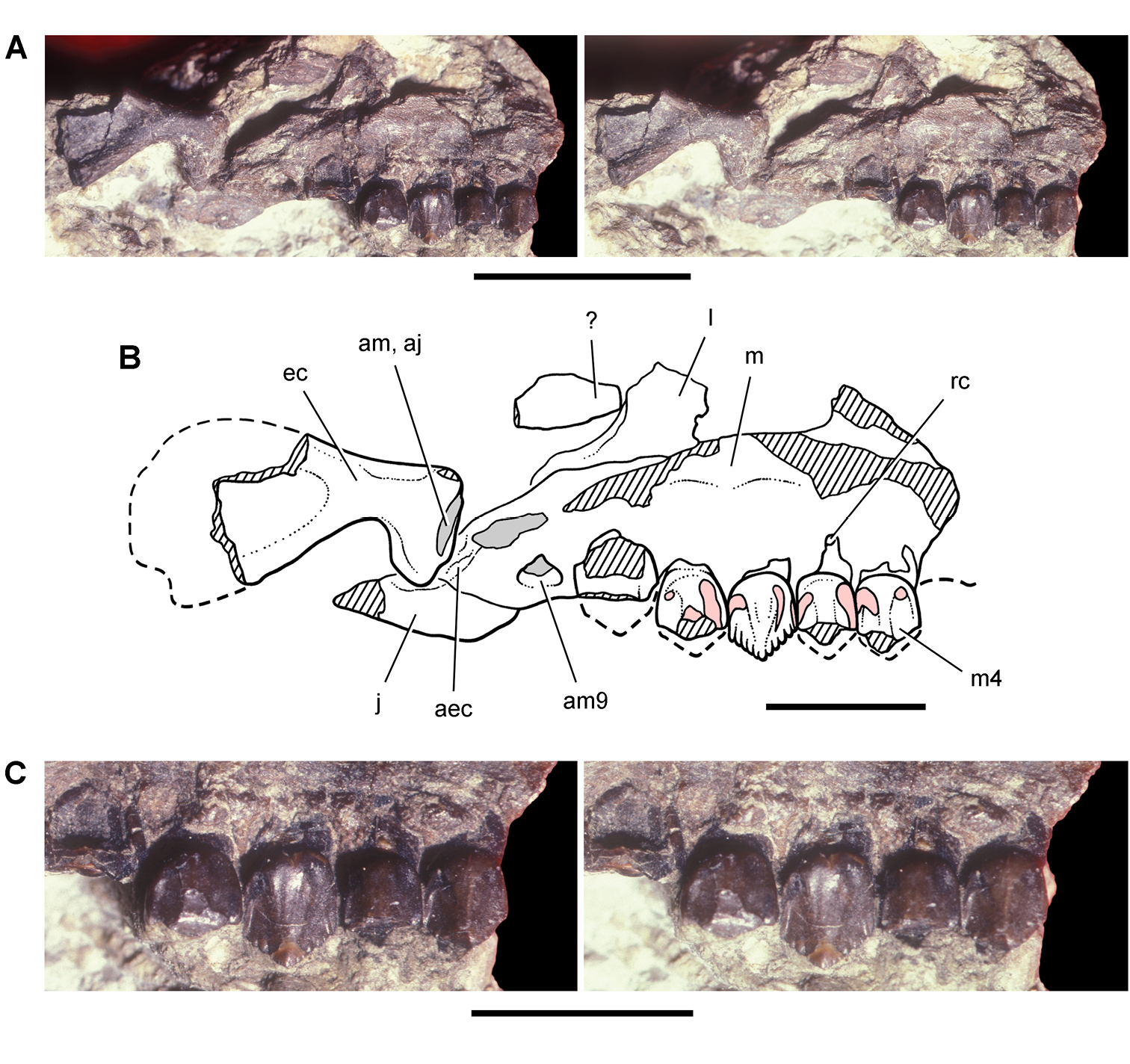
Partial skull of Echinodon becklesii from the Lower Cretaceous Purbeck Formation of England. Left maxilla and portions of the lacrimal, jugal and ectopterygoid in medial view (NHMUK 48210). Stereopairs of bones (A), line drawing of bones (B), and stereopairs of teeth (C). Hatching indicates broken bone; dashed lines indicate estimated edges; grey tone indicate matrix; pink tone indicates wear facets. Scale bar equals 1 cm in A and 5 mm in B and C. Abbreviations: aec articular surface for the ectopterygoid aj articular surface for the jugal am articular surface for the maxilla am9 alveolus for maxillary tooth 9 ec ectopterygoid j jugal l lacrimal m maxilla m4 maxillary tooth 4 rc replacement crown.
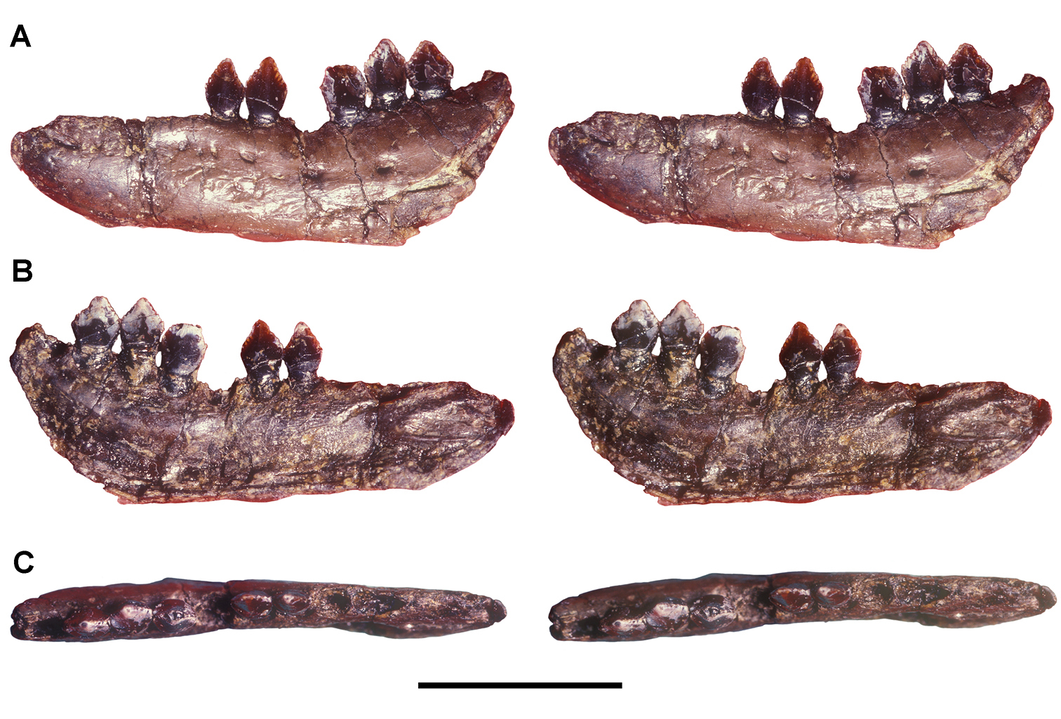
Dentary of Echinodon becklesii from the Lower Cretaceous Purbeck Formation of England. Stereopairs of left dentary (NHMUK 48215b) in lateral (A), medial (B), and dorsal (C) views. Scale bar equals 1 cm.
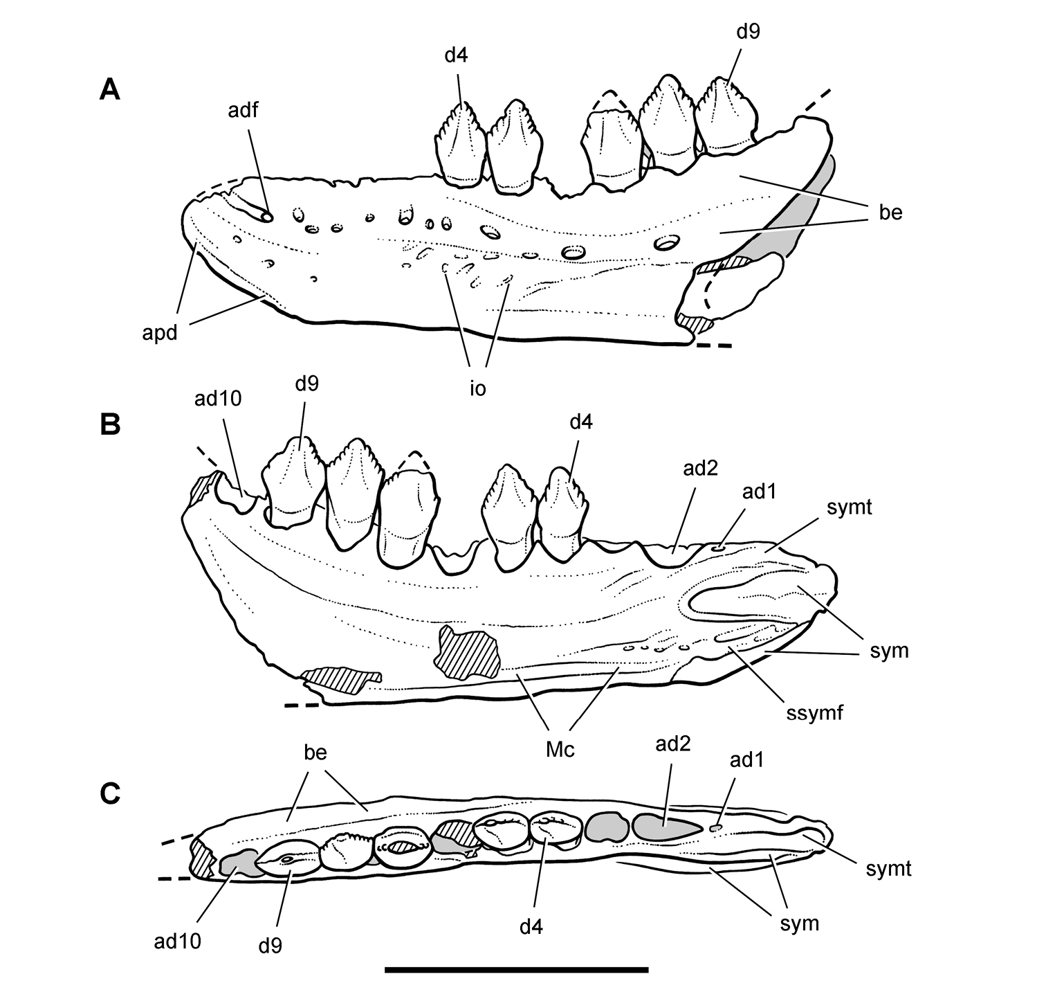
Dentary of Echinodon becklesii from the Lower Cretaceous Purbeck Formation of England. Line drawings of left dentary in lateral (A), medial (B), and dorsal (C) views (NHMUK 48215b). Hatching indicates broken bone; dashed lines indicate estimated edges; tone indicates matrix. Scale bar equals 1 cm. Abbreviations: ad1, 2, 10 alveolus for dentary tooth 1, 2, 10 adf anterior dentary foramen apd articular surface for the predentary be buccal emargination d4, 9 dentary tooth 4, 9 io impressed ornamentation Mc Meckel’s canal ssymf subsymphyseal flange sym symphysis symt symphyseal trough.
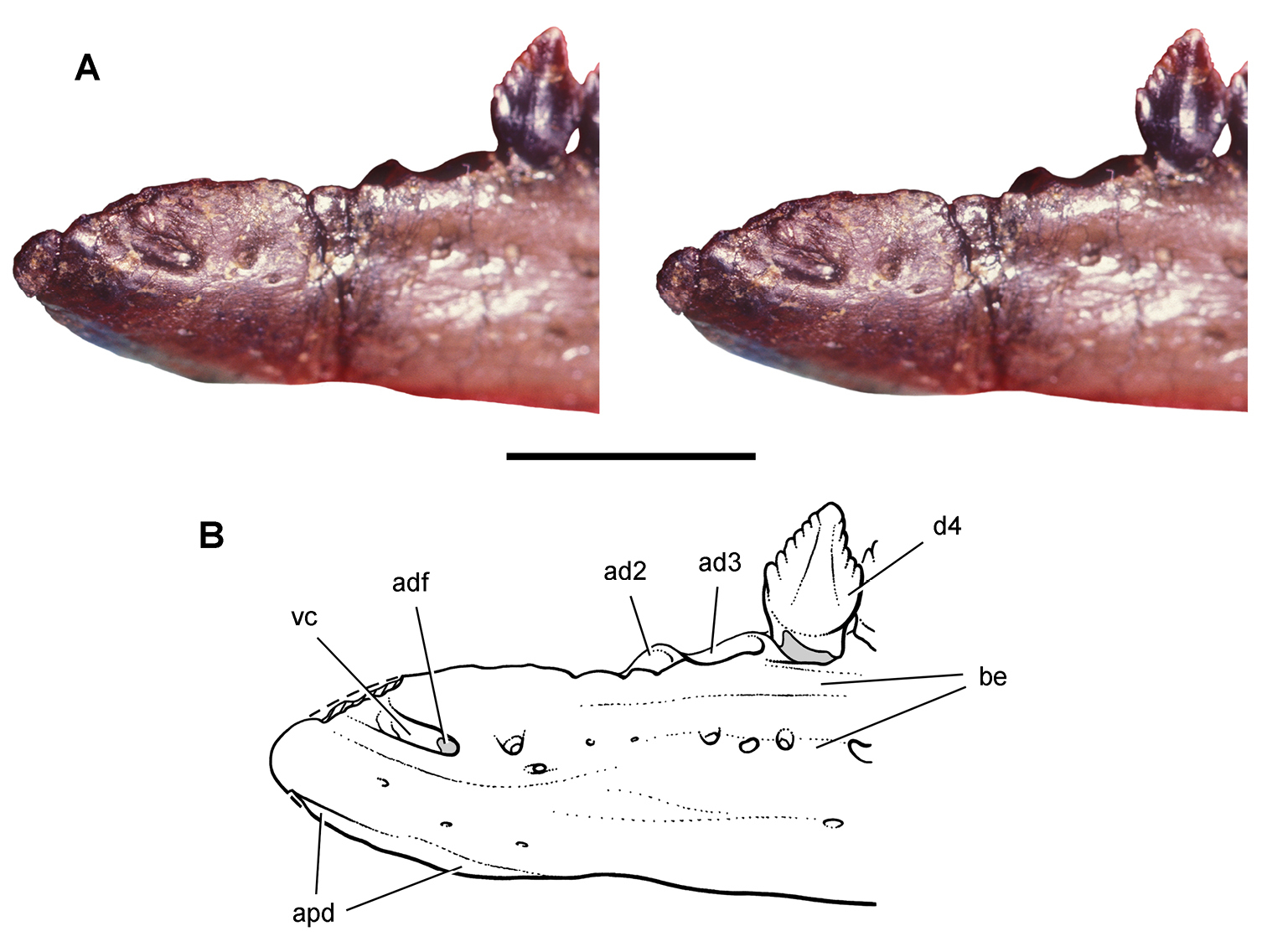
Dentary end of Echinodon becklesii from the Lower Cretaceous Purbeck Formation of England. Anterior end of the left dentary in lateral view (NHMUK 48215b). Stereopair (A) and line drawing (B). Scale bar equals 5 mm. Hatching indicates broken bone; dashed lines indicate estimated edges; tone indicates matrix. Abbreviations: ad2, 3 alveolus for dentary tooth 2, 3 adf anterior dentary foramen apd articular surface for the predentary be buccal emargination d4dentary tooth 4 vc vascular canal.
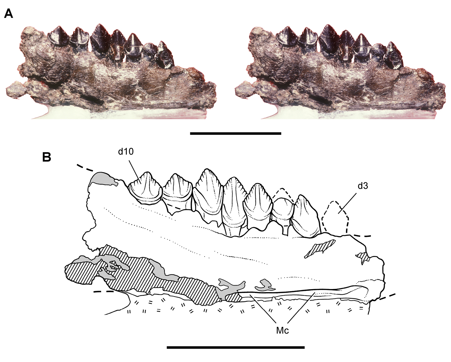
Dentary of Echinodon becklesii from the Lower Cretaceous Purbeck Formation of England. Portion of the left dentary (NHMUK 48213) in medial view. Stereopair (A) and line drawing (B). Hatching indicates broken bone; dashed lines indicate estimated edges; tone indicates matrix; hash marks indicate carbowax support. Scale bars equal 1 cm. Abbreviations: d3, 10 dentary tooth 3, 10 Mc Meckel’s canal.
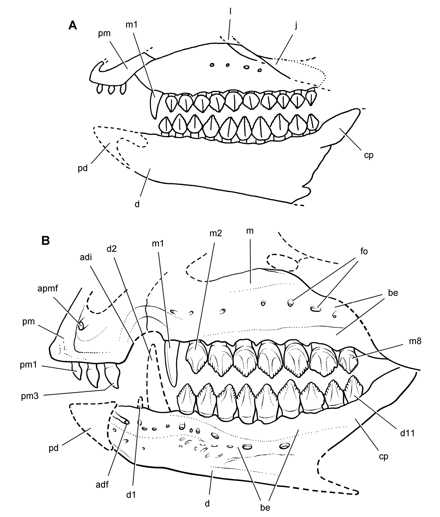
Skull of Echinodon becklesii from the Lower Cretaceous Purbeck Formation of England. Skull reconstructions in left lateral view. A From Galton (1978). B This study. Dashed lines indicate estimated edges and sutures. Abbreviations: adf anterior dentary foramen adi arched diastema apmf anterior premaxillary foramen be buccal emargination cp coronoid process d dentary d1, 2, 11 dentary tooth 1, 2, 11 fo foramen j jugal l lacrimal m maxilla m1, 2, 8 maxillary tooth 1, 2, 8 pd predentary pm premaxilla pm1, 3 premaxillary tooth 1, 3.
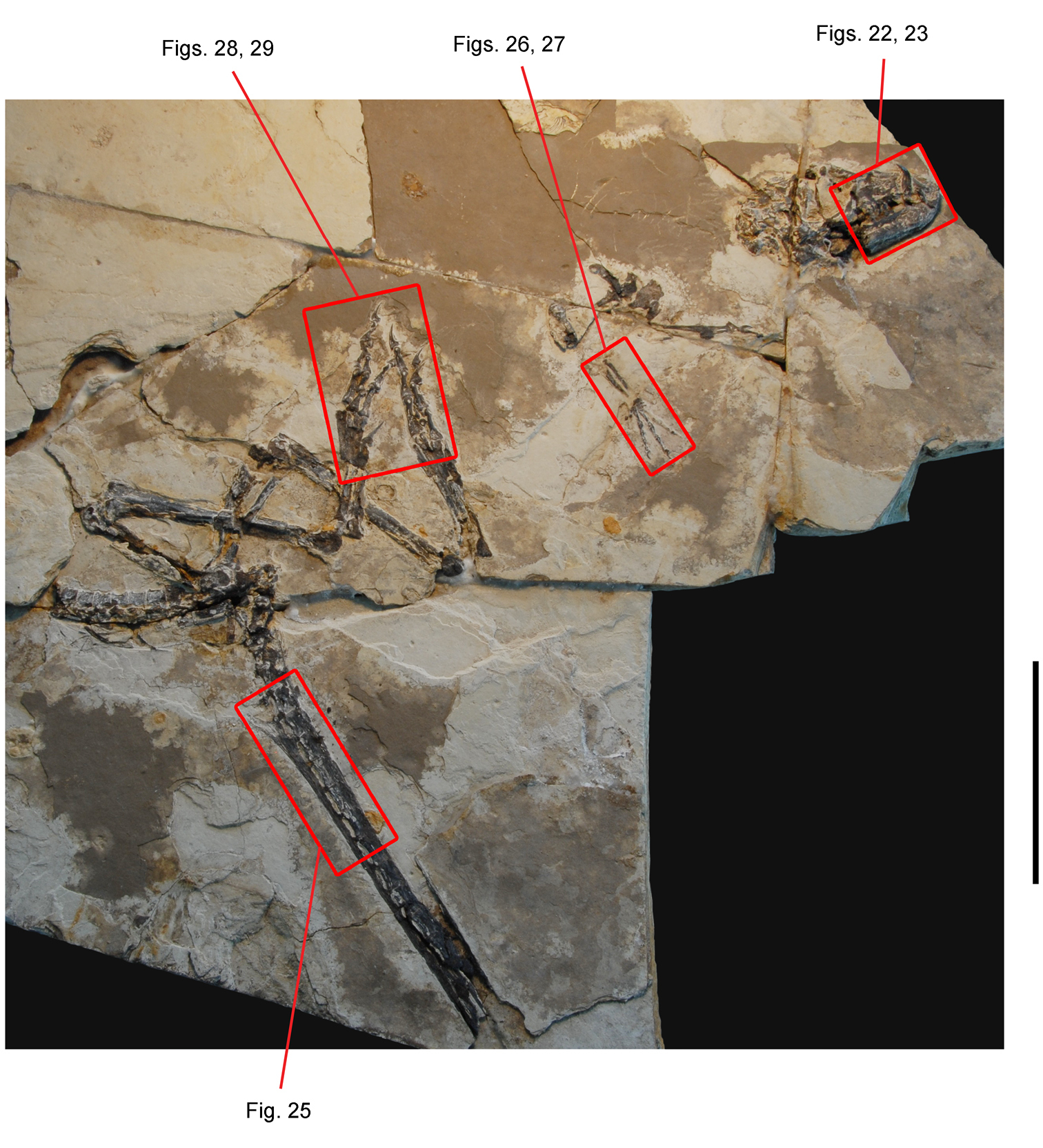
Partial skeleton of the heterodontosaurid Tianyulong confuciusi from the Lower Cretaceous Jehol Group of China. Partial skeleton mainly in right lateral view (IVPP V17090). Enlargements of subsequent figures are shown in red. Scale bar equals 10 cm.
Skull of the heterodontosaurid Tianyulong confuciusi from the Lower Cretaceous Jehol Group of China. Skull in right lateral view (IVPP V17090). Enlargements of subsequent figures are shown in red. Scale bar equals 2 cm.
Anterior portion of skull of the heterodontosaurid Tianyulong confuciusi from the Lower Cretaceous Jehol Group of China. Snout in right lateral view (IVPP V17090). Scale bar equals 5 mm.

Anterior portion of skull of the heterodontosaurid Tianyulong confuciusi from the Lower Cretaceous Jehol Group of China. Snout in right lateral view (IVPP V17090). Hatching indicates broken bone; dashed lines indicate estimated edges; tone indicates matrix. Scale bar equals 5 mm. Abbreviations: ad1 alveolus of dentary tooth 1 antfe antorbital fenestra be buccal emargination d dentary d1, 5, 9 dentary tooth 1, 5, 9 en external naris fo foramen l left m maxilla m4, 9 maxillary tooth 4, 9 n nasal nf narial fossa pd predentary pm premaxilla pm1, 2 premaxillary tooth 1, 2 r right.
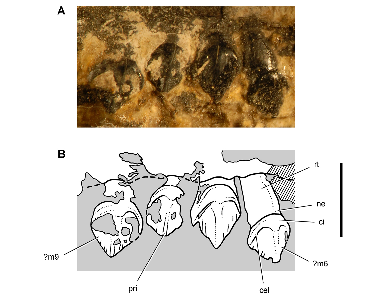
Maxillary dentition of the heterodontosaurid Tianyulong confuciusi from the Lower Cretaceous Jehol Group of China. Right maxillary teeth ?6-9 in lateral view (IVPP V17090). Hatching indicates broken bone; dashed lines indicate estimated edges; tone indicates matrix. Scale bar equals 3 mm. Abbreviations: cel cingular ectoloph ci cingulum m6 9 maxillary tooth 6 9 ne neck pri primary ridge rt root.
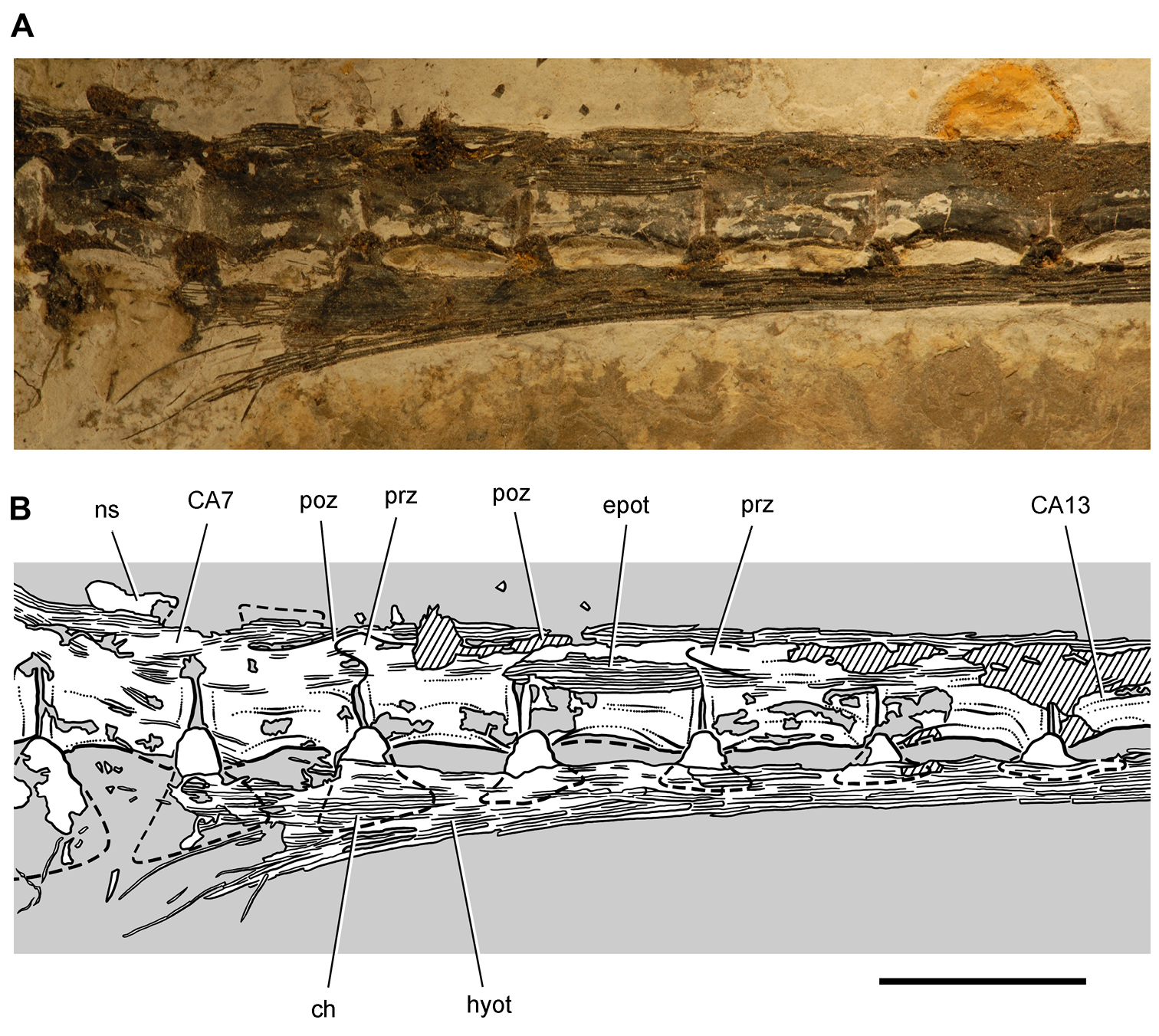
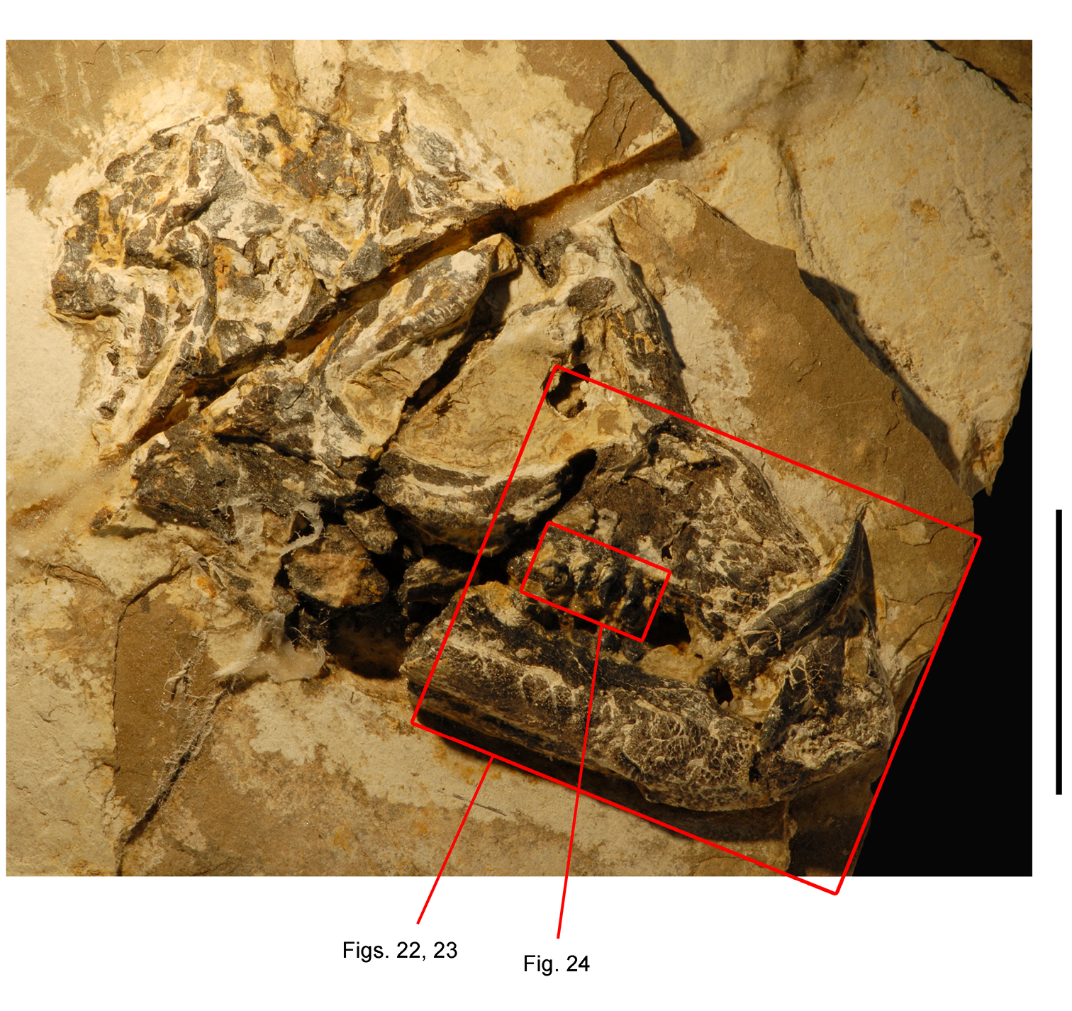
Tail of the heterodontosaurid Tianyulong confuciusi from the Lower Cretaceous Jehol Group of China. Anterior caudal vertebrae and ossified tendons in left lateral view, from the seventh to the thirteenth caudal vertebra. Hatching indicates broken bone; dashed lines indicate estimated edges; tone indicates matrix. Scale bar equals 1 cm. Abbreviations: CA caudal ch chevron epot epaxial ossified tendons hyot hypaxial ossified tendons ns neural spine poz postzygapophyses prz prezygapohpysis.
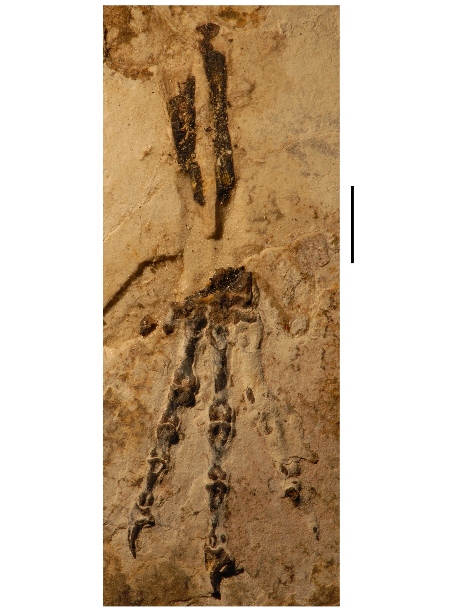
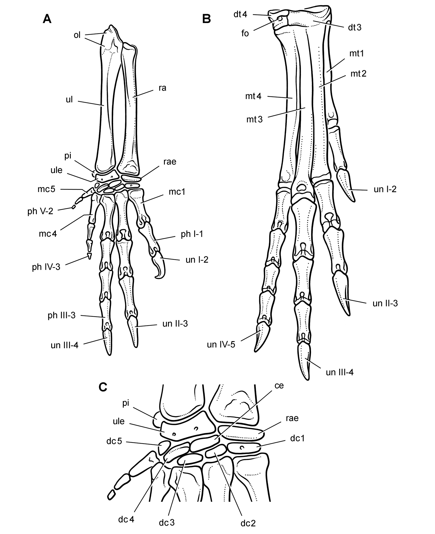
Forearm and manus of the heterodontosaurid Tianyulong confuciusi from the Lower Cretaceous Jehol Group of China. Left ulna, radius and manus for the most part in anterior or ventral view. Scale bar equals 5 mm.
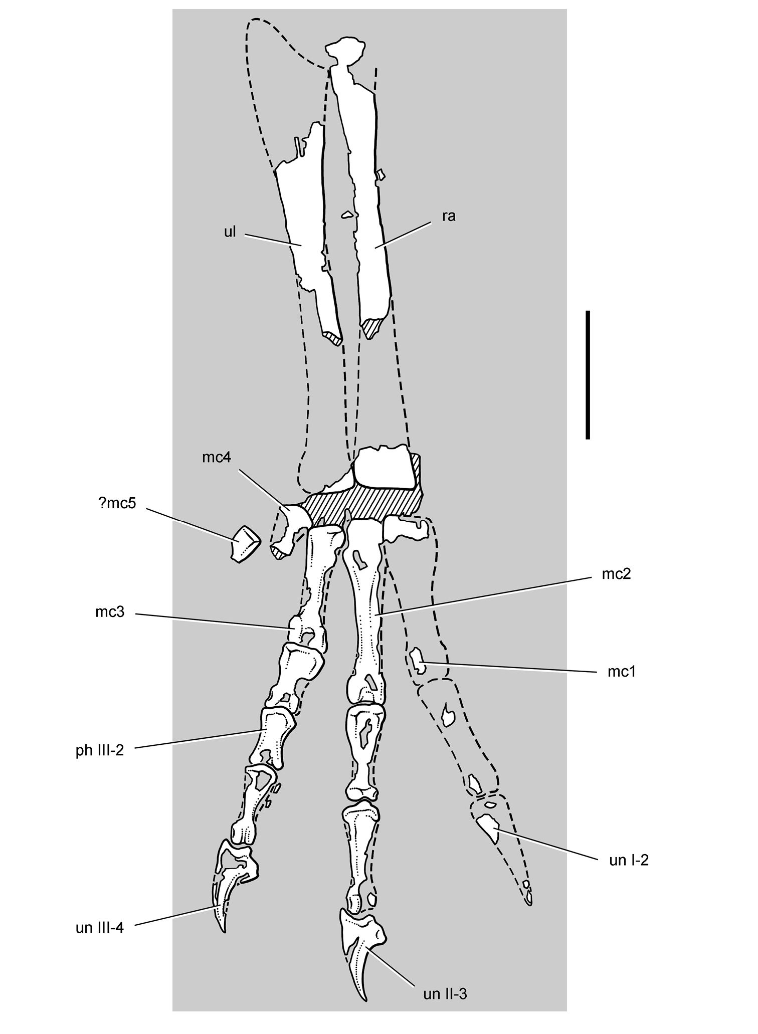
Forearm and manus of the heterodontosaurid Tianyulong confuciusi from the Lower Cretaceous Jehol Group of China. Left ulna, radius and manus for the most part in anterior or ventral view. Hatching indicates broken bone; dashed lines indicate estimated edges; tone indicates matrix. Scale bar equals 5 mm. Abbreviations: I-III digits I-III mc1-5 metacarpal 1-5 ph phalanx ra radius ul ulna un ungual.
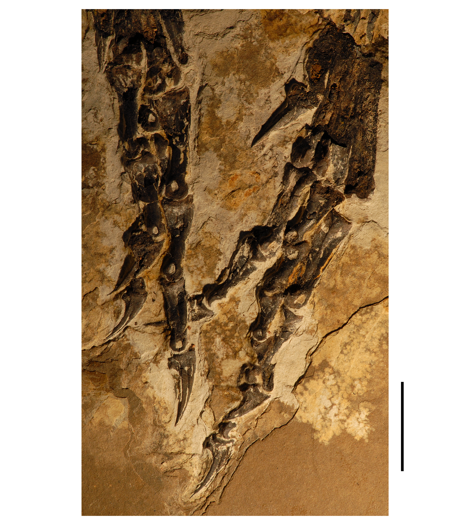
Pes of the heterodontosaurid Tianyulong confuciusi from the Lower Cretaceous Jehol Group of China. Left pedal phalanges (left) in dorsal and medial views; right pedal phalanges (right) in ventral and lateral views. Scale bar equals 1 cm.
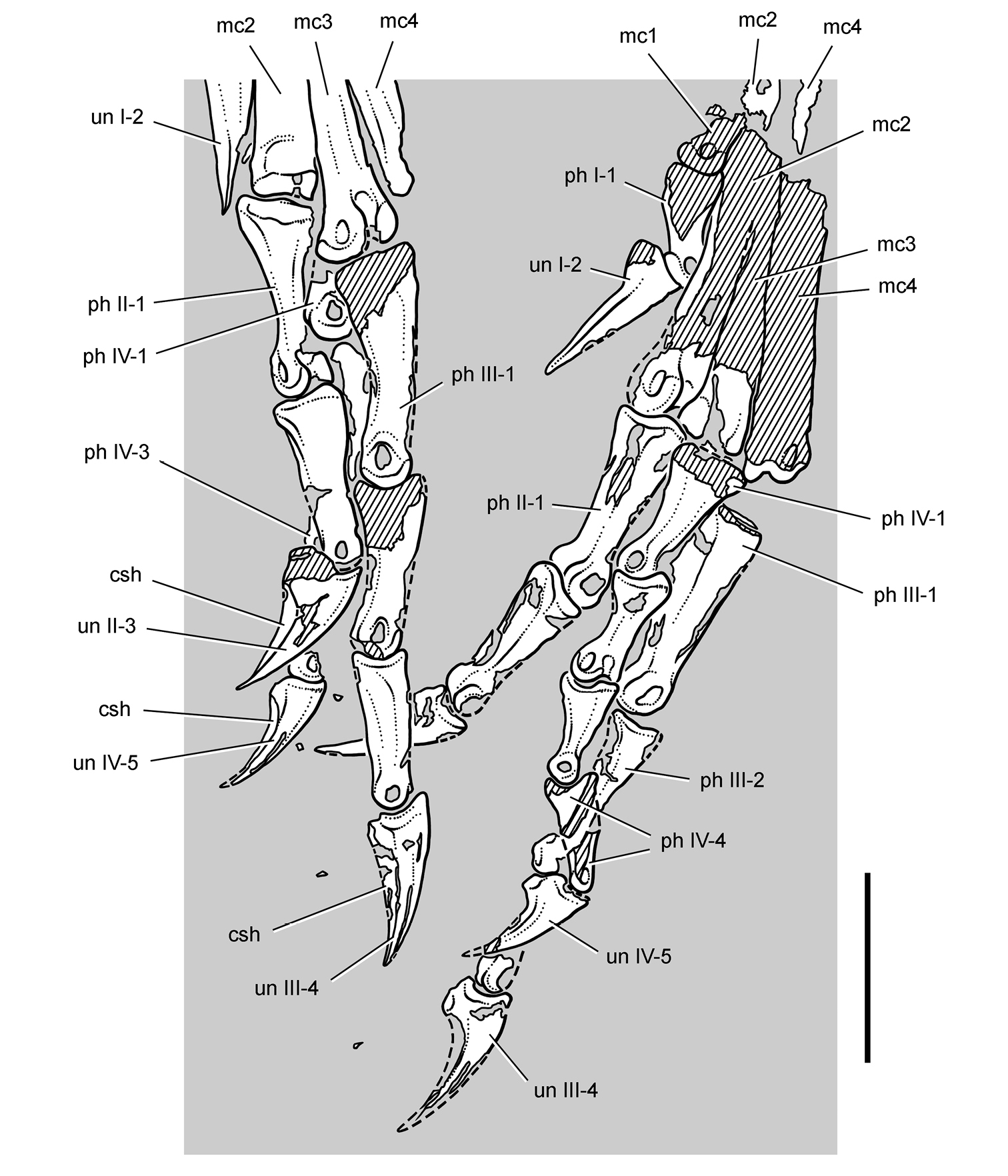
Pes of the heterodontosaurid Tianyulong confuciusi from the Lower Cretaceous Jehol Group of China. Left pedal phalanges (left) in dorsal and medial views; right pedal phalanges (right) in ventral and lateral views. Hatching indicates broken bone; dashed lines indicate estimated edges; tone indicates matrix. Scale bar equals 1 cm. Abbreviations: I-IV digits I-IV csh claw sheath mc1-4 metacarpal 1-4 ph phalanx un ungual.
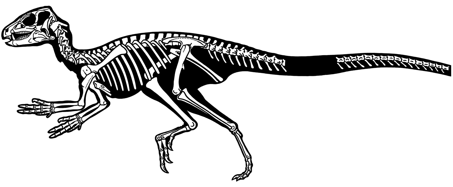
Skeleton of the heterodontosaurid Tianyulong confuciusi from the Lower Cretaceous Jehol Group of China. Silhouette skeletal reconstruction in lateral view showing preserved bones and integumental fibers (based on IVPP V17090). Distal most caudal vertebrae unknown.
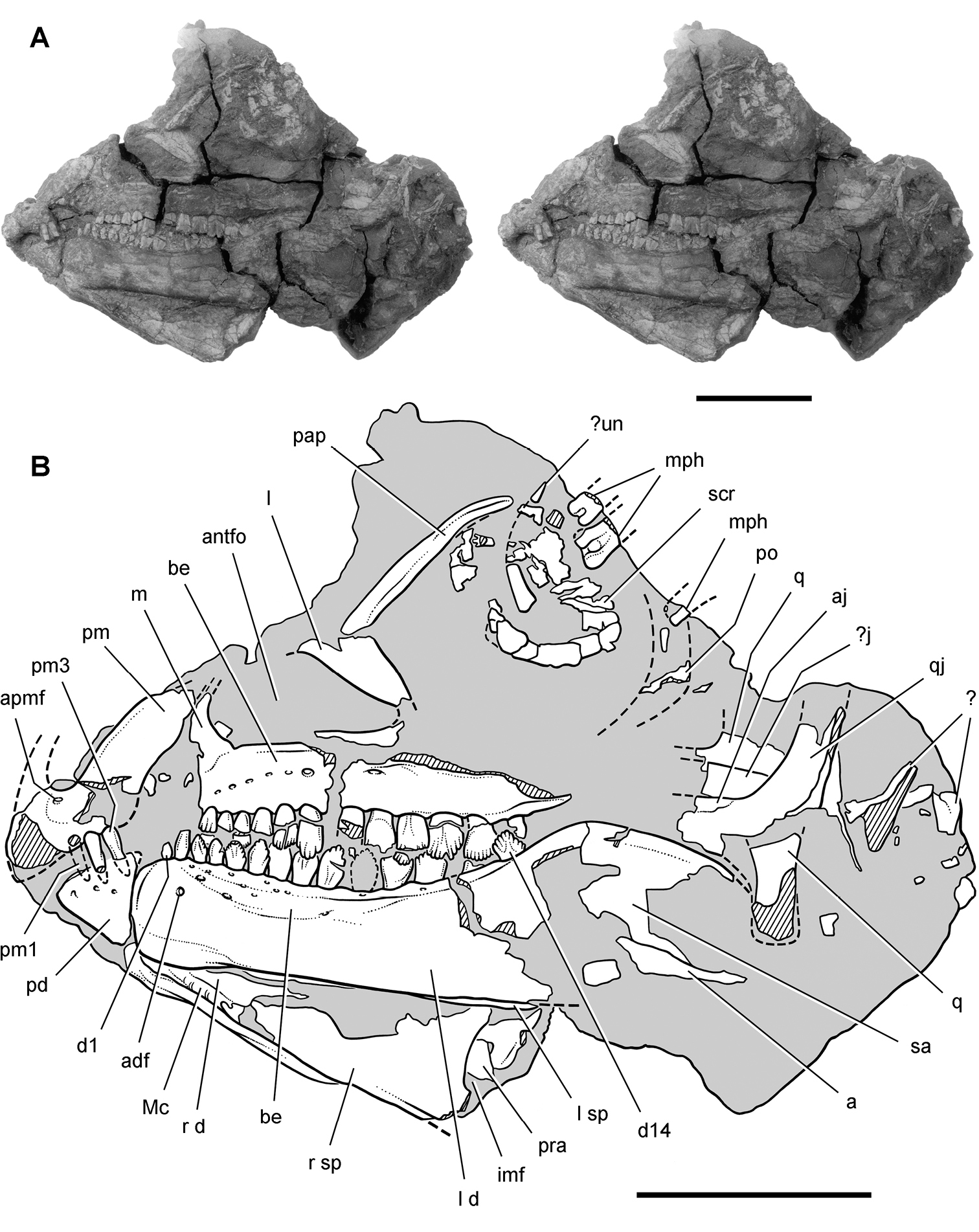
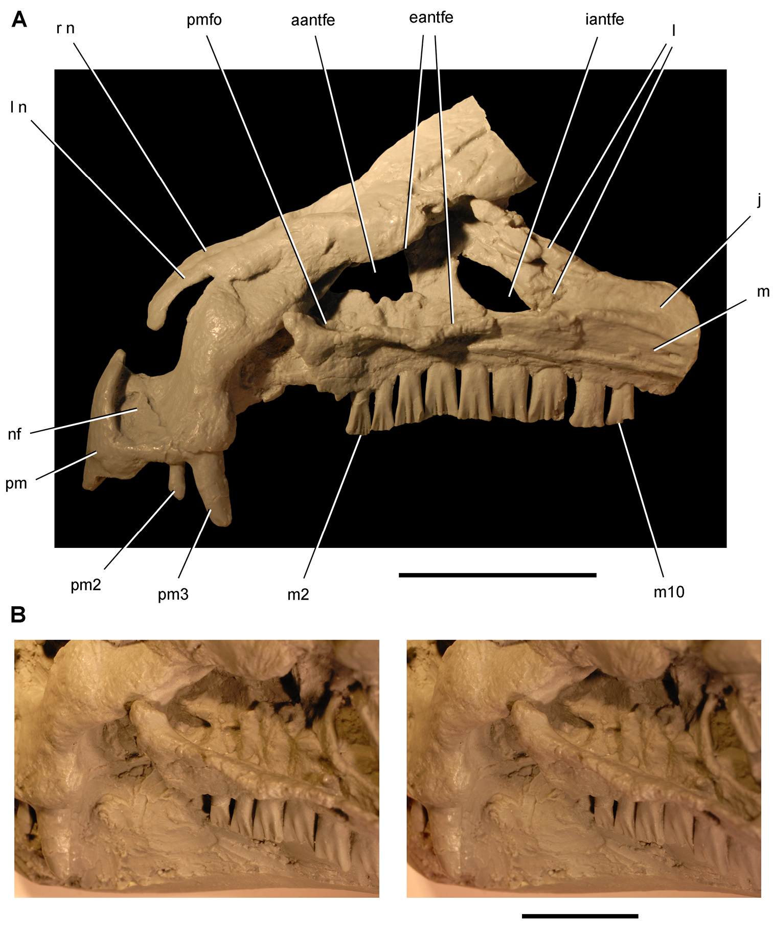
Skull of Abrictosaurus consors from the Lower Jurassic Upper Elliot Formation of South Africa. Stereopair (A) and line drawing (B) of skull in left lateral view (NHMUK RU B54). Hatching indicates broken bone; dashed lines indicate estimated edges; tone indicates matrix. Scale bars equal 2 cm. Abbreviations: a angular adf anterior dentary foramen aj articular scar for the jugal antfo antorbital fossa apmf anterior premaxillary foramen be buccal emargination d dentary d1, d14 dentary tooth 1, 14 imf internal mandibular fenestra j jugal l lacrimal or left m maxilla Mc Meckel’s canal mph manual phalanges pap palpebral pd predentary pm premaxilla pm1, 3 premaxillary tooth 1, 3 po postorbital pra prearticular q quadrate qj quadratojugal r right sa surangular scr scleral ring sp splenial un ungual.
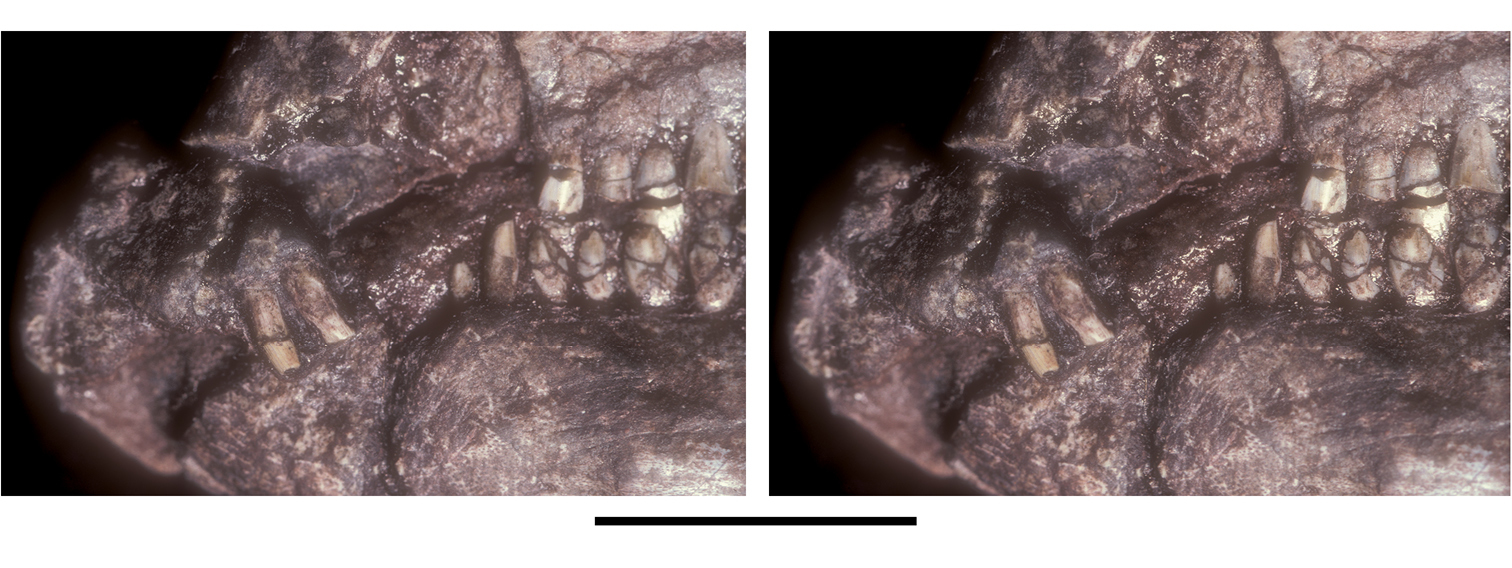
Snout of Abrictosaurus consors from the Lower Jurassic Upper Elliot Formation of South Africa. Stereopair of anterior end of skull in left lateral view (NHMUK RU B54). Scale bar equals 5 mm.
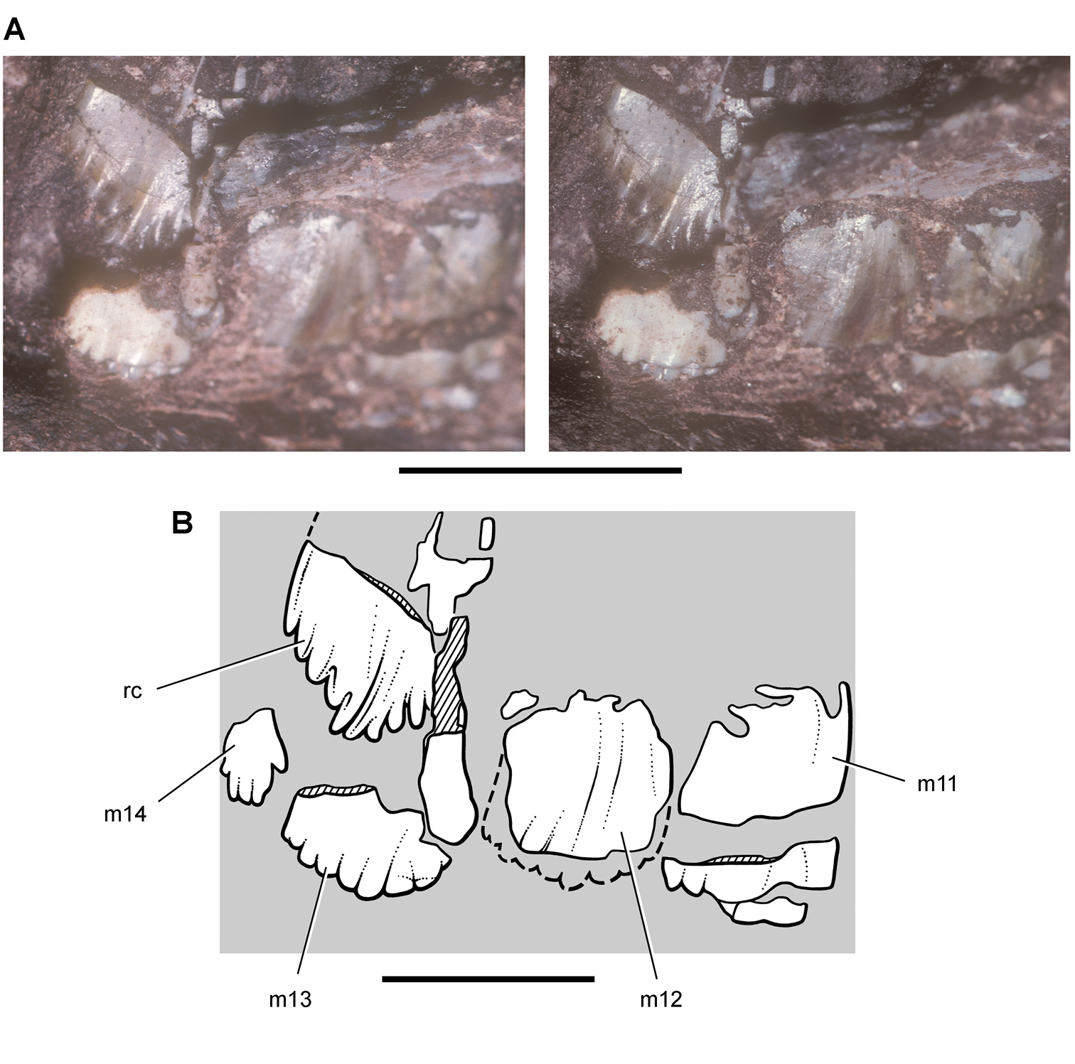
Maxillary teeth of Abrictosaurus consors from the Lower Jurassic Upper Elliot Formation of South Africa. Stereopair (A) and line drawing (B) of replacing crown medial to posterior right maxillary tooth in medial (lingual) view (NHMUK RU B54). Hatching indicates broken bone; dashed lines indicate estimated edges; tone indicates matrix. Scale bar in A equals 5 mm; scale bar in B equals 3 mm. Abbreviations: m11-14 maxillary tooth 11–14 rc replacement crown.
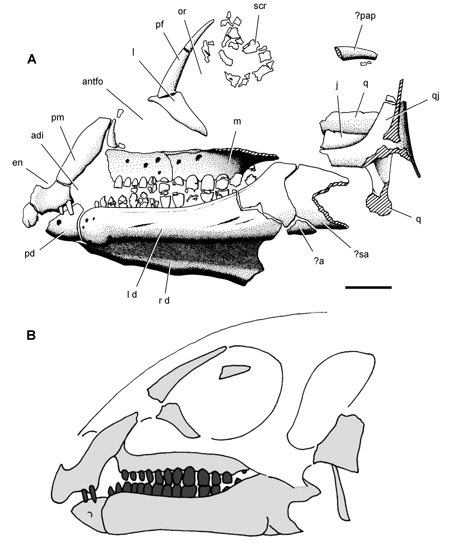
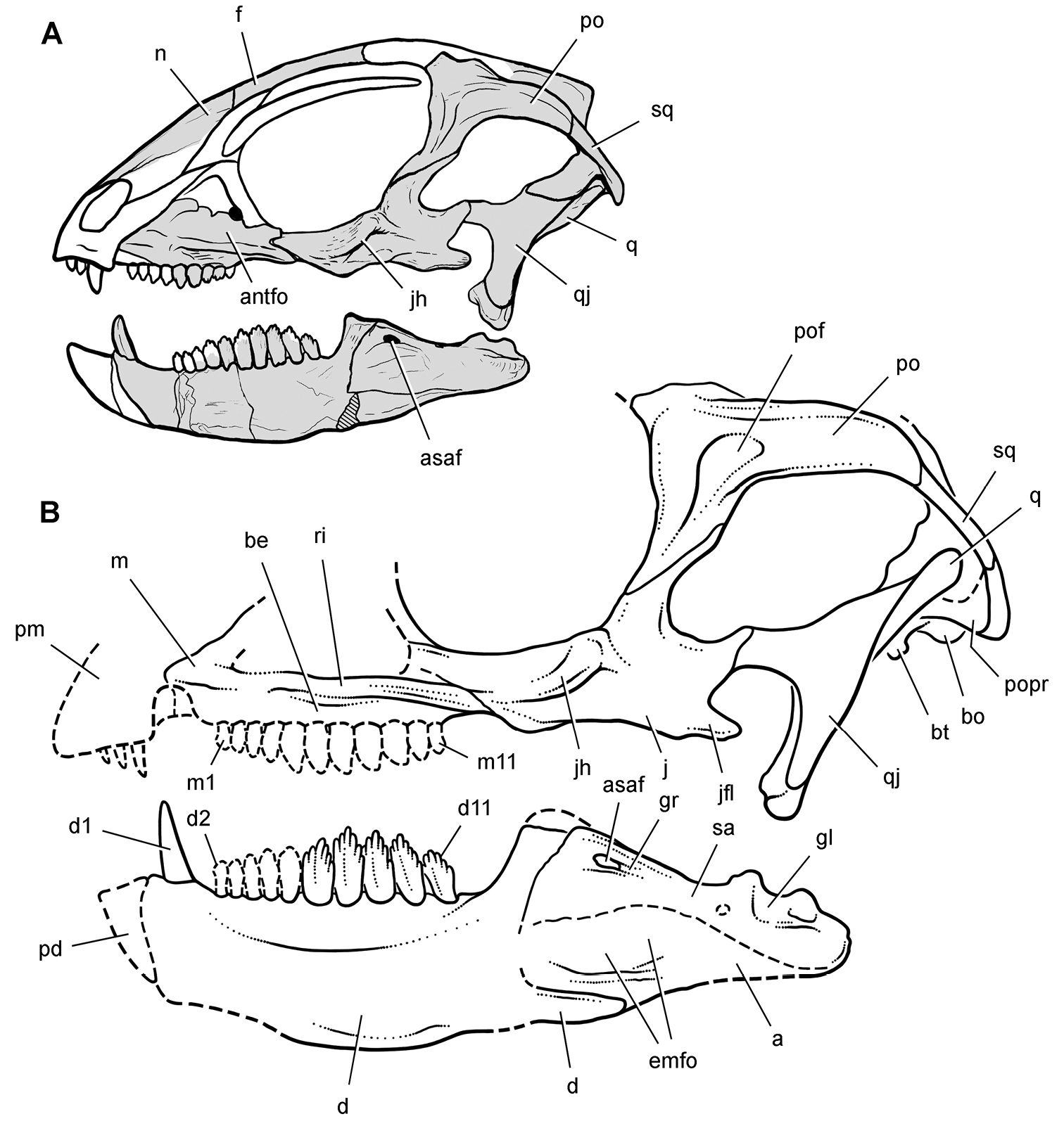
Skull of Abrictosaurus consors from the Lower Jurassic Upper Elliot Formation of South Africa. A Skull bones of NHMUK RU B54 in left lateral view as initially identified (from Thulborn 1974). B Diagrammatic skull reconstruction in left lateral view based on NHMUK RU B54 (from Norman et al. 2011). Scale bar equals 1 cm in A. Abbreviations: a angular adi arched diastema antfo antorbital fossa d dentary en external nares j jugal l lacrimal or left m maxilla or orbit pap palpebral pd predentary pf prefrontal pm premaxilla q quadrate qj quadratojugal r right sa surangular scr scleral ring.
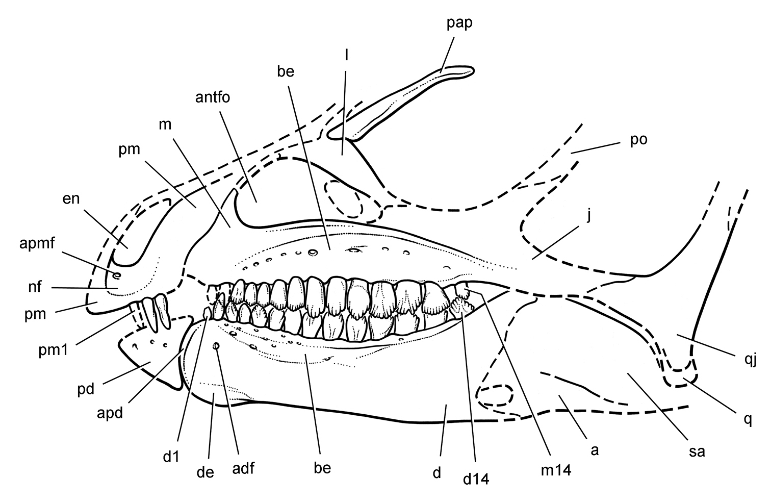
Skull of Abrictosaurus consors from the Lower Jurassic Upper Elliot Formation of South Africa. Skull reconstruction in left lateral view based on NHMUK RU B54. Dashed lines indicate estimated edges and sutures. Abbreviations: a angular adf anterior dentary foramen antfo antorbital fossa apd articular surface for the predentary apmf anterior premaxillary foramen be buccal emargination d dentary d1, d14 dentary tooth 1, 14 de dentary expansion en external nares j jugal l lacrimal m maxilla m14 maxillary tooth 14 nf narial fossa pap palpebral pd predentary pm premaxilla pm1 premaxillary tooth 1 po postorbital q quadrate qj quadratojugal sa surangular.
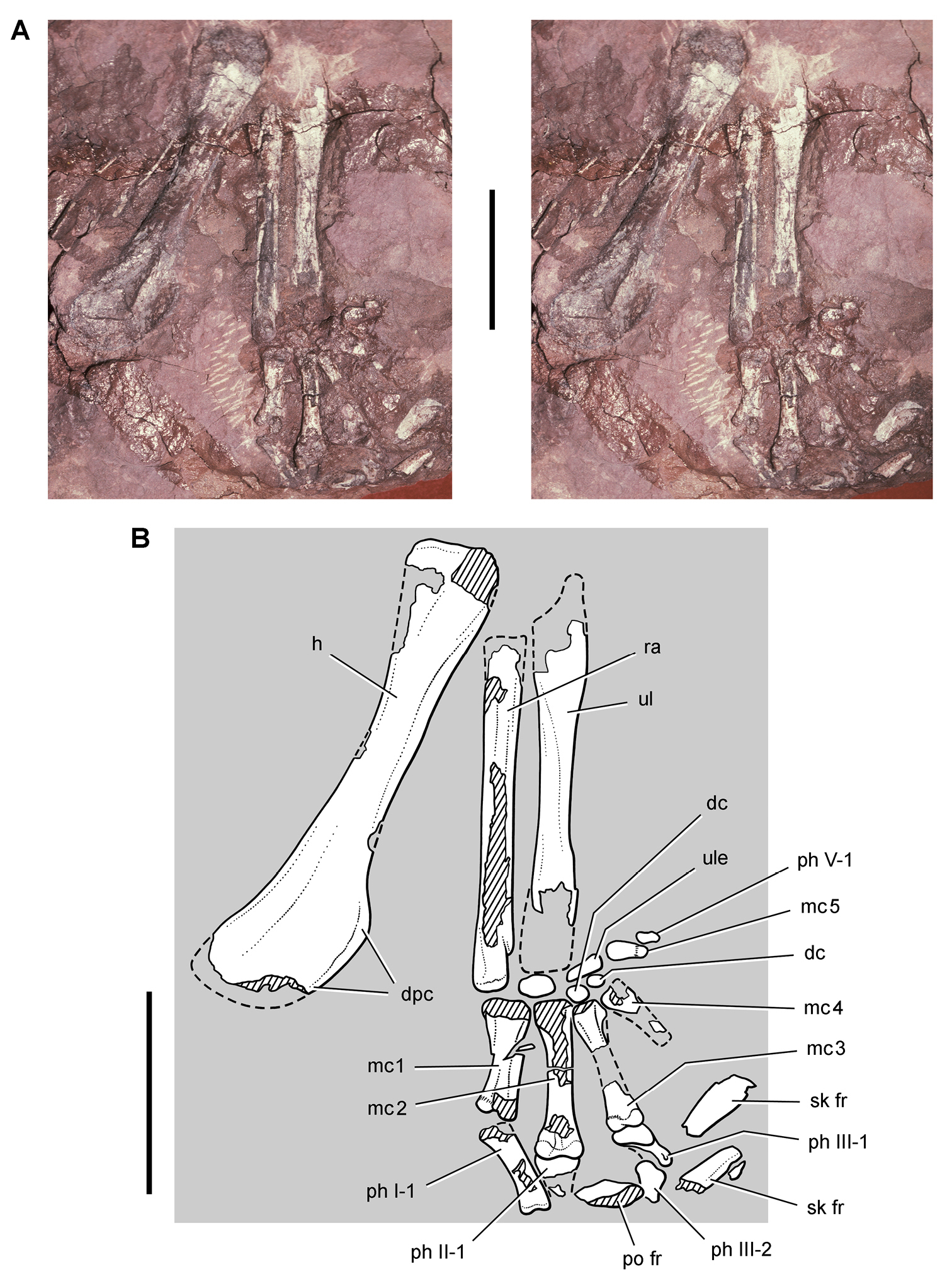
Forelimb of Abrictosaurus consors from the Lower Jurassic Upper Elliot Formation of South Africa. Stereopair (A) and line drawing (B) of the right forelimb in anterior and ventral views (NHMUK RU B54). Hatching indicates broken bone; dashed lines indicate estimated edges; tone indicates matrix. Scale bars equal 2 cm in A and B. Abbreviations: dc distal carpal dpc deltopectoral crest fr fragment h humerus mc1-5 metacarpals 1-5 ph I-1 phalanx 1 of manual digit I ph II-1 phalanx 1 of manual digit II ph III-1 phalanx 1 of manual digit III ph III-2 phalanx 2 of manual digit III ph V-1 phalanx 1 of manual digit V po postorbital ra radius sk skull ul ulna ule ulnare.
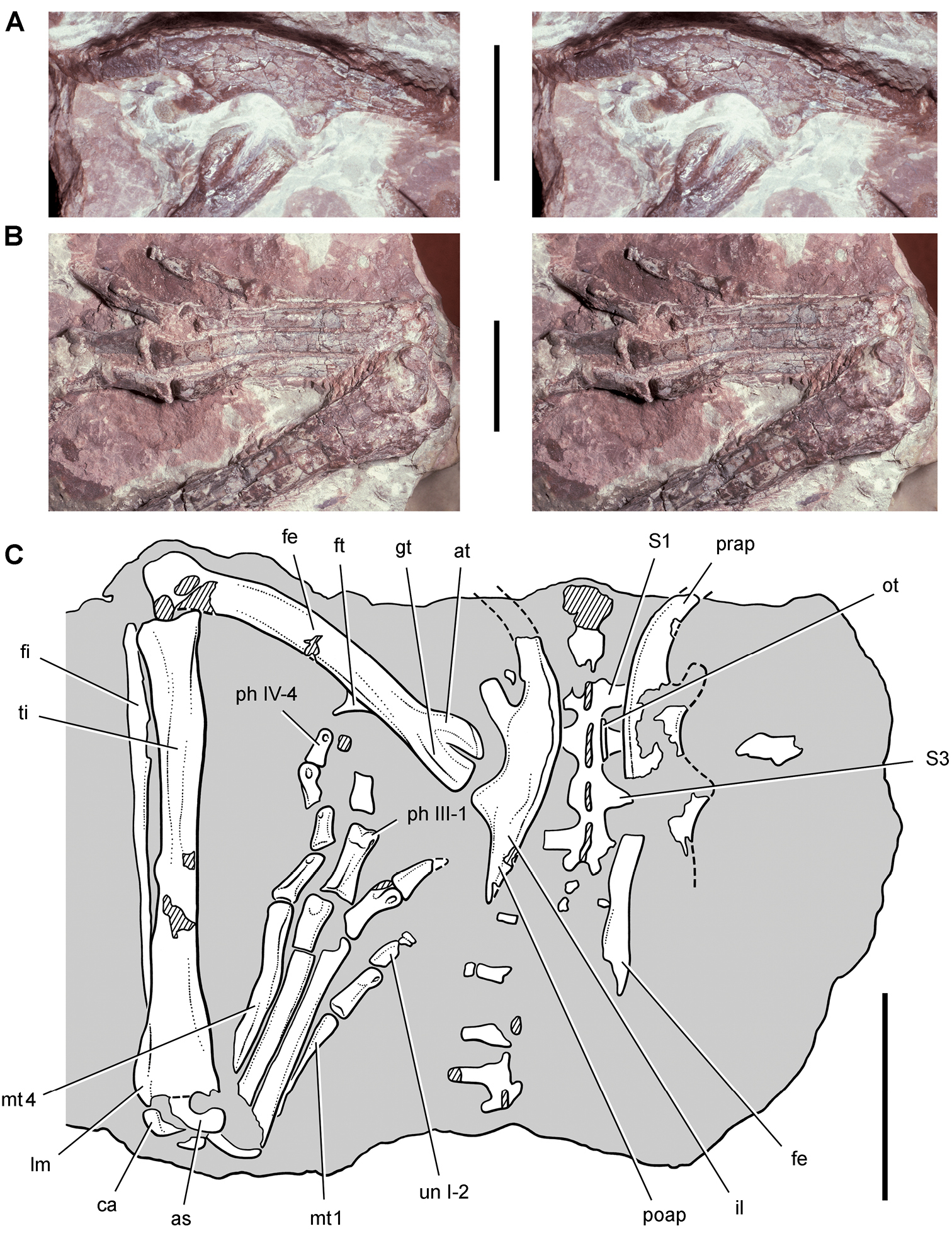
Ilium and hindlimb of Abrictosaurus consors from the Lower Jurassic Upper Elliot Formation of South Africa. Stereopairs of the left ilium in lateral view (A) and left pes in dorsal view (B) and line drawing of the sacrum, ilia and left hindlimb mostly in dorsal view (C) (NHMUK RU B54). Hatching indicates broken bone; dashed lines indicate estimated edges; tone indicates matrix. Scale bars equal 2 cm in A and B, 4 cm in C. Abbreviations: I, III, IV digits I, III, IV as astragalus at anterior trochanter ca calcaneum fe femur fi fibula ft fourth trochanter gt greater trochanter il ilium lm lateral malleolus mt1, 4 metatarsals 1, 4 ot ossified tendon ph phalanx poap postacetabular process prap preacetabular process S1, 3 sacral vertebra 1, 3 ti tibia un ungual.
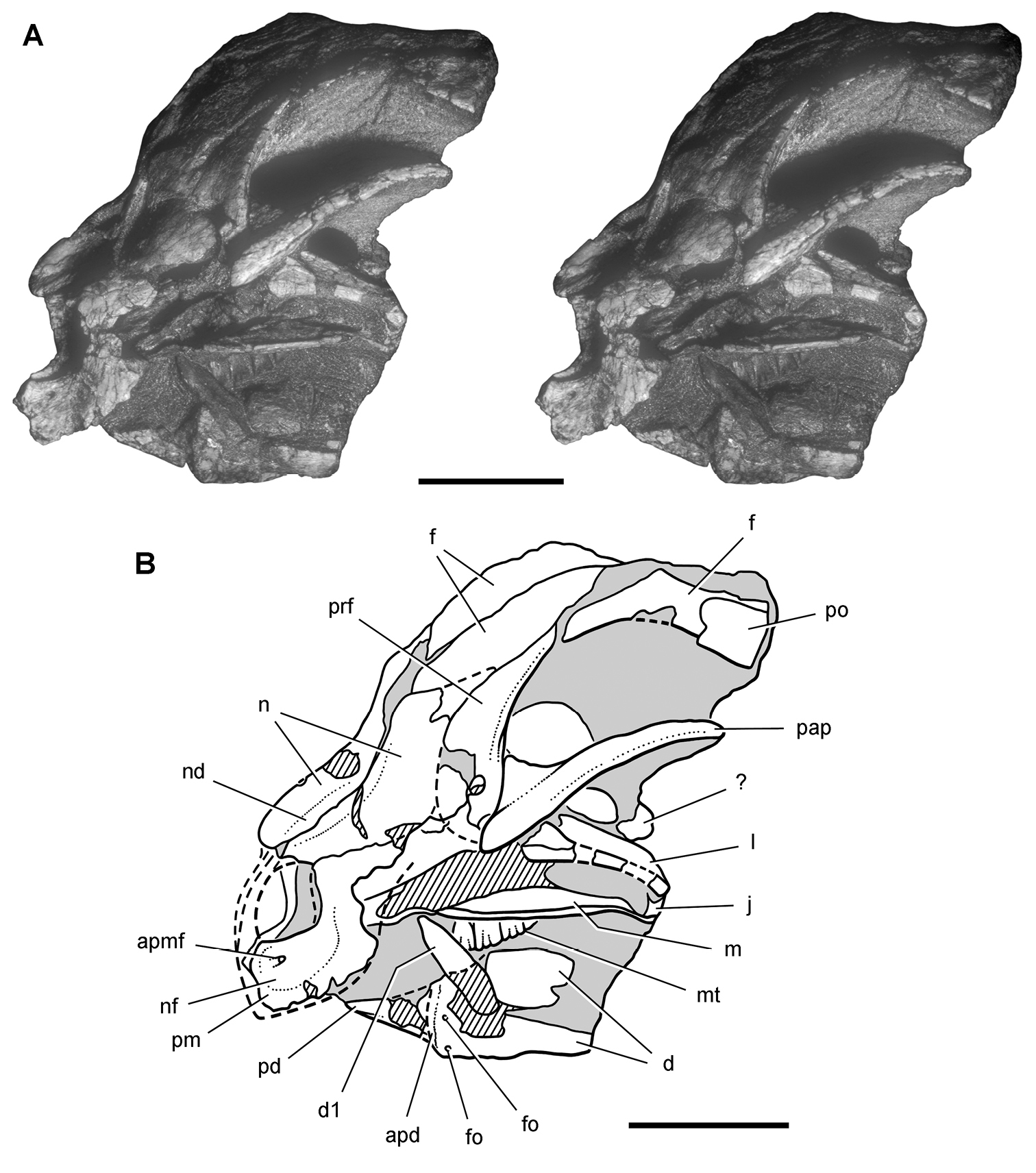
Snout of the heterodontosaurid Heterodontosaurus tucki from from the Lower Jurassic Upper Elliot and Clarens formations of South Africa. Anterior one-half of a juvenile skull (SAM-PK-K10487). Stereopair (A) and line drawing (B) in anterolateral view. Hatching indicates broken bone; dashed lines indicate estimated edges; tone indicates matrix. Scale bars equal 1 cm in A and B. Abbreviations: apd articular surface for predentary apmf anterior premaxillary foramen d dentary d1 dentary tooth 1 f frontal fo foramen j jugal l lacrimal m maxilla mt maxillary teeth n nasal nd nasal depression nf narial fossa pap palpebral pd predentary pm premaxilla po postorbital prf prefrontal.
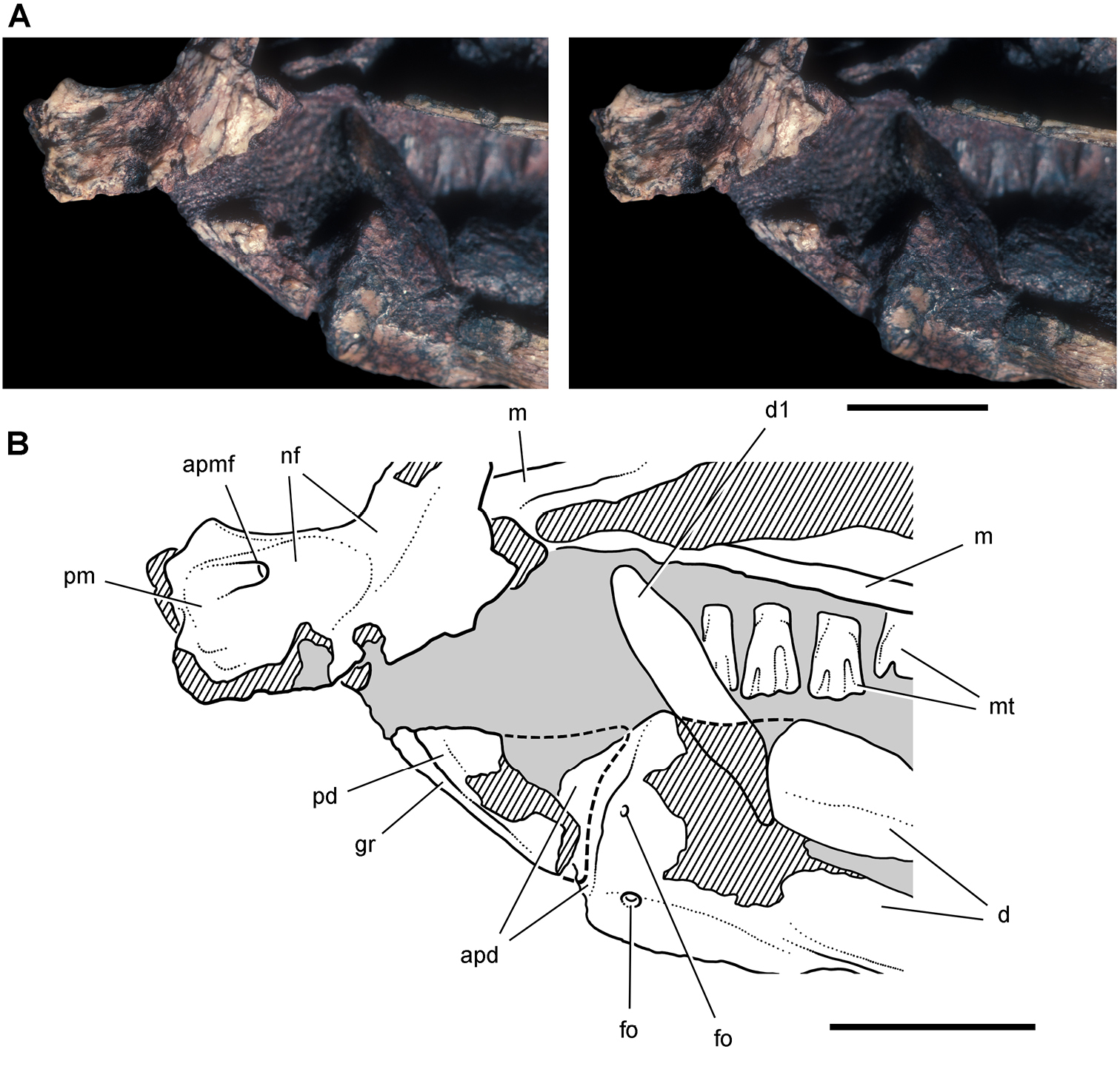
Snout end of Heterodontosaurus tucki from from the Lower Jurassic Upper Elliot and Clarens formations of South Africa. Close-up view of the anterior end of a juvenile skull (SAM-PK-K10487). Stereopair (A) and line drawing (B) in left lateral view. Hatching indicates broken bone; dashed lines indicate estimated edges; tone indicates matrix. Scale bars equal 5 mm in A and B. Abbreviations: apd articular surface for the predentary apmf anterior premaxillary foramen d dentary d1 dentary tooth 1 fo, foramen gr groove m maxilla mt maxillary teeth nf narial fossa pd predentary pm premaxilla.
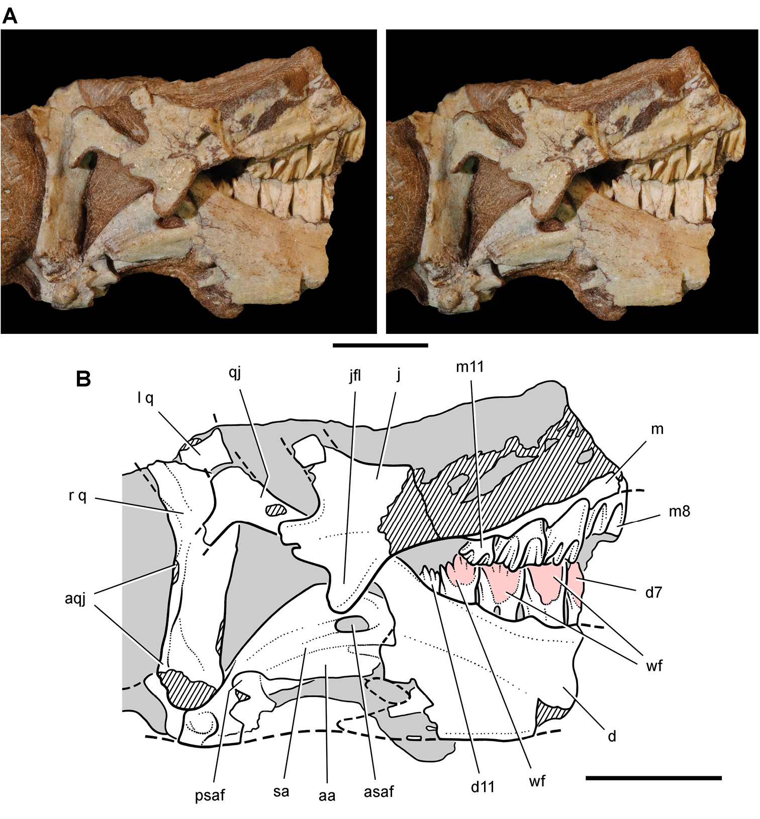
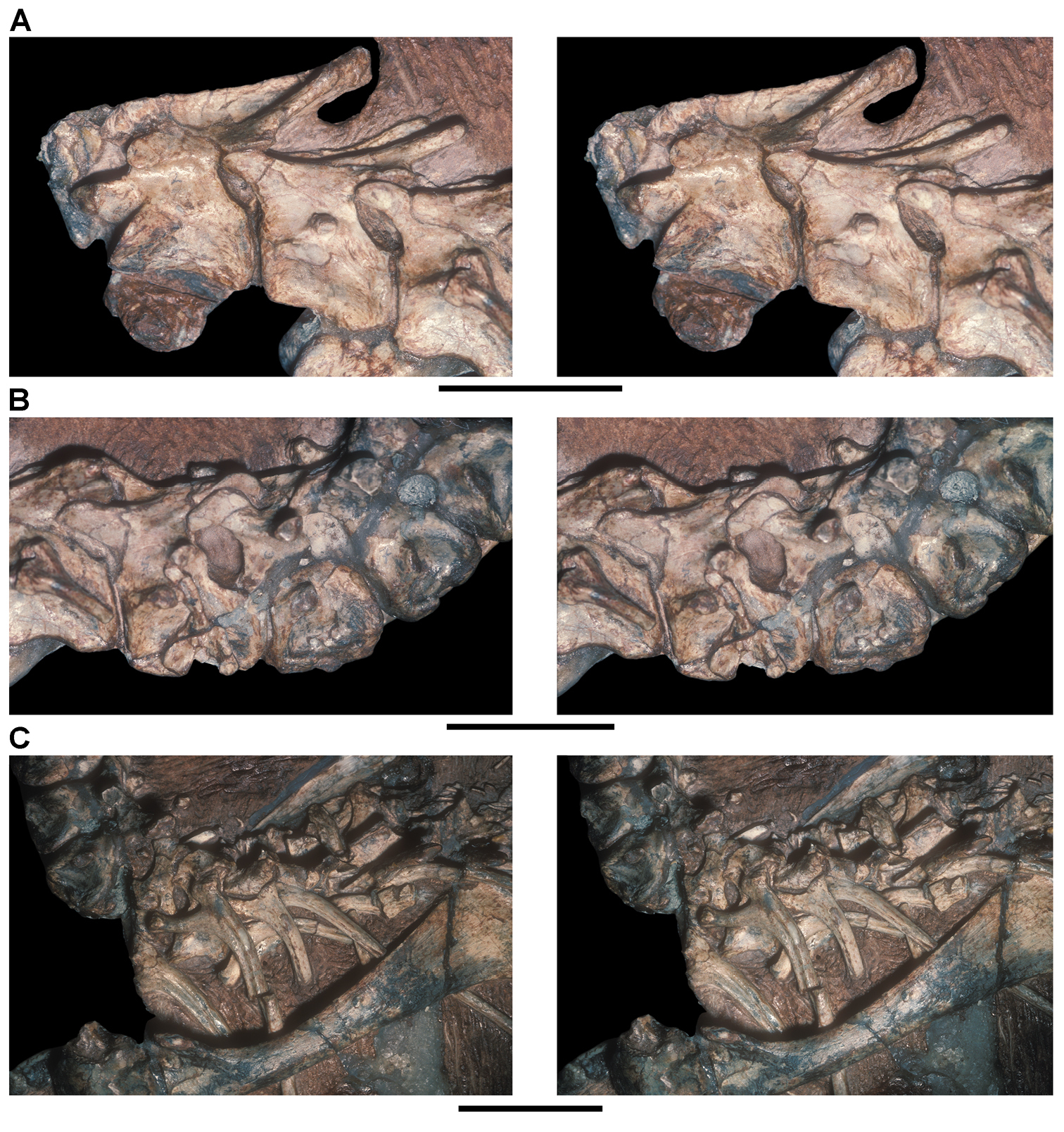
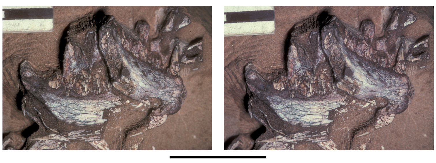
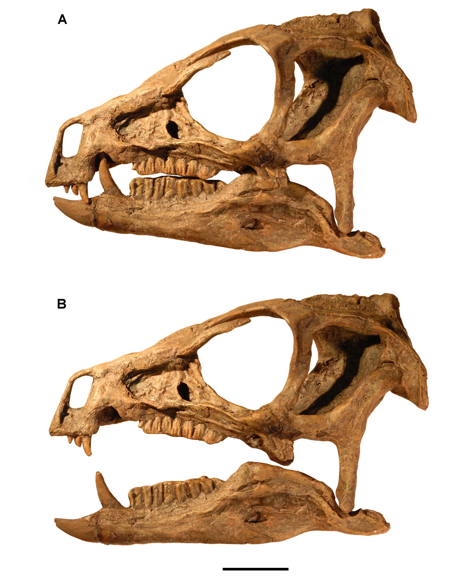
Posterior end of the skull of Heterodontosaurus tucki from from the Lower Jurassic Upper Elliot and Clarens formations of South Africa. Posterior portion of a juvenile skull (AMNH 24000). Stereopair (A) and line drawing (B) in right lateral view. Hatching indicates broken bone; dashed lines indicate estimated edges; grey tone indicates matrix; pink tone indicates wear facets. Scale bars equal 1 cm in A and B. Abbreviations: aa articular surface for angular aqj articular surface for quadratojugal asaf anterior surangular foramen d dentary d7, 11 dentary tooth 7, 11 j jugal jfl jugal flange l left m maxilla m8, 11 maxillary tooth 8, 11 psaf posterior surangular foramen q quadrate qj quadratojugal r right sa surangular wf wear facet.
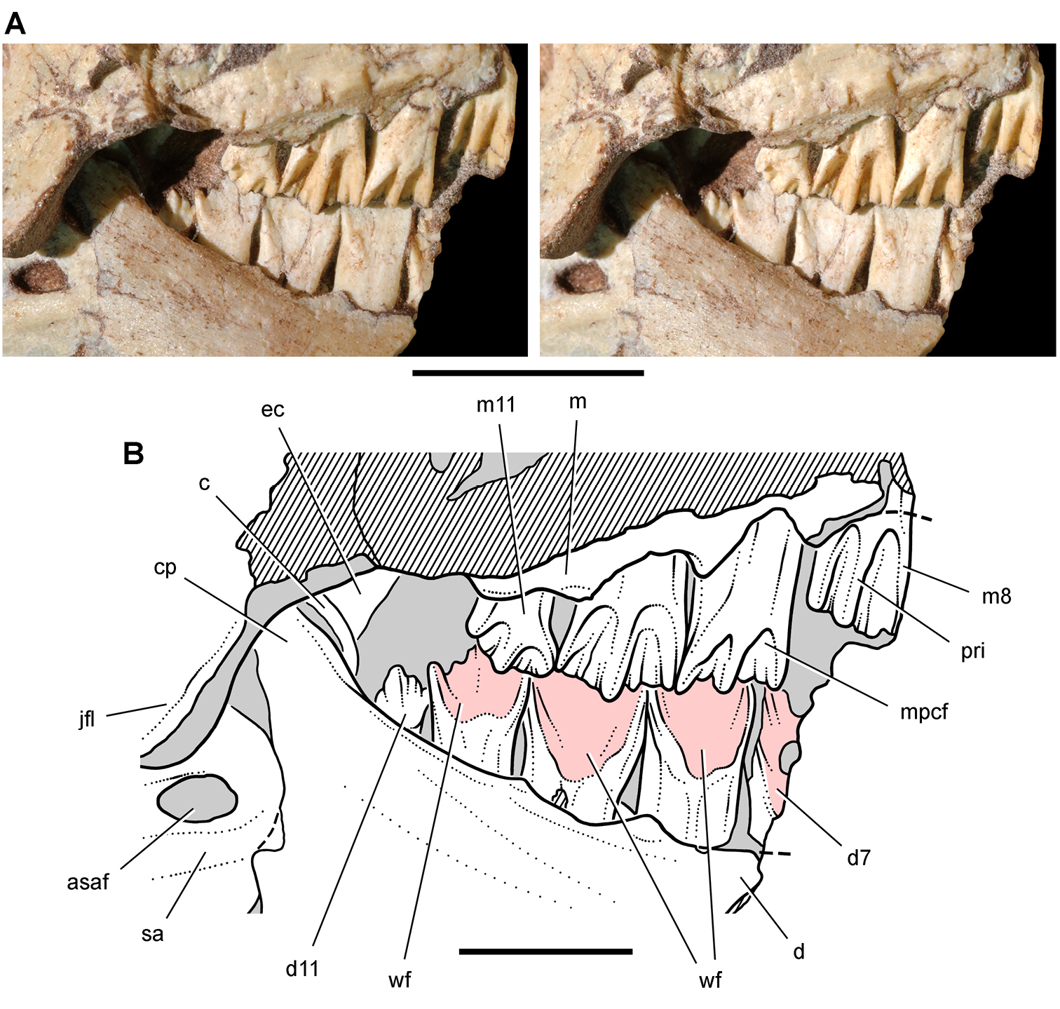
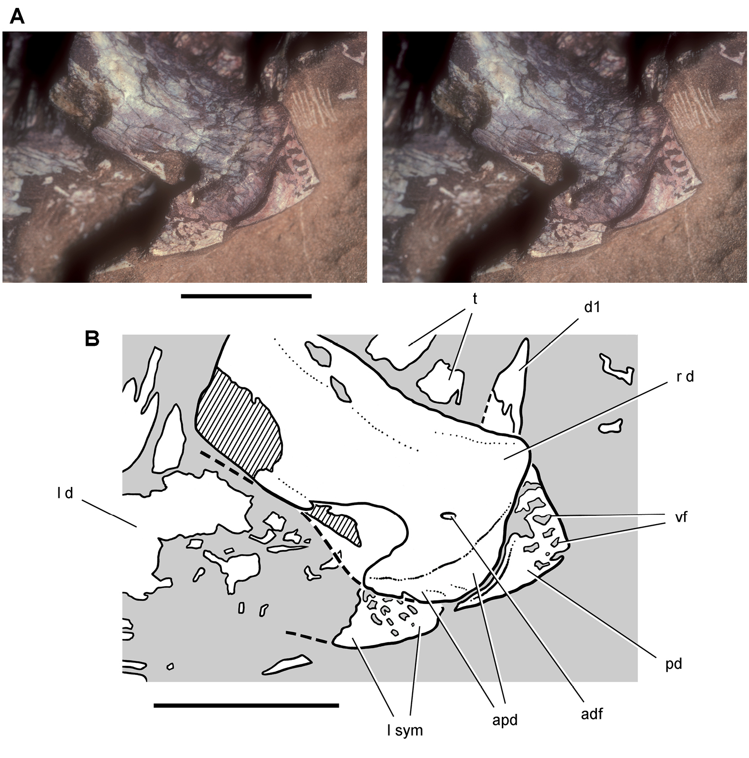
Posterior dentition of Heterodontosaurus tucki from from the Lower Jurassic Upper Elliot and Clarens formations of South Africa. Tooth wear and replacement in posterior maxillary and dentary teeth of a juvenile skull (AMNH 24000). Stereopair (A) and line drawing (B) in right lateral view. Hatching indicates broken bone; dashed lines indicate estimated edges; grey tone indicates matrix; pink tone indicates wear facets. Scale bars equal 2 cm in A and 1 cm in B. Abbreviations: asaf anterior surangular foramen c coronoid cp coronoid process d dentary d7, 11 dentary tooth 7, 11 ec ectopterygoid jfl jugal flange m maxilla m8, 11 maxillary tooth 8, 11 mpcf mesial paracingular fossa pri primary ridge sa surangular wf wear facet.
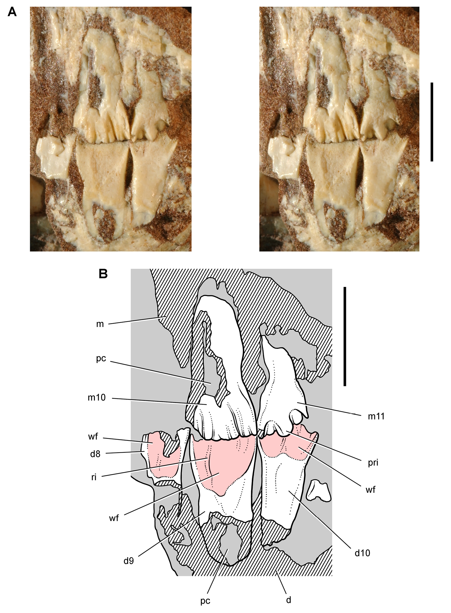
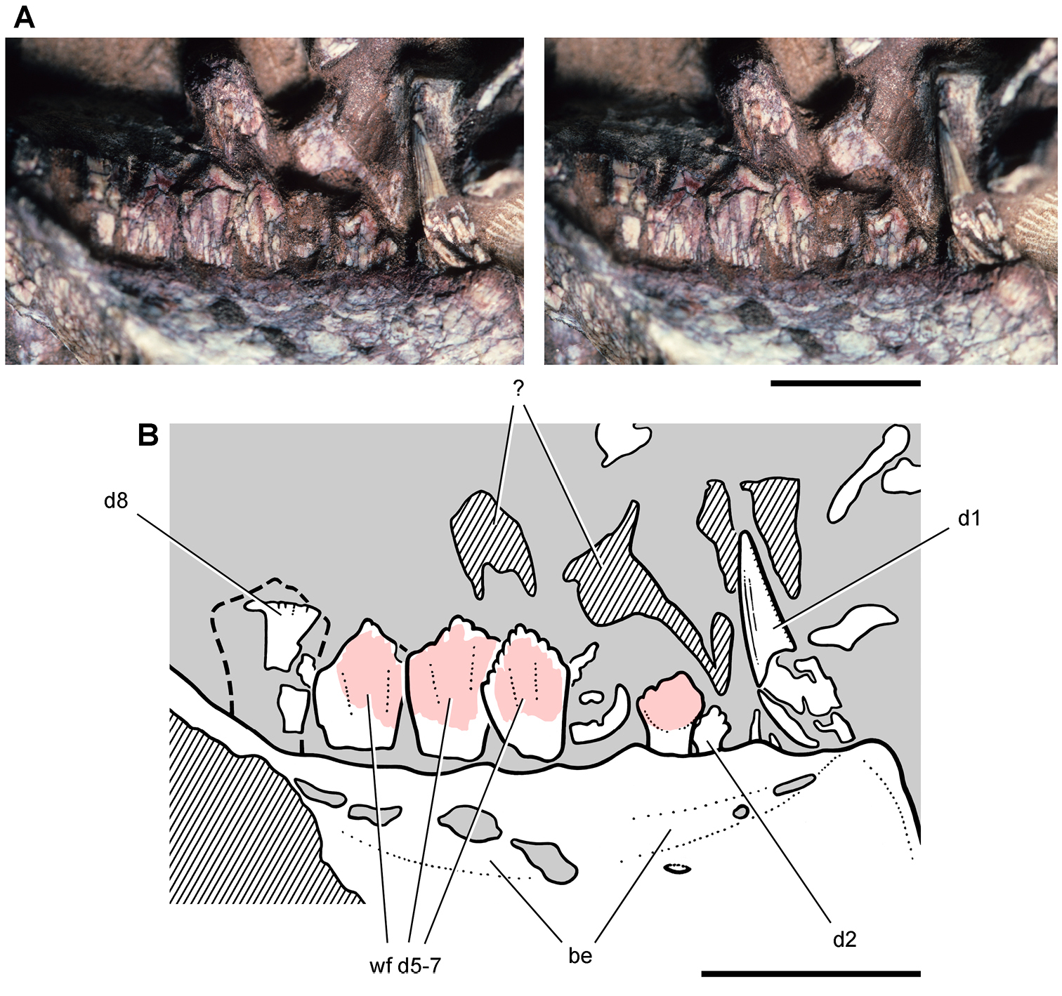
Posterior dentition of Heterodontosaurus tucki from the Lower Jurassic Upper Elliot and Clarens formations of South Africa. Tooth wear and replacement in posterior maxillary and dentary teeth in a juvenile skull (AMNH 24000). Stereopair (A) and line drawing (B) in left lateral view. Hatching indicates broken bone; grey tone indicates matrix; pink tone indicates wear facets. Scale bars equal 5 mm in A and B. Abbreviations: d dentary d8-10 dentary tooth 8–10 m maxilla m10, 11 maxillary tooth 10, 11 pc pulp cavity pri primary ridge ri ridge wf wear facet.
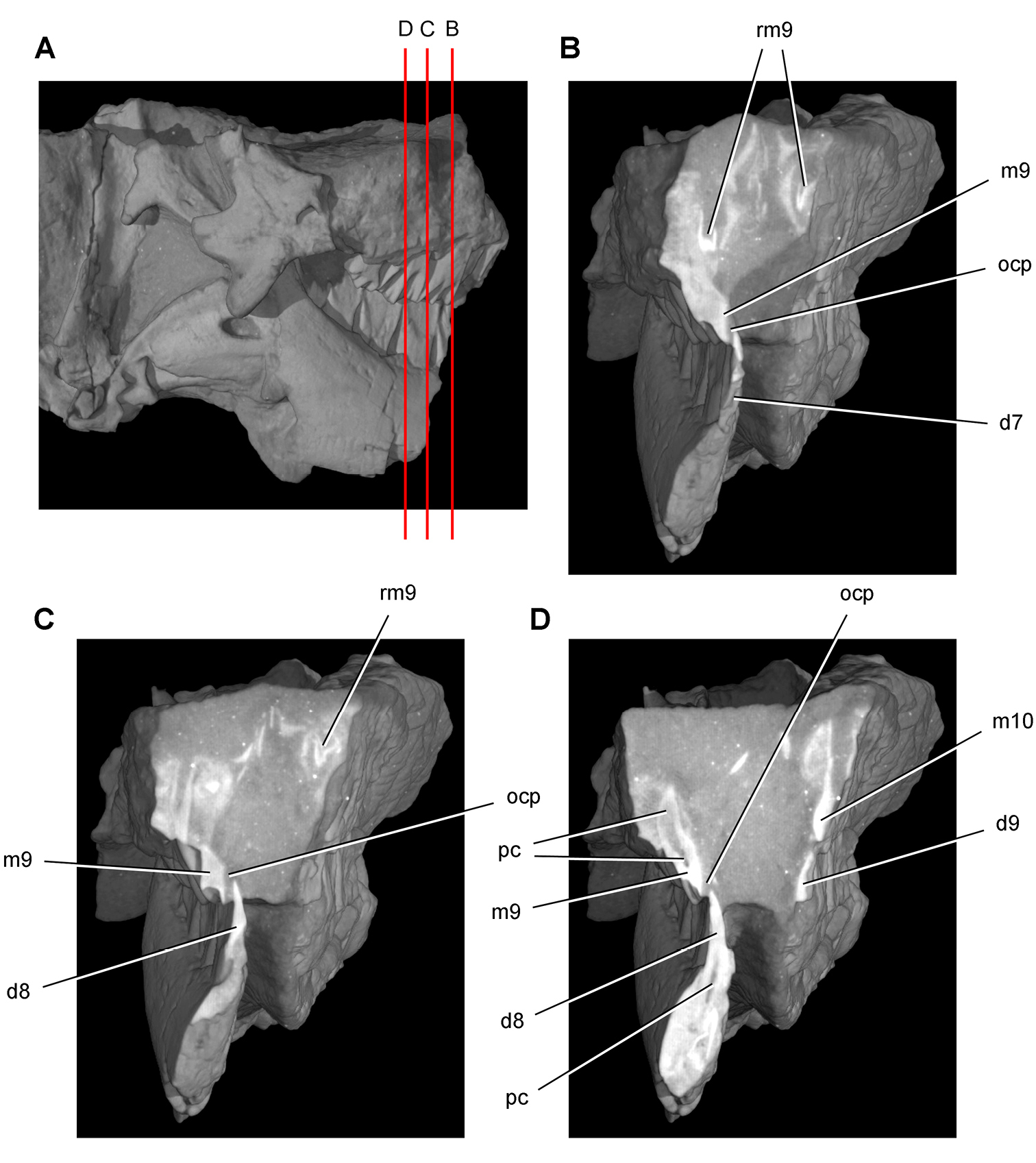
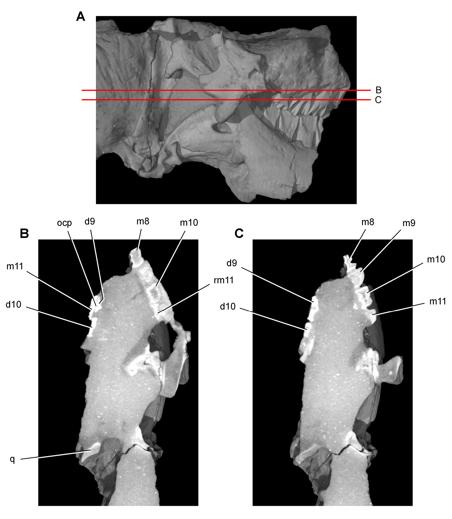
Tooth structure, occlusion, and replacement in Heterodontosaurus tucki from the Lower Jurassic Elliot and Clarens Formations of South Africa. Successive coronal computed-tomographic sections in cutaway view of a subadult skull (AMNH 24000). A Posterior portion of skull in right lateral view showing the location of coronal cross-sections B Cross-section through maxillary tooth 9 and dentary tooth 7 C, D Cross-sections through maxillary tooth 9 and dentary tooth 8. Abbreviations: d7-9 dentary teeth 7-9 m9, 10 maxillary tooth 9, 10 ocp occlusal plane pc pulp cavity rm9 replacement maxillary tooth 9.
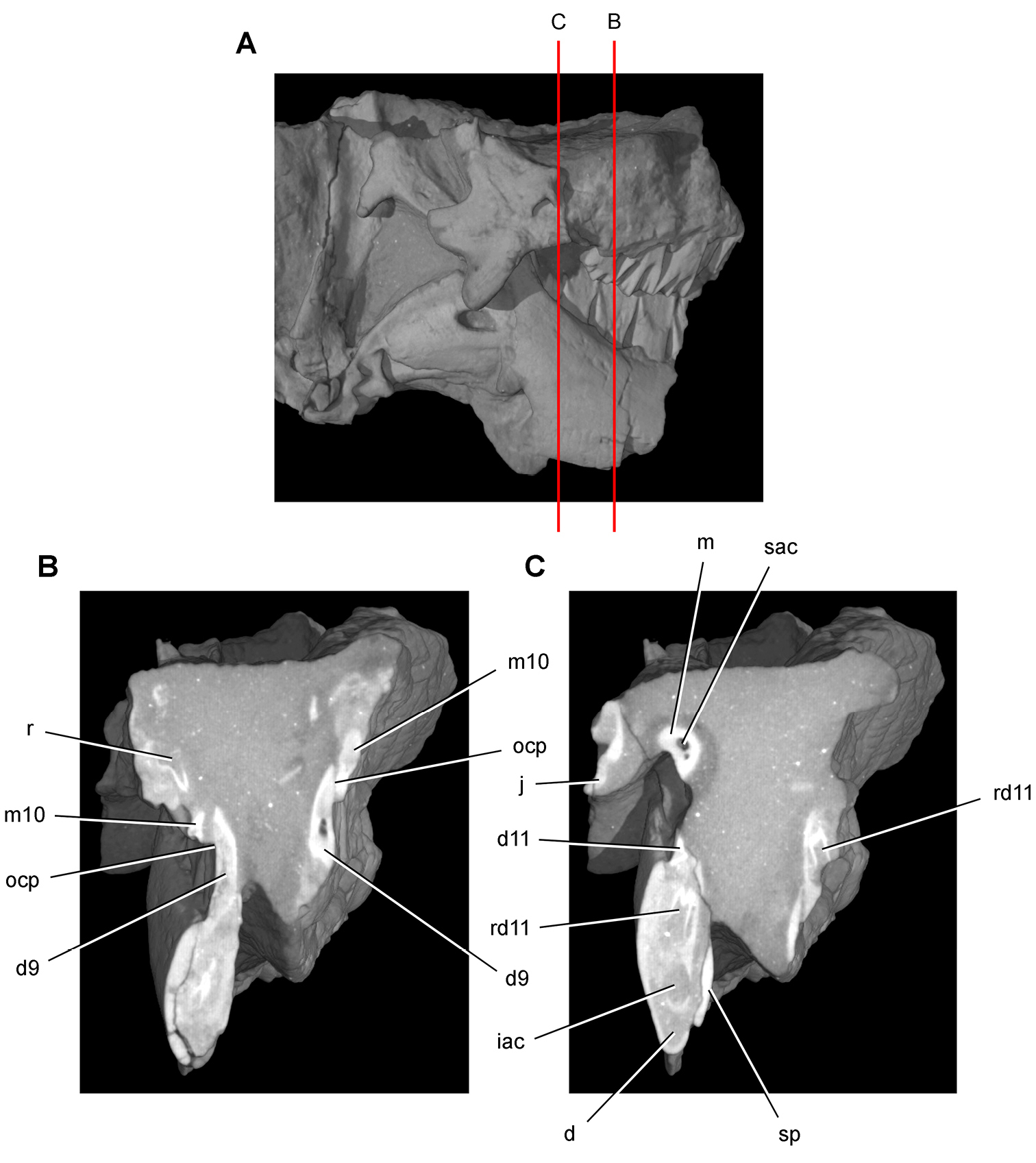
Tooth structure, occlusion, and replacement in Heterodontosaurus tucki from the Lower Jurassic Elliot and Clarens Formations of South Africa.Successive coronal computed-tomographic sections in cutaway view of a subadult skull (AMNH 24000). A Posterior portion of skull in right lateral view showing the location of coronal cross-sections B Cross-section through maxillary tooth 10 and dentary tooth 9 C Cross-section through dentary tooth 11. Abbreviations: d dentary d9, 11 dentary tooth 9, 11 iac inferior alveolar canal j jugal m maxilla m10 maxillary tooth 10 ocp occlusal plane r replacement tooth rd11 replacement dentary tooth 11 sac superior alveolar canal sp splenial.
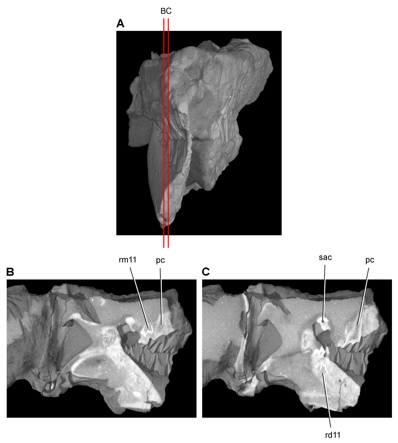
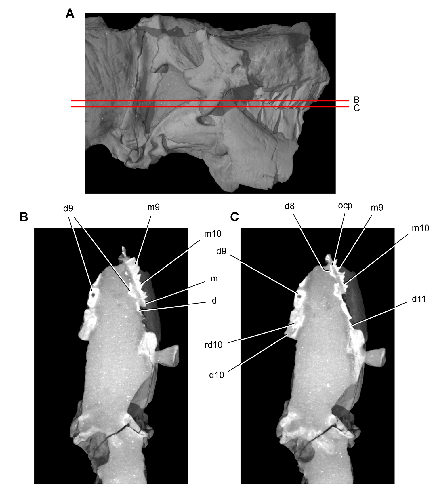
Tooth structure, occlusion, and replacement in Heterodontosaurus tucki from the Lower Jurassic Elliot and Clarens Formations of South Africa.Successive sagittal computed-tomographic sections in cutaway view of a subadult skull (AMNH 24000). A Posterior portion of skull in anterior view showing the location of sagittal cross-sections B, C Cross-sections in right lateral view through right maxillary and dentary rami. Abbreviations: pc pulp cavity rd11 replacement dentary tooth 11 rm11 replacement maxillary tooth 11 sac superior alveolar canal.
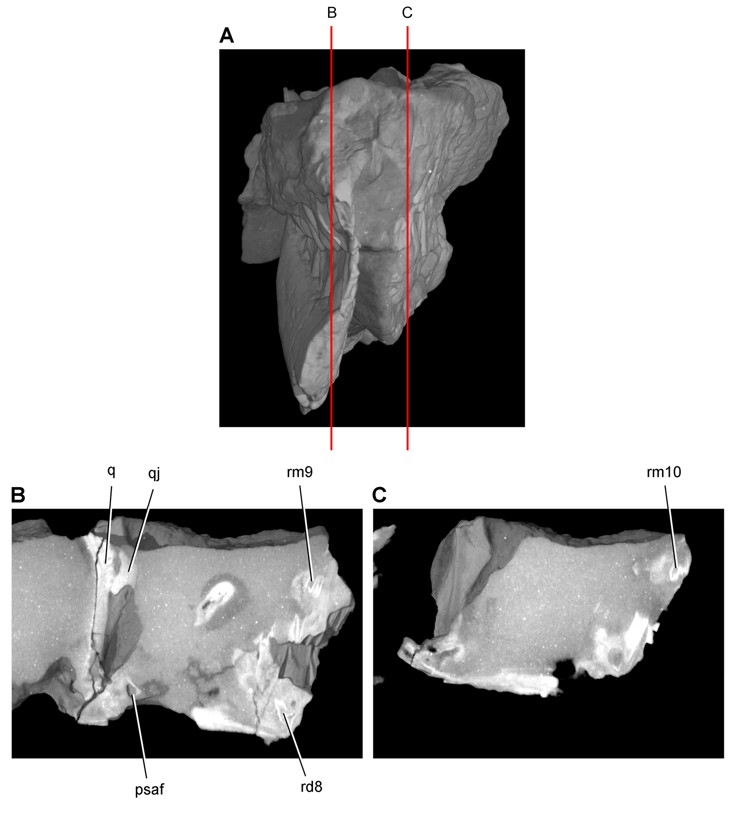
Tooth structure, occlusion, and replacement in Heterodontosaurus tucki from the Lower Jurassic Elliot and Clarens Formations of South Africa. Successive sagittal computed-tomographic sections in cutaway view of a subadult skull (AMNH 24000). A Posterior portion of skull in anterior view showing the location of sagittal cross-sections B Cross-section in right lateral view through right maxillary and dentary rami C Cross-section in right lateral view through left maxillary and dentary rami. Abbreviations: psaf posterior surangular foramen q quadrate qj quadratojugal rd8 replacement dentary tooth 8 rm9, 10 replacement maxillary teeth 9, 10.
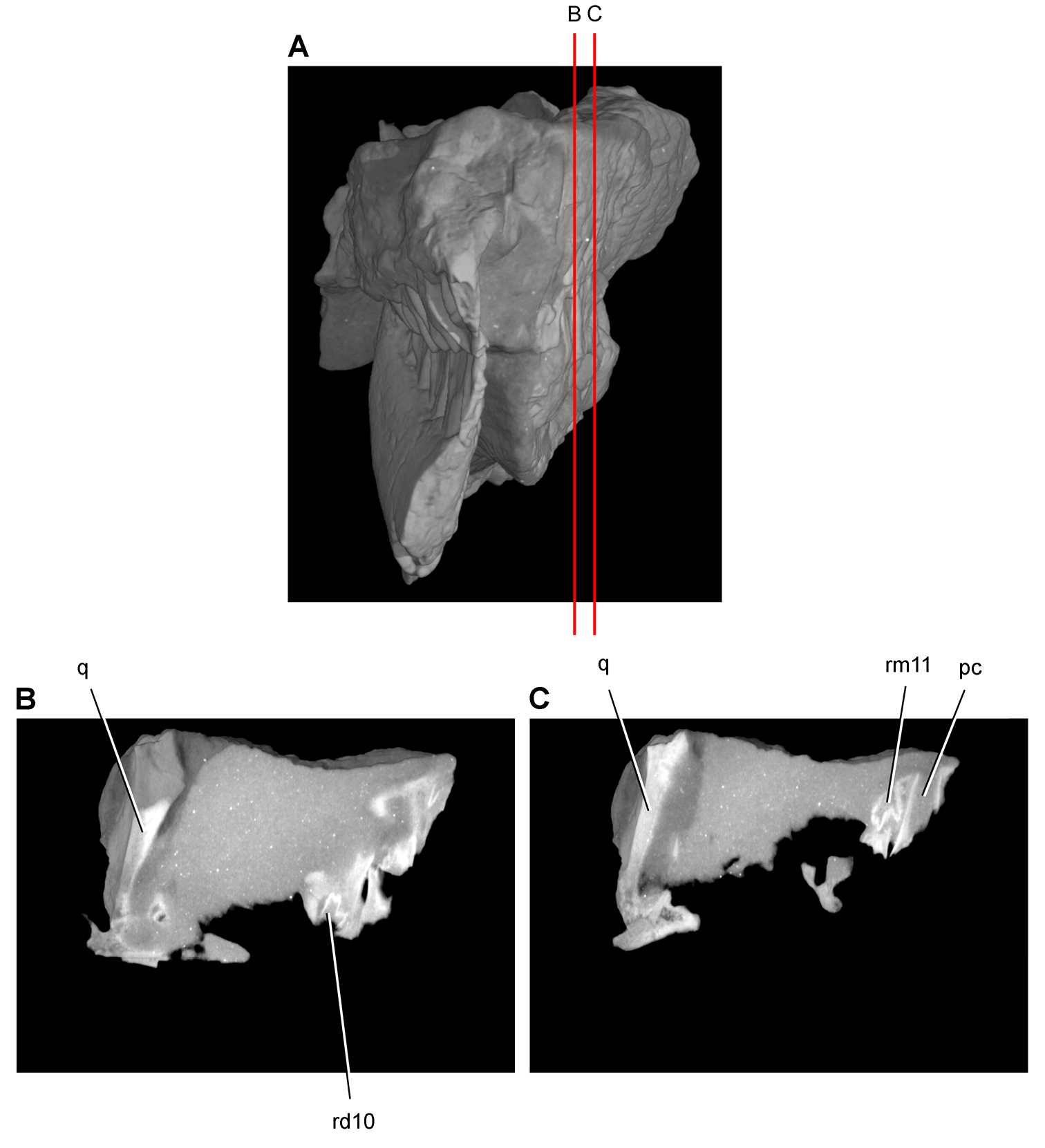
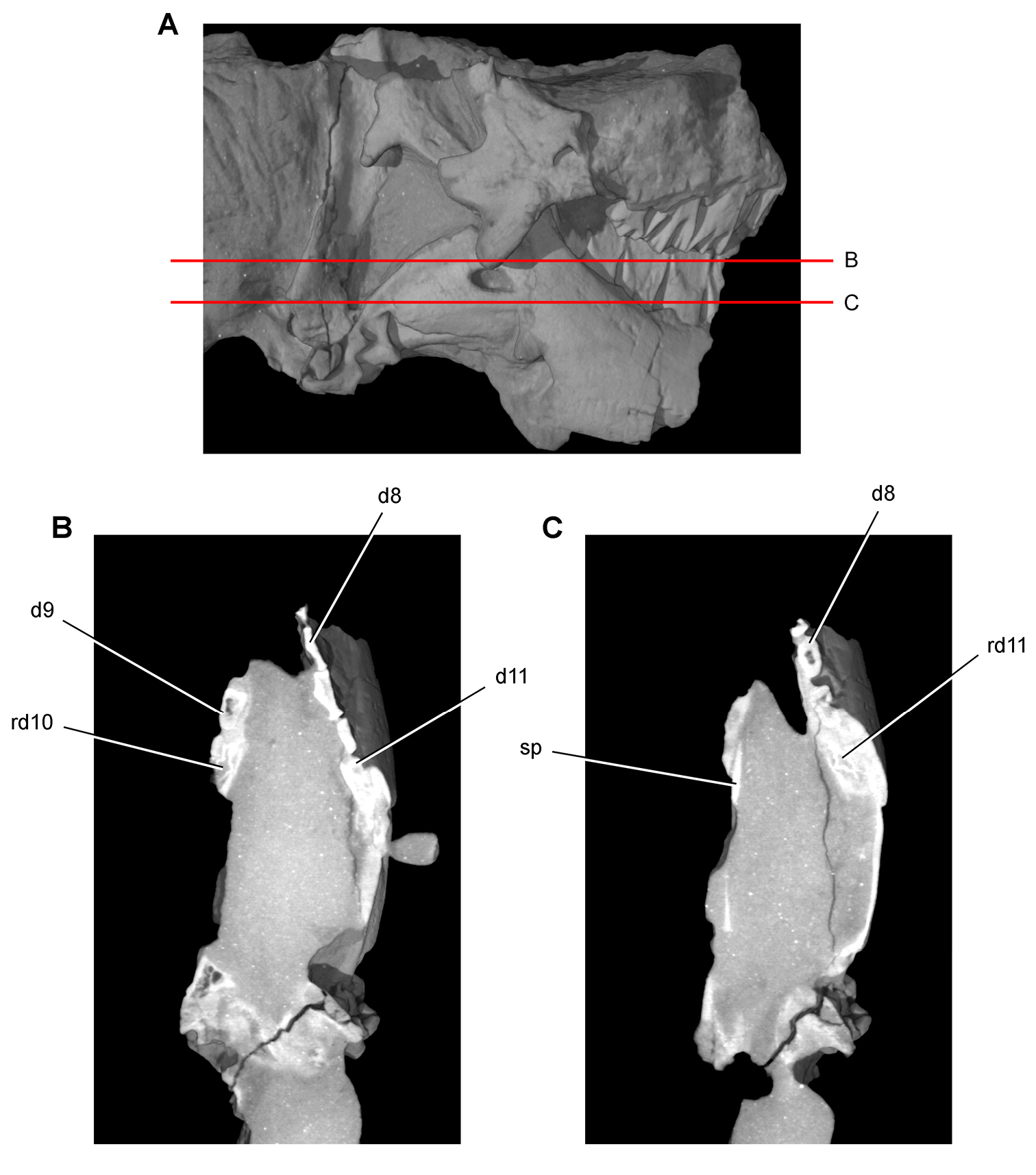
Tooth structure, occlusion, and replacement in Heterodontosaurus tucki from the Lower Jurassic Elliot and Clarens Formations of South Africa. Successive sagittal computed-tomographic sections in cutaway view of a subadult skull (AMNH 24000). A Posterior portion of skull in anterior view showing the location of sagittal cross-sections B, C Cross-sections in right lateral view through left maxillary and dentary rami. Abbreviations: pc pulp cavity q quadrate rd10 replacement dentary tooth 10 rm11 replacement maxillary tooth 11.
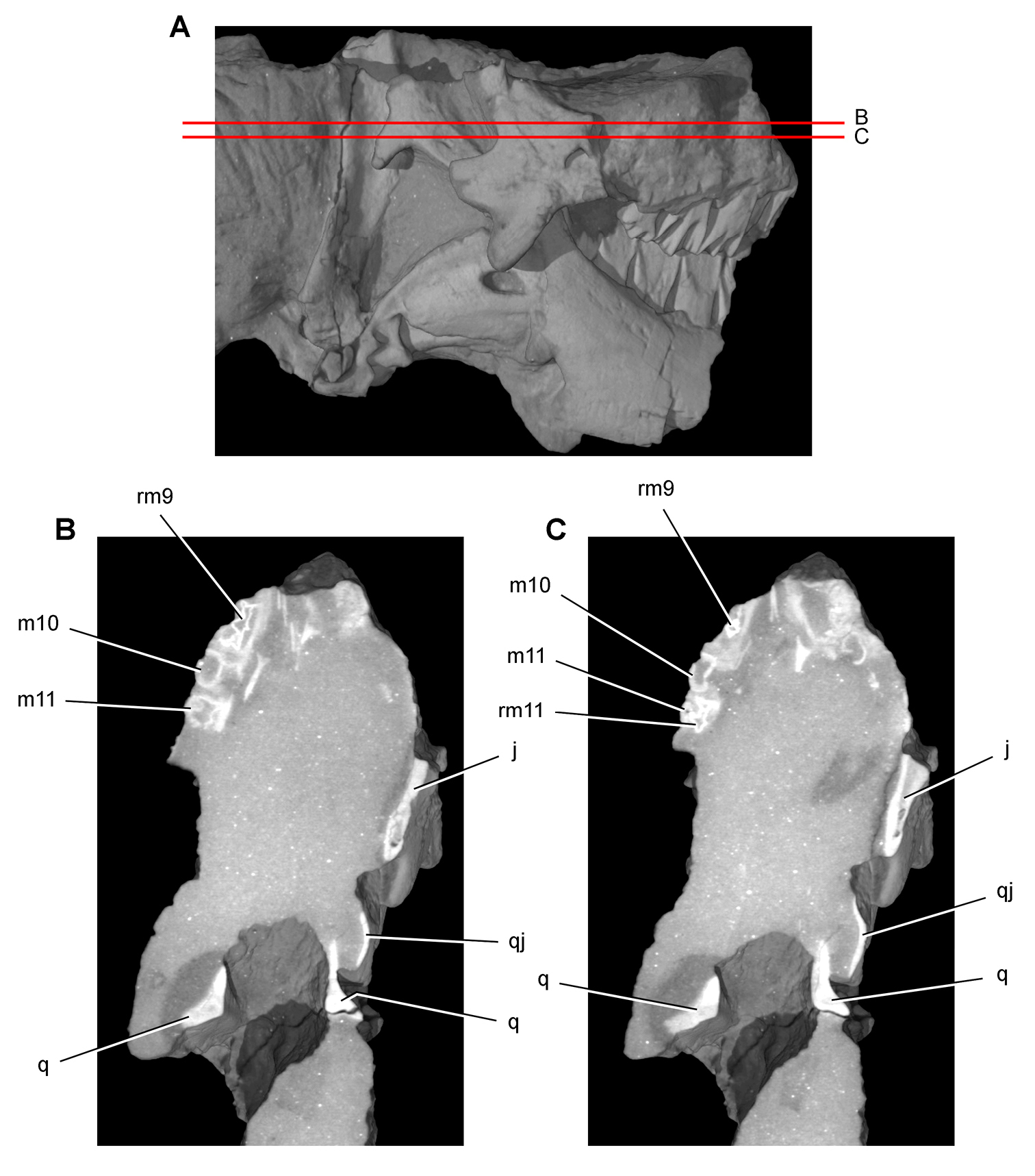
Tooth structure, occlusion, and replacement in Heterodontosaurus tucki from the Lower Jurassic Elliot and Clarens Formations of South Africa.Successive horizontal computed-tomographic sections in cutaway view of a subadult skull (AMNH 24000). A Posterior portion of skull in right lateral view showing the location of horizontal cross-sections B, C Cross-sections (anterior toward top of page) through the maxilla. Abbreviations: j jugal m10, 11 maxillary tooth 10, 11 q quadrate qj quadratojugal rm9, 11 replacement maxillary tooth 9, 11.
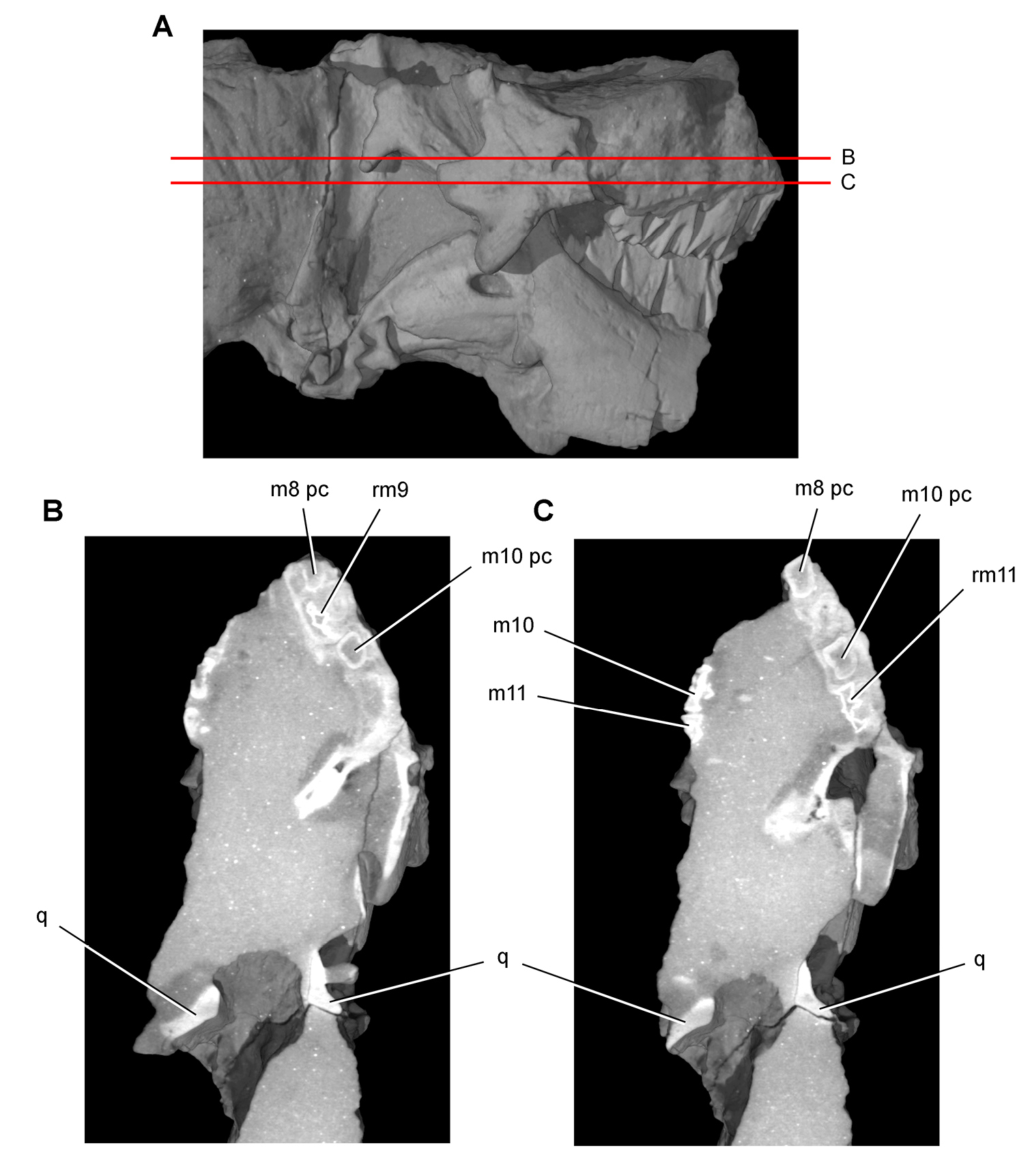
Tooth structure, occlusion, and replacement in Heterodontosaurus tucki from the Lower Jurassic Elliot and Clarens Formations of South Africa. Successive horizontal computed-tomographic sections in cutaway view of a subadult skull (AMNH 24000). A Posterior portion of skull in right lateral view showing the location of horizontal cross-sections B Cross-section (anterior toward top of page) through the maxilla C Cross-section (anterior toward top of page) through occluding portions of maxillary tooth rows.Abbreviations: m8, 10, 11 maxillary tooth 8, 10, 11 pc pulp cavity q quadrate rm9, 11 replacement maxillary tooth 9, 11.
Tooth structure, occlusion, and replacement in Heterodontosaurus tucki from the Lower Jurassic Elliot and Clarens Formations of South Africa.Successive horizontal computed-tomographic sections in cutaway view of a subadult skull (AMNH 24000). A Posterior portion of skull in right lateral view showing the location of horizontal cross-sections B, C Cross-sections (anterior toward top of page) through occluding portions of maxillary and dentary tooth rows.Abbreviations: d9, 10, dentary tooth 9, 10 m8-11 maxillary teeth 8-11 ocp occlusal plane q quadrate rm11 replacement maxillary tooth 11.
Tooth structure, occlusion, and replacement in Heterodontosaurus tucki from the Lower Jurassic Elliot and Clarens Formations of South Africa. Successive horizontal computed-tomographic sections in cutaway view of a subadult skull (AMNH 24000). A Posterior portion of skull in right lateral view showing the location of horizontal cross-sections B, C Cross-sections (anterior toward top of page) through occluding portions of maxillary and dentary tooth rows.Abbreviations: d dentary d8-11 dentary teeth 8-11 m maxilla m9, 10 maxillary tooth 9, 10 ocp occlusal plane rd10 replacement dentary tooth 10.
Tooth structure, occlusion, and replacement in Heterodontosaurus tucki from the Lower Jurassic Elliot and Clarens Formations of South Africa. Successive horizontal computed-tomographic sections in cutaway view of a subadult skull (AMNH 24000). A Posterior portion of skull in right lateral view showing the location of horizontal cross-sections B, C Cross-sections (anterior toward top of page) through the dentary. Abbreviations: d8, 9, 11 dentary tooth 8, 9, 11 rd10, 11 replacement dentary tooth 10, 11 sp splenial.
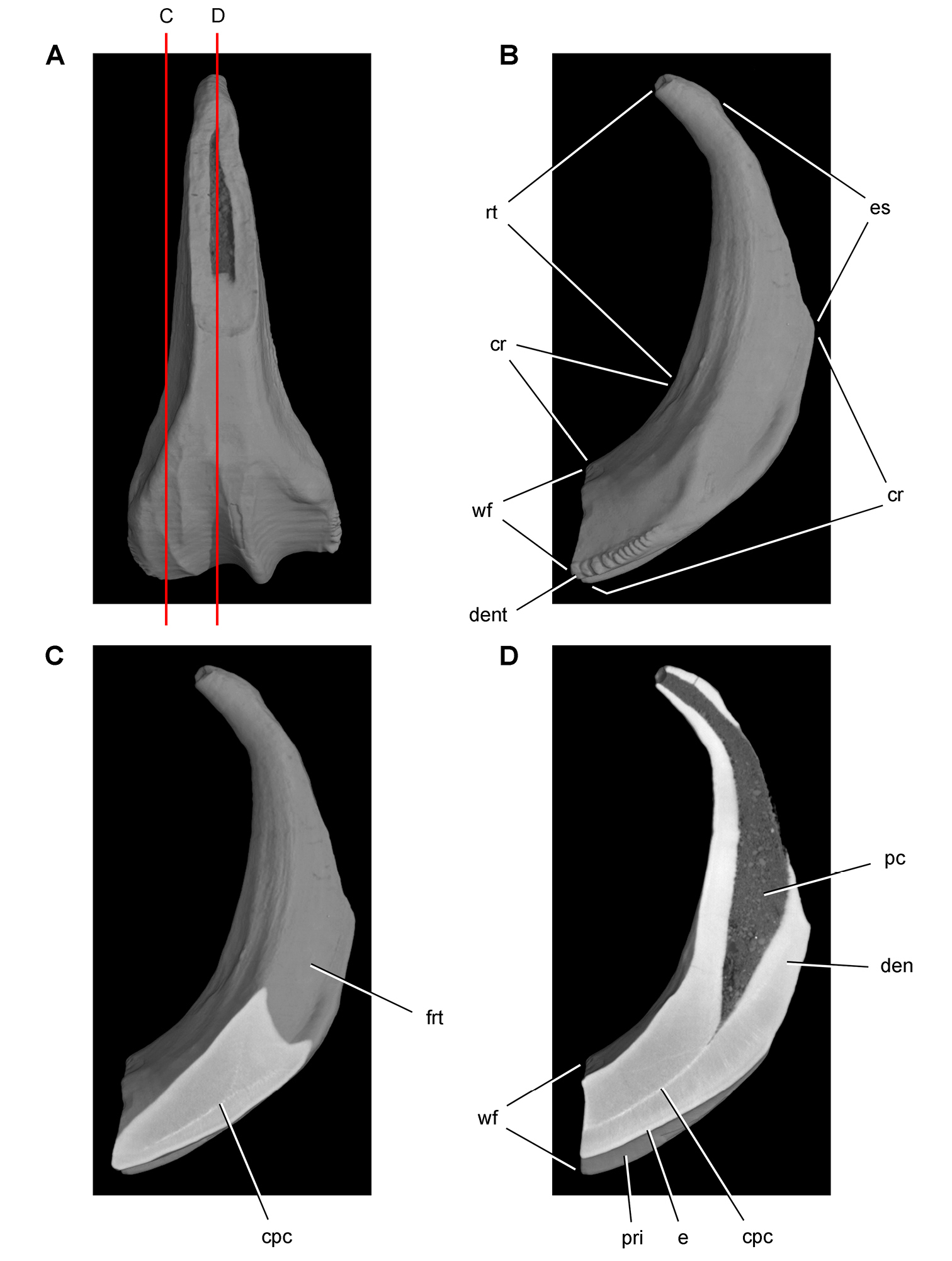
Tooth structure in Ouranosaurus nigeriensis from the mid-Cretaceous Elrhaz Formation of Niger. Successive coronal computed-tomographic sections in cutaway view of a worn maxillary tooth showing internal structure (MNBH GAD28) A Maxillary tooth in labial view showing the location of coronal cross-sections B Maxillary tooth in distal view showing the division between crown and root and the fossa for an adjacent replacement tooth C Section through mesial portion of crown D Section through mid section of crown. Abbreviations: cpc collapsed pulp cavity cr crown den dentine dent denticle e enamel es erosional surface frt fossa for replacement tooth pc pulp cavity pri primary ridge rt root wf wear facet.
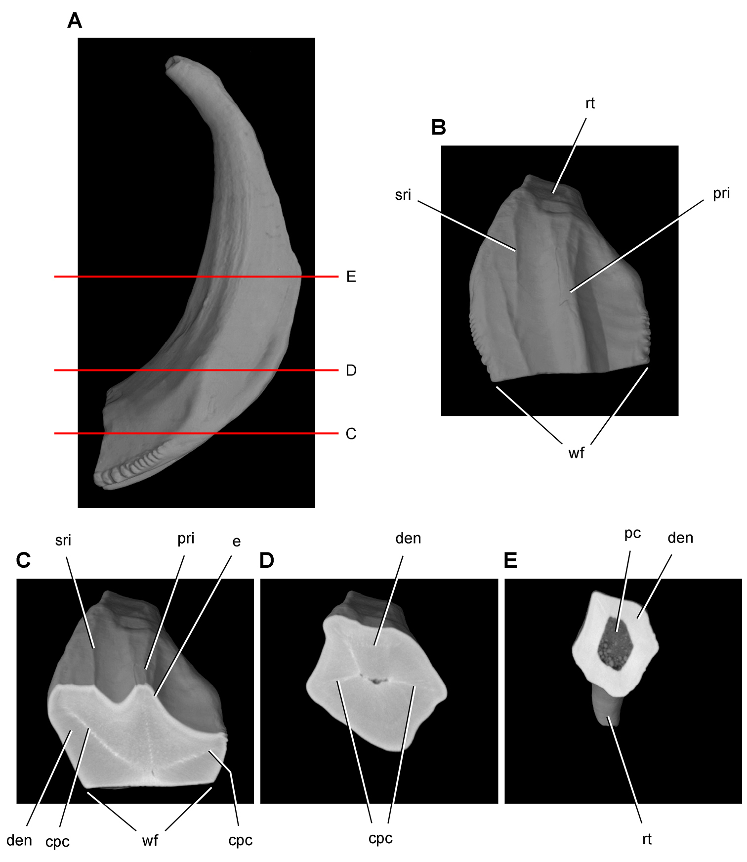
Tooth structure in Ouranosaurus nigeriensis from the mid-Cretaceous Elrhaz Formation of Niger. Successive horizontal computed-tomographic sections in cutaway view of a worn maxillary tooth showing internal structure (MNBH GAD28) A Maxillary tooth in distal view showing the location of horizontal cross-sections B Maxillary tooth in apical view (labial toward top of page) C Cross-section (labial toward top of page) through mid crown D Cross-section (labial toward top of page) through base of the crown E Cross-section (labial toward top of page) through proximal portion of the root. Abbreviations: cpc collapsed pulp cavity den dentine e enamel pc pulp cavity pri primary ridge rt root sri secondary ridge wf wear facet.
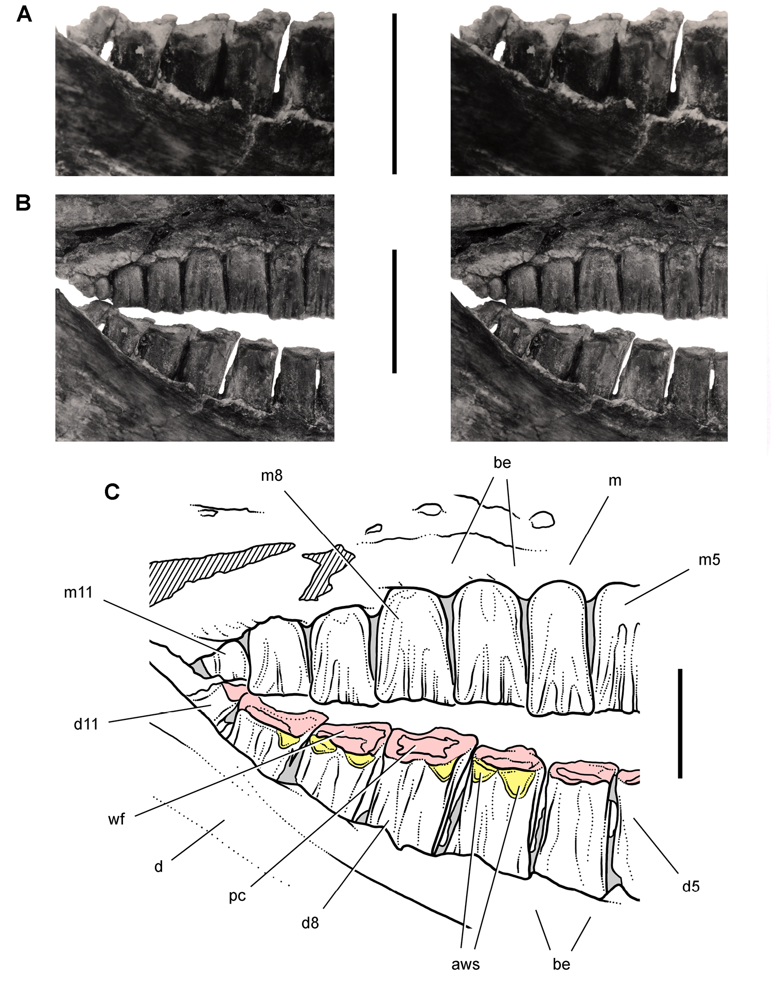
Posterior dentition of Heterodontosaurus tucki from the Lower Jurassic Upper Elliot and Clarens formations of South Africa. Posterior half of worn maxillary and dentary tooth row in an adult skull in right lateral view (SAM-PK-K1332). Stereopair (A) of right posterior dentary tooth row tipped labially (laterally) exposing the wear facets in dorsolateral view. Stereopair (B) and line drawing (C) of the posterior half of the tooth rows in natural articulation in lateral view. Hatching indicates broken bone; grey tone indicates matrix; pink tone indicates wear facets; yellow tone indicates accessory wear surfaces. Scale bars equal 1 cm in A and B, 5 mm in C. Abbreviations: aws accessory wear surface be buccal emargination d dentary d5, 8, 11 dentary tooth 5, 8, 11 m maxilla m5, 8, 11 maxillary tooth 5, 8, 11 pc pulp cavity wf wear facet.

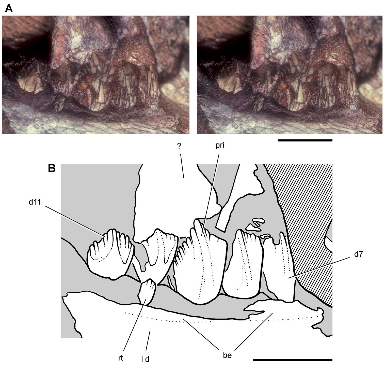
Postcaniniform dentary tooth row of Heterodontosaurus tucki from the Lower Jurassic Upper Elliot and Clarens formations of South Africa. Worn right dentary tooth row in an adult skull (SAM-PK-K1332). Stereopair (A) and line drawing (B) in medial view. Dashed lines indicate estimated edges; tone indicates matrix. Scale bars equal 1 cm in A and B. Abbreviations: c coronoid d dentary d2, 8, 11 dentary tooth 2, 8, 11 fo foramen imf internal mandibular fenestra; Mc Meckel’s canal sp splenial.
Skull of Heterodontosaurus tucki from the Lower Jurassic Upper Elliot and Clarens formations of South Africa. Previous skull reconstructions in left lateral view A From Charig and Crompton (1974) B Reversed from Weishampel (1984).
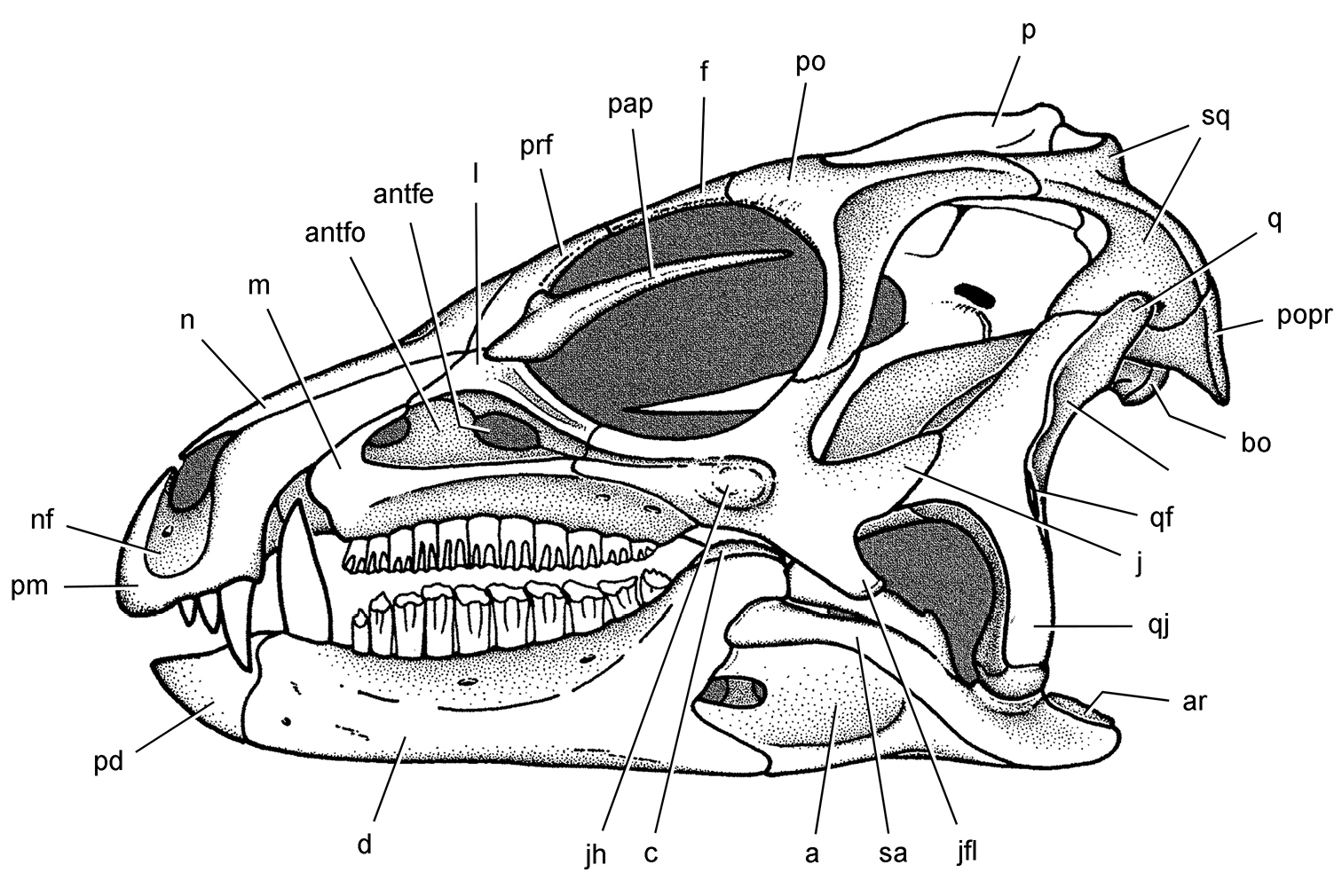
Skull of Heterodontosaurus tucki from the Lower Jurassic Upper Elliot and Clarens formations of South Africa. Skull reconstruction in left lateral view from Norman et al. (2011). Abbreviations: a angular antfe antorbital fenestra antfo antorbital fossa ar articular bo basioccipital c coronoid d dentary f frontal j jugal jfl jugal flange jh jugal horn l lacrimal m maxilla n nasal nf narial fossa p parietal pap palpebral pd predentary pm premaxilla po postorbital popr paroccipital process prf prefrontal q quadrate qf quadrate foramen qj quadratojugal sa surangular sq squamosal.
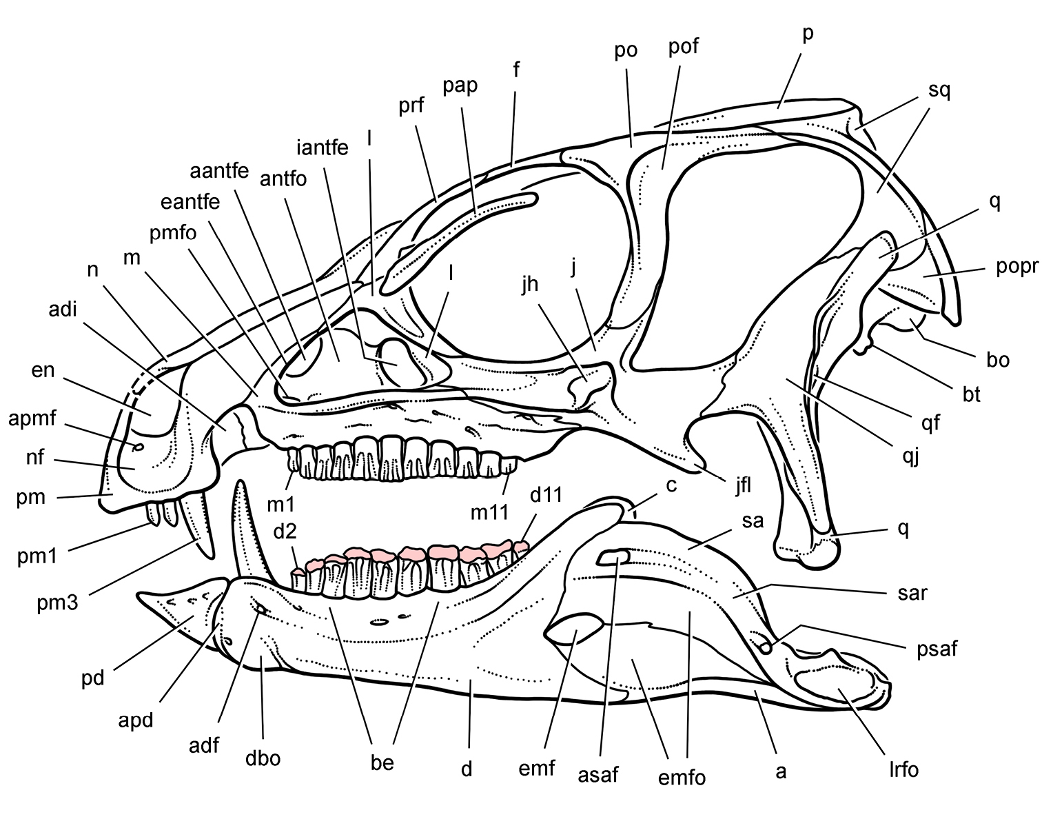
Skull of Heterodontosaurus tucki from the Lower Jurassic Upper Elliot and Clarens formations of South Africa. New skull reconstruction in lateral view showing the dentition with intermediate wear (scleral ring not shown). Pink tone indicates wear facets. Abbreviations: a angular aantfe accessory antorbital fenestra adf anterior dentary foramen adi arched diastema antfo antorbital fossa apd articular surface for the predentary apmf anterior premaxillary foramen asaf anterior surangular foramen be buccal emargination bo basioccipital bt basal tubera c coronoid d dentary d2, d11 dentary tooth 2, 11 dbo dentary boss eantfe external antorbital fenestra emf external mandibular fenestra emfo external mandibular fossa en external naris f frontal iantfe internal antorbital fenestra j jugal jfl jugal flange jh jugal horn l lacrimal lrfo lateral retroarticular fossa m maxilla m1, 11 maxillary tooth 1, 11 n nasal nf narial fossa p parietal pap palpebral pd predentary pm premaxilla pm1, 3 premaxillary tooth 1, 3 pmfo promaxillary fossa po postorbital pof postorbital fossa popr paroccipital process prf prefrontal psaf posterior surangular foramen q quadrate qf quadrate foramen qj quadratojugal sa surangular sar surangular ridge sq squamosal.
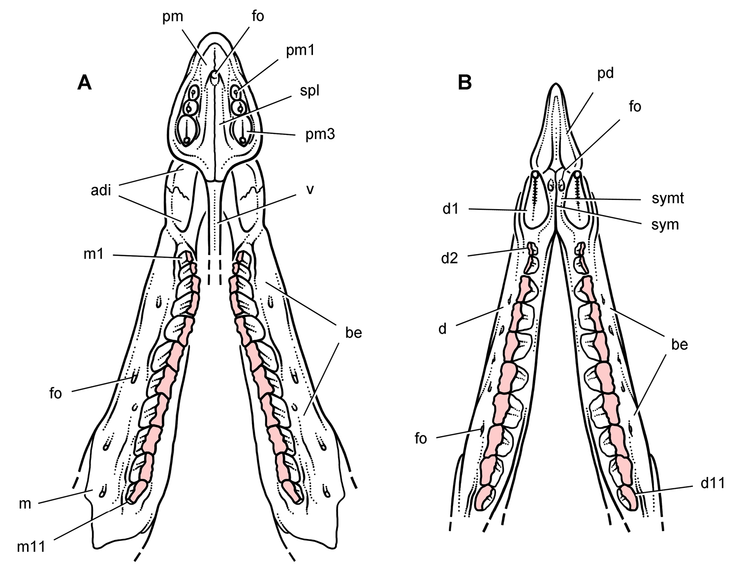
Jaws of Heterodontosaurus tucki from the Lower Jurassic Upper Elliot and Clarens formations of South Africa. Reconstruction of upper and lower jaws showing the dentition with heavy wear (based on SAM-PK-K1332). Dashes indicate estimated edges; pink tone indicates wear facets A Upper dentition and anterior palate in ventral view B Lower dentition, dentary symphysis and predentary in dorsal view. Abbreviations: adi arched diastema be buccal emargination d dentary d1, 2, 11 dentary tooth 1, 2, 11 fo foramen m maxilla m1, 11 maxillary tooth 1, 11 pd predentary pm premaxilla pm1, 3 premaxillary tooth 1, 3 spl secondary palate sym symphysis symt symphyseal trough v vomer.
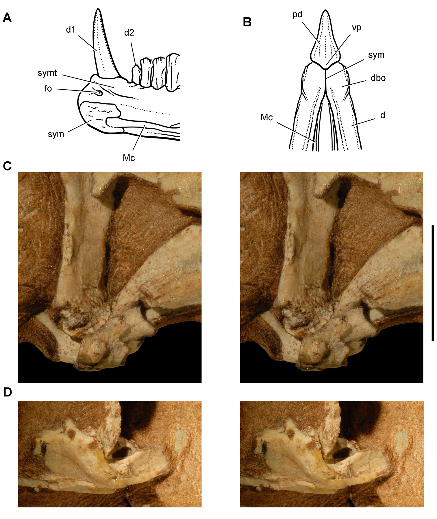
Jaw joints of Heterodontosaurus tucki from the Lower Jurassic Upper Elliot and Clarens formations of South Africa. Dentary symphysis and quadrate-articular jaw joint A Reconstruction of the anterior portion of the right dentary in medial view (based on SAM-PK-K1332) B Reconstruction of the anterior end of the lower jaws in ventral view (based on SAM-PK-K1332) C Stereopair of the right lower jaw joint in lateral view (the lateral edge of the quadrate condyle and articular are broken away exposing the tight jaw articulation) in a subadult skull (AMNH 24000) D Stereopair of the left lower jaw joint in lateral view (the lateral edge of the quadrate condyle is broken away exposing the inclined trough of the jaw articulation) in a subadult skull (AMNH 24000). Scale bar equals 1 cm in C and D. Abbreviations: d dentary d1, 2 dentary tooth 1, 2 dbo dentary boss fo foramen Mc Meckel’s canal pd predentary sym symphysis symt symphyseal trough vp ventral process.
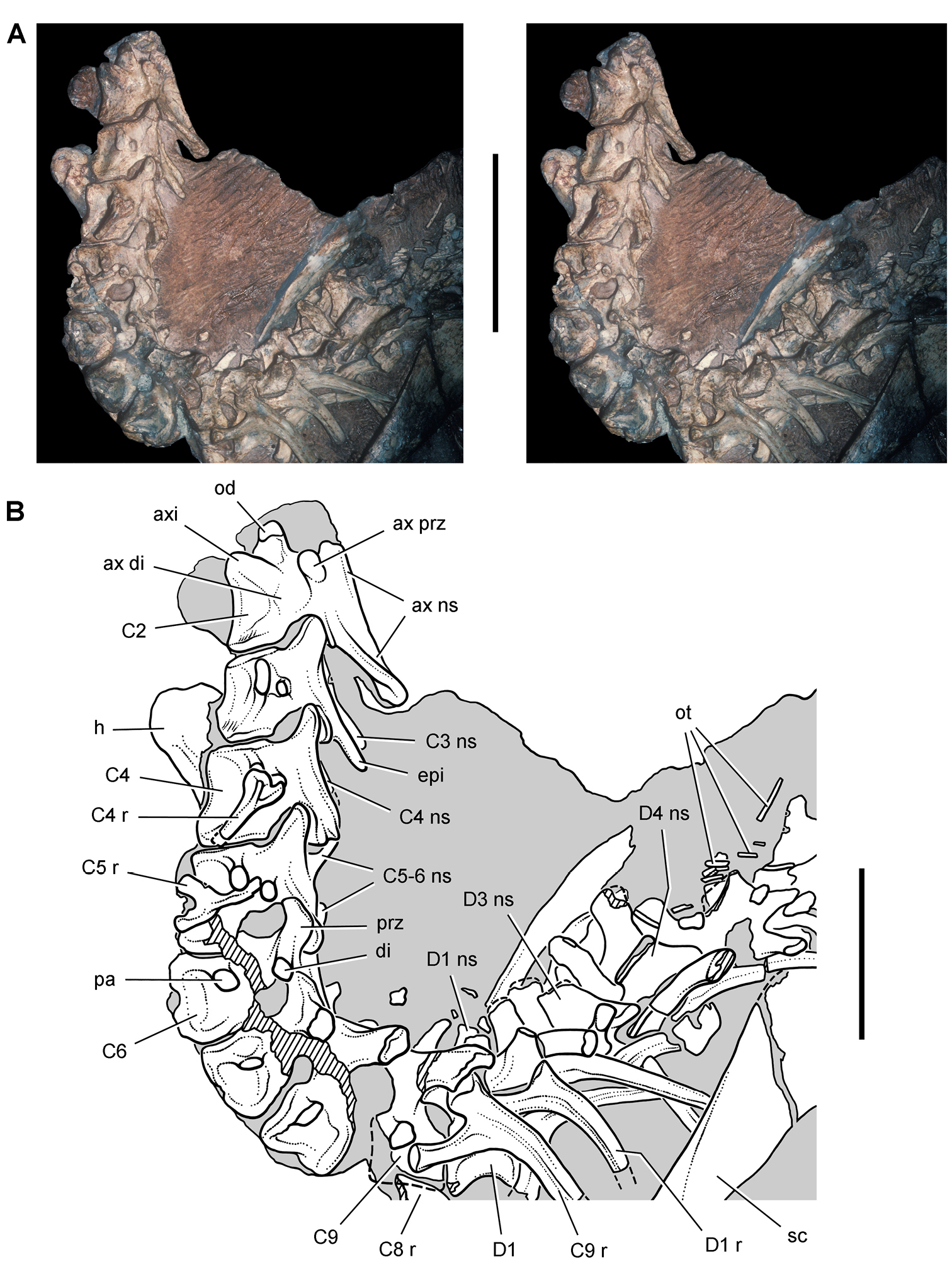
Presacral vertebrae of Heterodontosaurus tucki from the Lower Jurassic Upper Elliot and Clarens formations of South Africa. Cervical and anterior dorsal vertebrae and ribs of an adult skeleton (SAM-PK-K1332). Stereopair (A) and line drawing (B) in left lateral view. Hatching indicates broken bone; dashed lines indicate estimated edges; tone indicates matrix. Scale bars equal 5 cm in A and 3 cm in B. Abbreviations: ax axial axi axial intercentrum C2-6, 8, 9 cervical vertebra 2–6, 8, 9 D1, 3, 4 dorsal vertebra 1, 3, 4 di diapophysis epi epipophysis h humerus ns neural spine od odontoid ot ossified tendons pa parapophysis prz prezygapohpysis r rib sc scapula.
Presacral vertebrae of Heterodontosaurus tucki from the Lower Jurassic Upper Elliot and Clarens formations of South Africa. Cervical and anterior dorsal vertebrae and ribs of an adult skeleton (SAM-PK-K1332). Stereopairs of C1-3 (A), C4-6 (B), and C7-D4 (C) in left lateral view. Scale bars equal 2 cm in A and B, 3 cm in C.
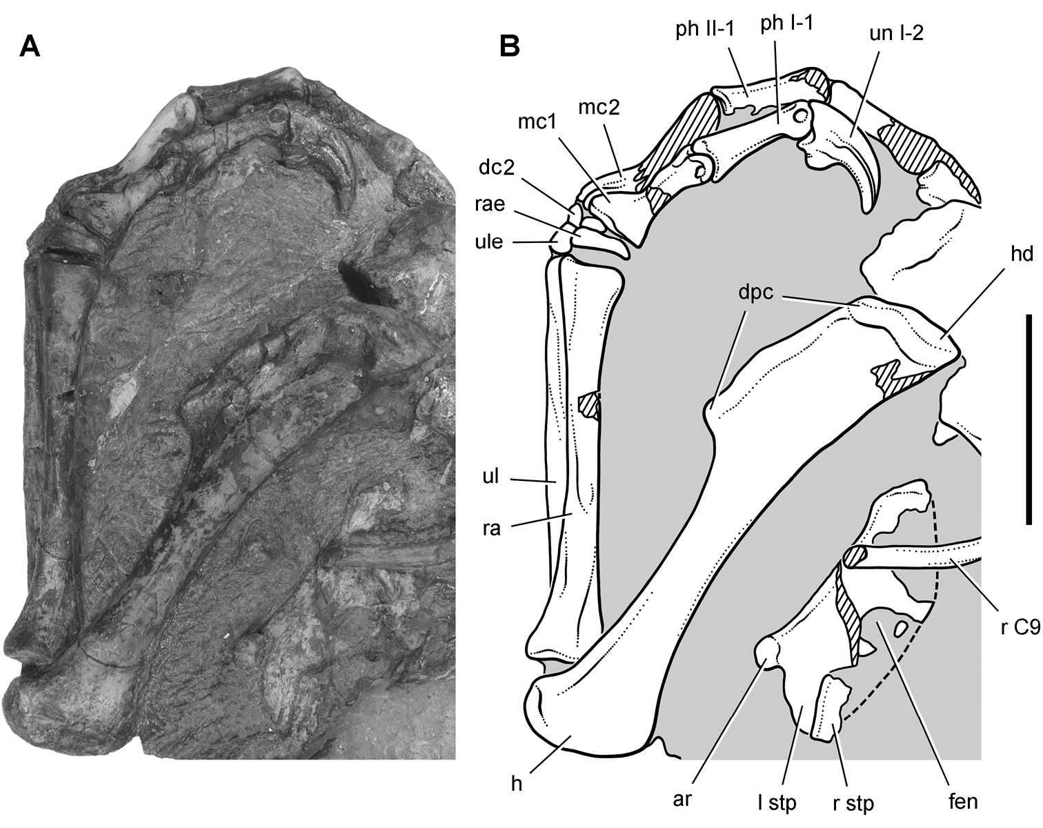
Pectoral girdle and forelimb of Heterodontosaurus tucki from the Lower Jurassic Upper Elliot and Clarens formations of South Africa. Pectoral girdle and forelimb of an adult skeleton (SAM-PK-K1332). Photograph (A) and line drawing (B) of the sternal plates and left forelimb in dorsal and medial view, respectively. Hatching indicates broken bone; dashed lines indicate estimated edges; tone indicates matrix. Scale bar equals 3 cm. Abbreviations: I, II digit I, II ar articular surface for a sternal rib C9 cervical 9 dc2 distal carpal 2 dpc deltopectoral crest fen fenestra h humerus hd head l left mc1, 2 metacarpal 1, 2 ph phalanx r rib or right ra radius rae radiale stp sternal plate ul ulna ule ulnare un ungual.
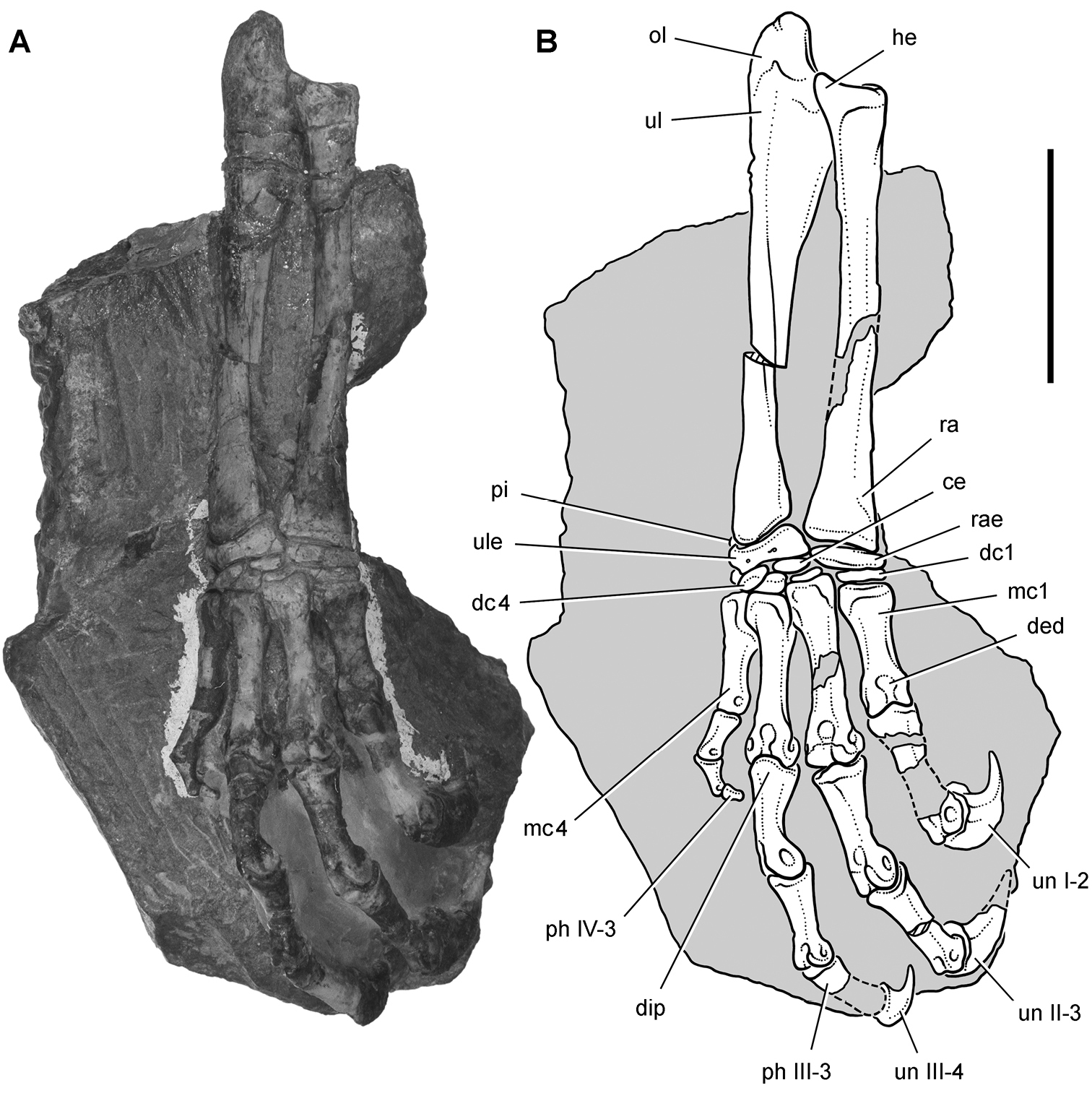
Forelimb of Heterodontosaurus tucki from the Lower Jurassic Upper Elliot and Clarens formations of South Africa. Antebrachium, carpus and manus of an adult skeleton (SAM-PK-K1332). Photograph (A) and line drawing (B) of the right radius, ulna, carpus and manus in dorsal view. Hatching indicates broken bone; dashed lines indicate estimated edges; tone indicates matrix. Scale bar equals 3 cm. Abbreviations: I-IV digits I-IV ce centrale dc1, 4 distal carpal 1, 4 ded dorsal extensor depression dip dorsal intercondylar process he heel mc1, 4 metacarpal 1, 4 ol olecranon ph, phalanx pi pisiform ra radius rae radiale ul ulna ule ulnare un ungual.
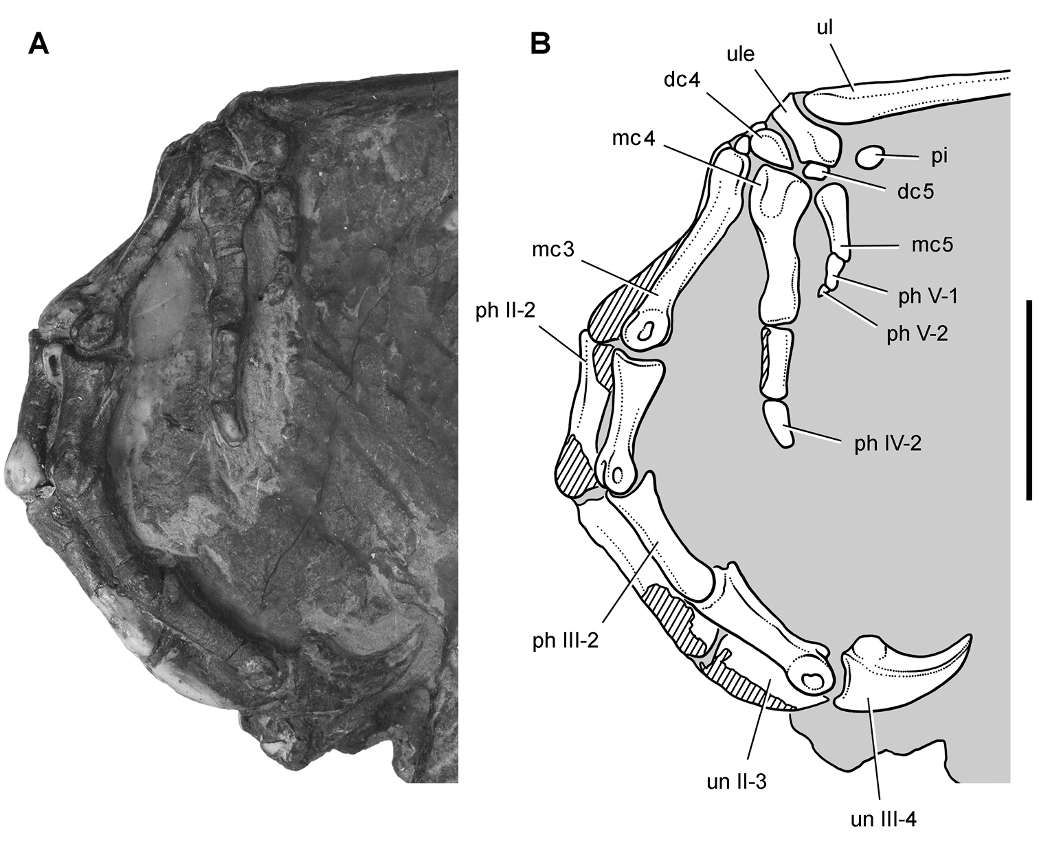
Carpus and manus of Heterodontosaurus tucki from the Lower Jurassic Upper Elliot and Clarens formations of South Africa. Carpus and manus of an adult skeleton (SAM-PK-K1332). Photograph (A) and line drawing (B) of the left carpus and manus in lateral view. Hatching indicates broken bone; tone indicates matrix. Scale bar equals 2 cm. Abbreviations: II-V digits II-V dc4, 5 distal carpal 4, 5 mc3-5 metacarpals 3–5 ph phalanx pi pisiform ul ulna ule ulnare un ungual.
Limbs of Heterodontosaurus tucki from the Lower Jurassic Upper Elliot and Clarens formations of South Africa. Reconstructions based on an adult skeleton (SAM-PK-K1332). Right forearm, carpus and manus (A), right distal tarsals and pes (B), and right carpus (C) in dorsal view. Abbreviations: I-V digits I-V ce centrale dc1-5 distal carpals 1–5 dt3, 4 distal tarsal 3, 4 fo foramen mc1, 4, 5 metacarpal 1, 4, 5 mt1-4 metatarsals 1–4 ol olecranon ph phalanx pi pisiform ra radius rae radiale ul ulna ule ulnare un ungual.
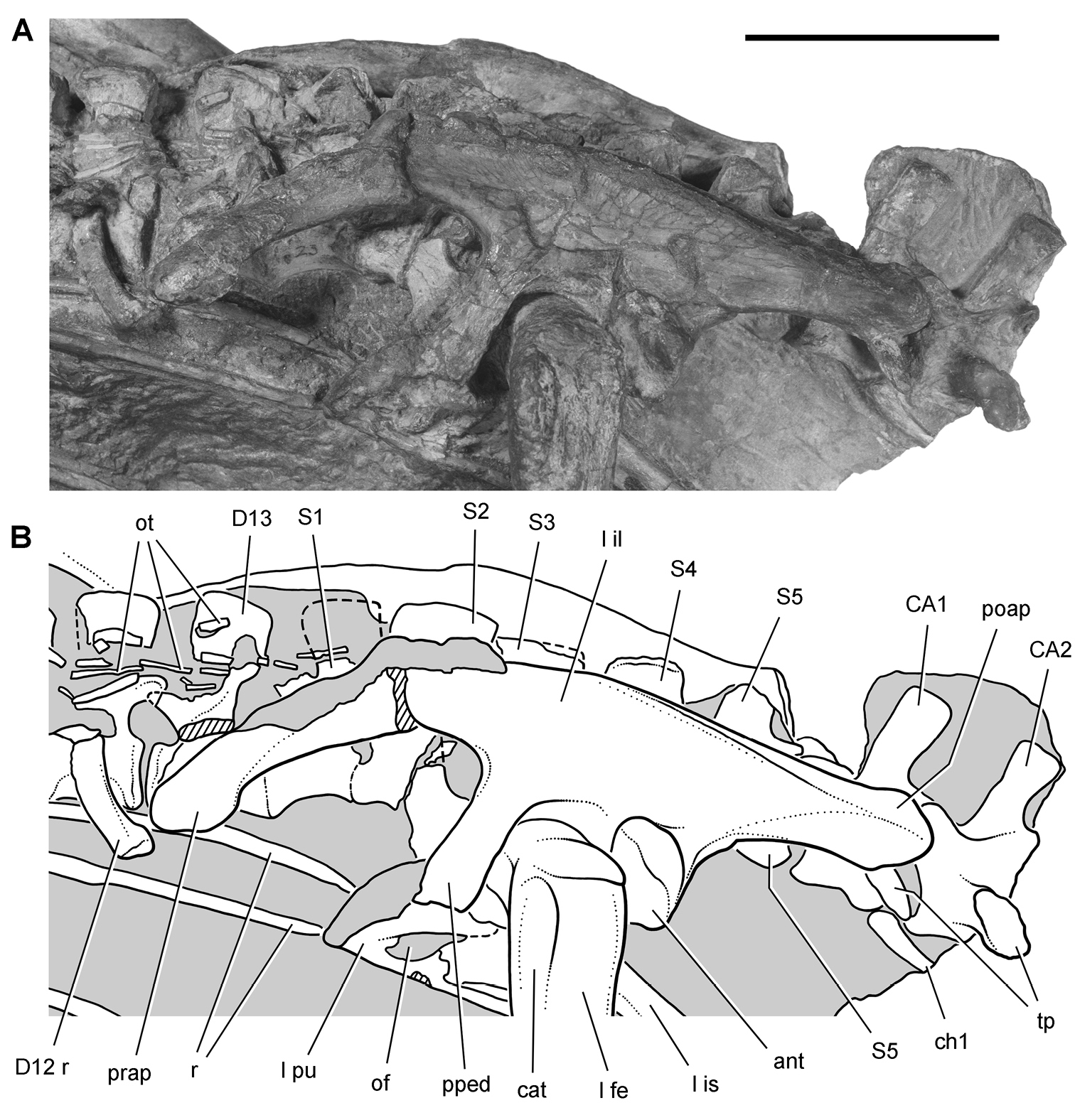
Hip and posterior trunk of Heterodontosaurus tucki from the Lower Jurassic Upper Elliot and Clarens formations of South Africa. Hip and posterior trunk in an adult skeleton (SAM-PK-K1332). Photograph (A) and line drawing (B) of the left ilium and pubis and sacral vertebrae in left lateral view. Hatching indicates broken bone; dashed lines indicate estimated edges; tone indicates matrix. Scale bar equals 3 cm. Abbreviations: ant antitrochanter CA caudal vertebra cat coossified anterior trochanter ch chevron D dorsal vertebra fe femur il ilium is ischium l left of obturator foramen ot ossified tendon poap postacetabular process pped pubic peduncle prap preacetabular process pu pubis r rib S sacral vertebra tp transverse process.
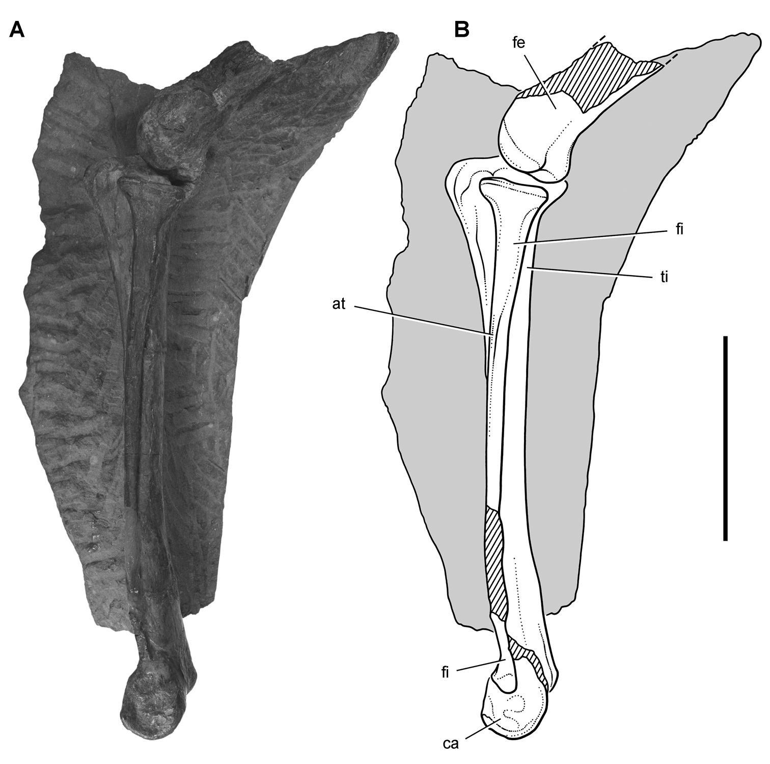
Hindlimb of Heterodontosaurus tucki from from the Lower Jurassic Upper Elliot and Clarens formations of South Africa. Tibiotarsus in an adult skeleton (SAM-PK-K1332). Photograph (A) and line drawing (B) of the left distal femur and tibiotarsus in lateral view. Hatching indicates broken bone; dashed lines indicate estimated edges; tone indicates matrix. Scale bar equals 5 cm. Abbreviations: at anterior trochanter ca calcaneum fe femur fi fibula ti tibia.
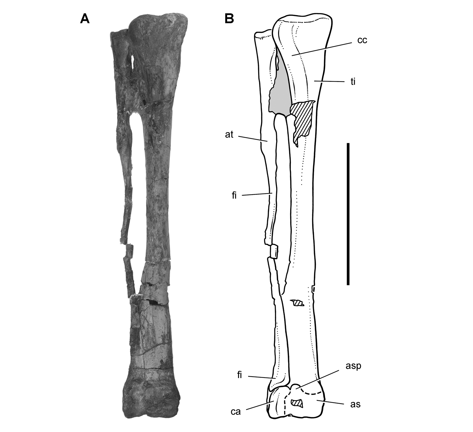
Hindlimb of Heterodontosaurus tucki from the Lower Jurassic Upper Elliot and Clarens formations of South Africa. Tibiotarsus in an adult skeleton (SAM-PK-K1328). Photograph (A) and line drawing (B) of the right tibiotarsus in anterior view. Hatching indicates broken bone; dashed lines indicate estimated edges; tone indicates matrix. Scale bar equals 5 cm. Abbreviations: as astragalus asp ascending process at anterior trochanter ca calcaneum cc cnemial crest fi fibula ti tibia.
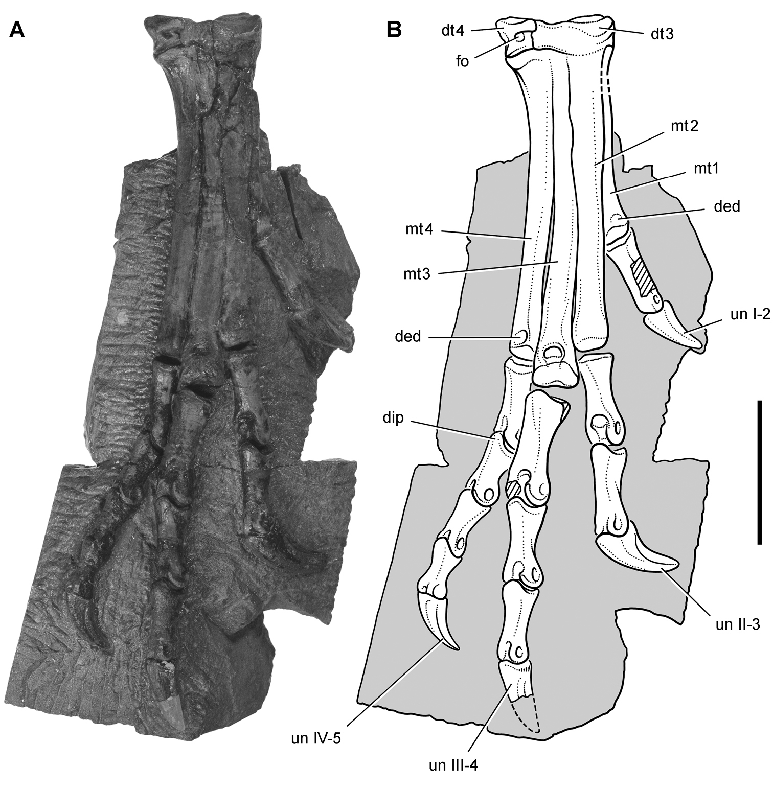
Pes of Heterodontosaurus tucki from the Lower Jurassic Upper Elliot and Clarens formations of South Africa. Tarsometatarsus and pes of an adult skeleton (SAM-PK-K1328). Photograph (A) and line drawing (B) of the right tarsometatarsus and pes in anterior view. Hatching indicates broken bone; dashed lines indicate estimated edges; tone indicates matrix. Scale bar equals 3 cm. Abbreviations: I-IV digits I-IV ded dorsal extensor depression dip dorsal intercondylar process dt3, 4 distal tarsals 3, 4 fo foramen mt1-4 metatarsals 1-4 un ungual.
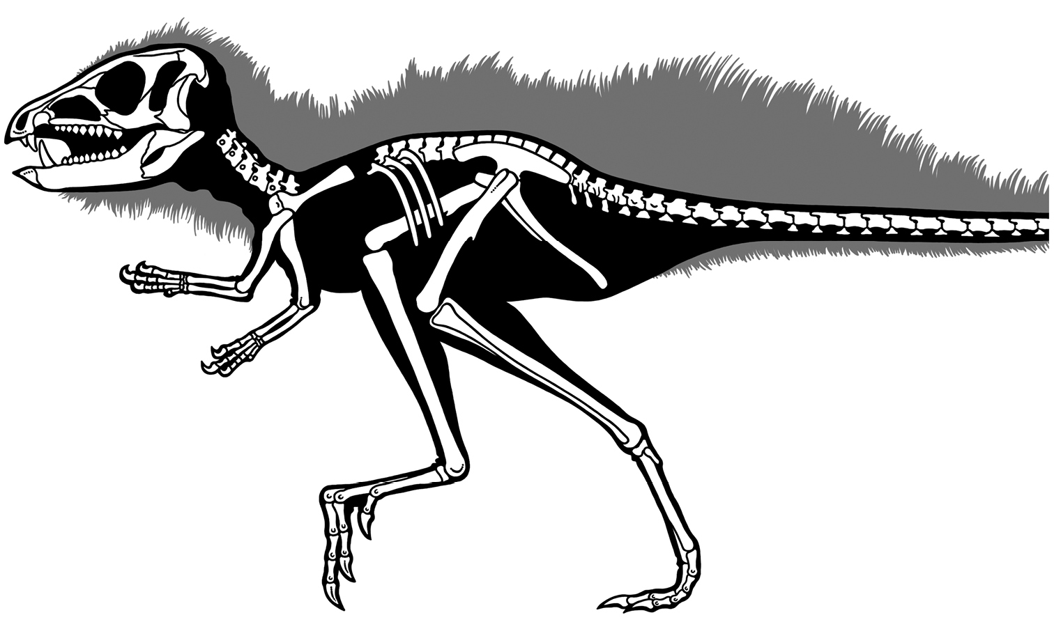
Skeleton of Heterodontosaurus tucki from the Lower Jurassic Upper Elliot and Clarens formations of South Africa. Silhouette skeletal reconstruction in lateral view showing preserved bones (based on SAM-PK-K1332). Distal most caudal vertebrae unknown.
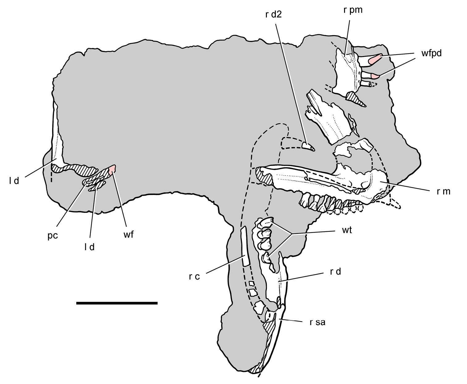
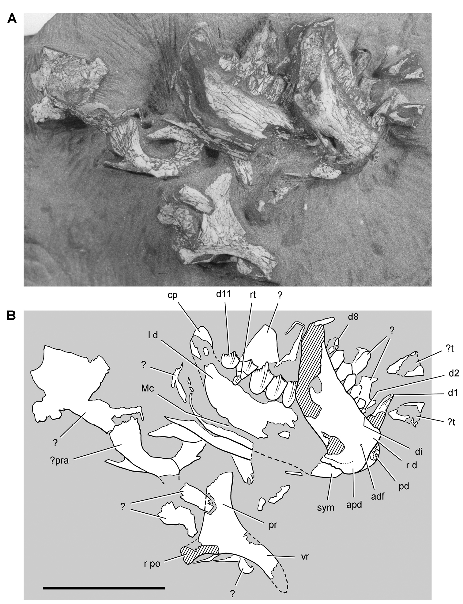
Block with remains of the heterodontosaurid Lycorhinus angustidens from the Lower Jurassic Upper Elliot Formation of South Africa. Top view of block showing the right premaxilla and maxilla and portions of the right and left lower jaws (NHMUK RU A100). Hatching indicates broken bone; dashed lines indicate estimated edges; grey tone indicates matrix; pink tone indicates wear facets. Scale bar equals 3 cm. Abbreviations: c coronoid d dentary d2 dentary tooth 2 l left m maxilla pc pulp cavity pm premaxilla r right sa surangular wf wear facet wfpd wear facet from predentary bill wt worn teeth.
Block with remains of the heterodontosaurid Lycorhinus angustidens from the Lower Jurassic Upper Elliot Formation of South Africa. Bottom view of block (NHMUK RU A100) showing portions of the right and left lower jaws. Hatching indicates broken bone; dashed lines indicate estimated edges; tone indicates matrix. Scale bar equals 3 cm. Abbreviations: a angular d dentary d1, 2 dentary tooth 1, 2 emfo external mandibular fossa imf internal mandibular fenestra l left r right sa surangular sp splenial sym symphysis.
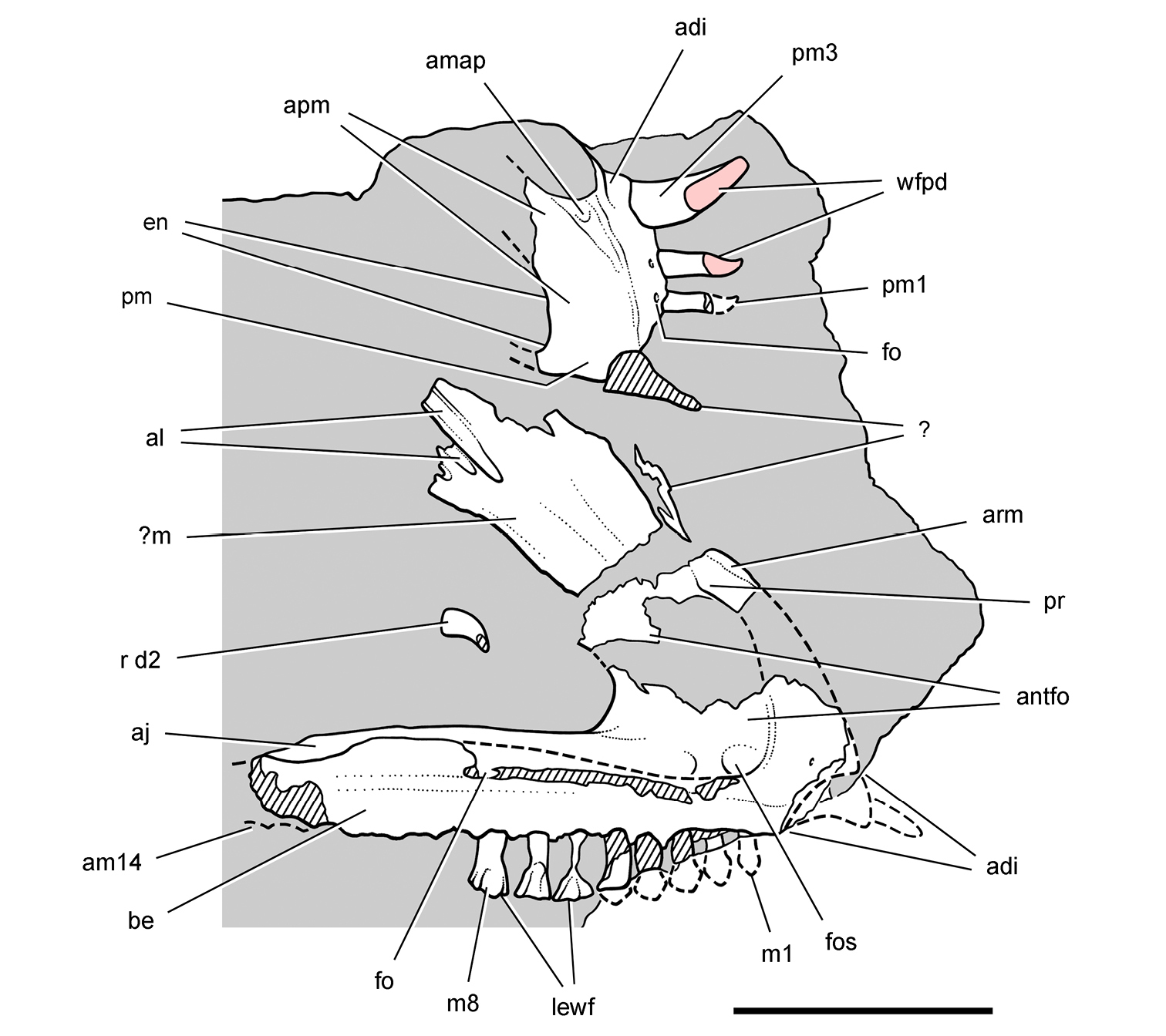
Upper jaw of the heterodontosaurid Lycorhinus angustidens from the Lower Jurassic Upper Elliot Formation of South Africa. Premaxilla and maxilla of an adult individual with worn teeth (NHMUK RU A100). Right premaxilla and maxilla in medial and lateral views, respectively. Hatching indicates broken bone; dashed lines indicate estimated edges; grey tone indicates matrix; pink tone indicates wear facets. Scale bar equals 2 cm. Abbreviations: adi arched diastema aj articular surface for the jugal al articular surface for the lacrimal am14 alveolus for maxillary tooth 14 amap articular surface for the maxillary anterior process antfo antorbital fossa apm articular surface for the opposing premaxilla arm ascending ramus of the maxilla be buccal emargination d2 dentary tooth 2 en external nares fo foramen fos fossa lewf leading edge of the wear facet m maxilla m1, 8 maxillary tooth 1, 8 pm premaxilla pm1, 3 premaxilla tooth 1, 3 pr process r right wfpd wear facet from predentary bill.
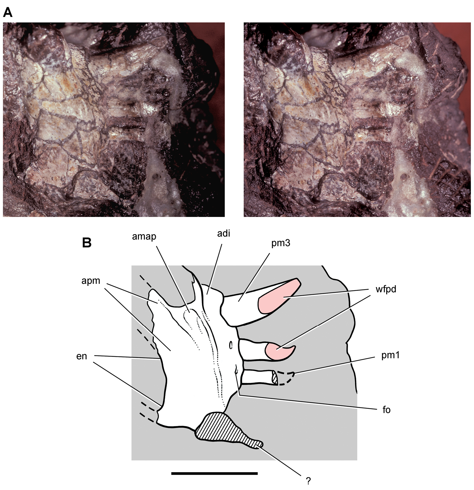
Premaxilla of the heterodontosaurid Lycorhinus angustidens from the Lower Jurassic Upper Elliot Formation of South Africa. Premaxilla of an adult individual with worn teeth (NHMUK RU A100). Stereopair (A) and line drawing (B) of the right premaxilla in medial view. Hatching indicates broken bone; dashed lines indicate estimated edges; grey tone indicates matrix; pink tone indicates wear facets. Scale bar equals 1 cm. Abbreviations: adi arched diastema amap articular surface for the maxillary anterior process apm articular surface for the opposing premaxilla en external nares fo foramen pm1, 3 premaxilla tooth 1, 3 wfpd wear facet from predentary bill.
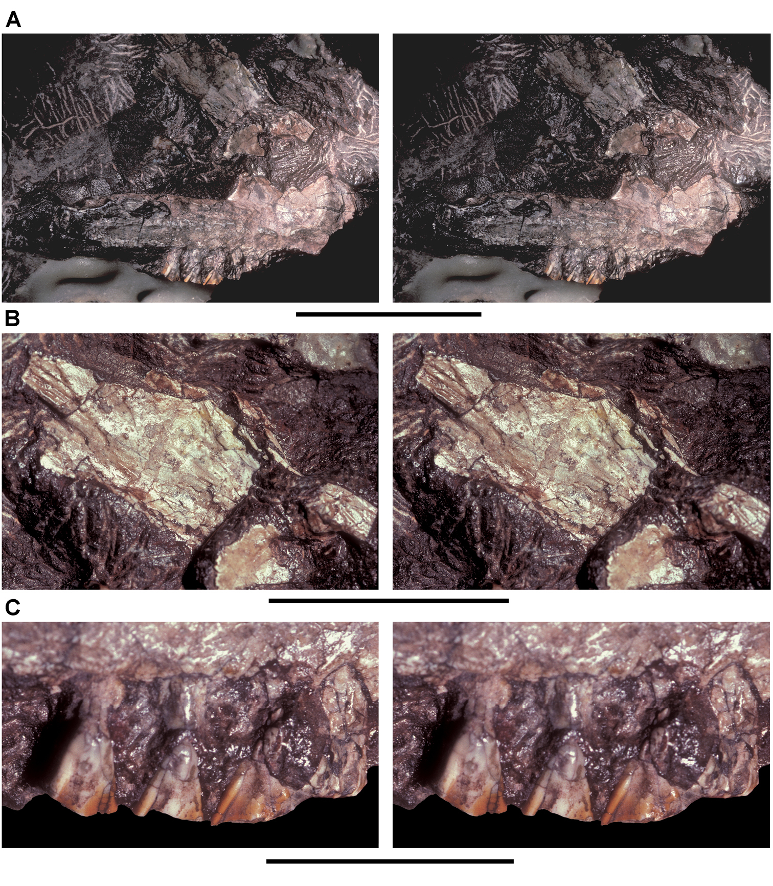
Maxilla of the heterodontosaurid Lycorhinus angustidens from the Lower Jurassic Upper Elliot Formation of South Africa. Maxilla of an adult individual with worn teeth (NHMUK RU A100). A Stereopair of the right maxilla in lateral view B Close-up view of the ascending ramus C Close-up view of the worn crowns of maxillary teeth 6–8. Scale bars equal 2 cm in A, 3 cm in B, and 1 cm in C.
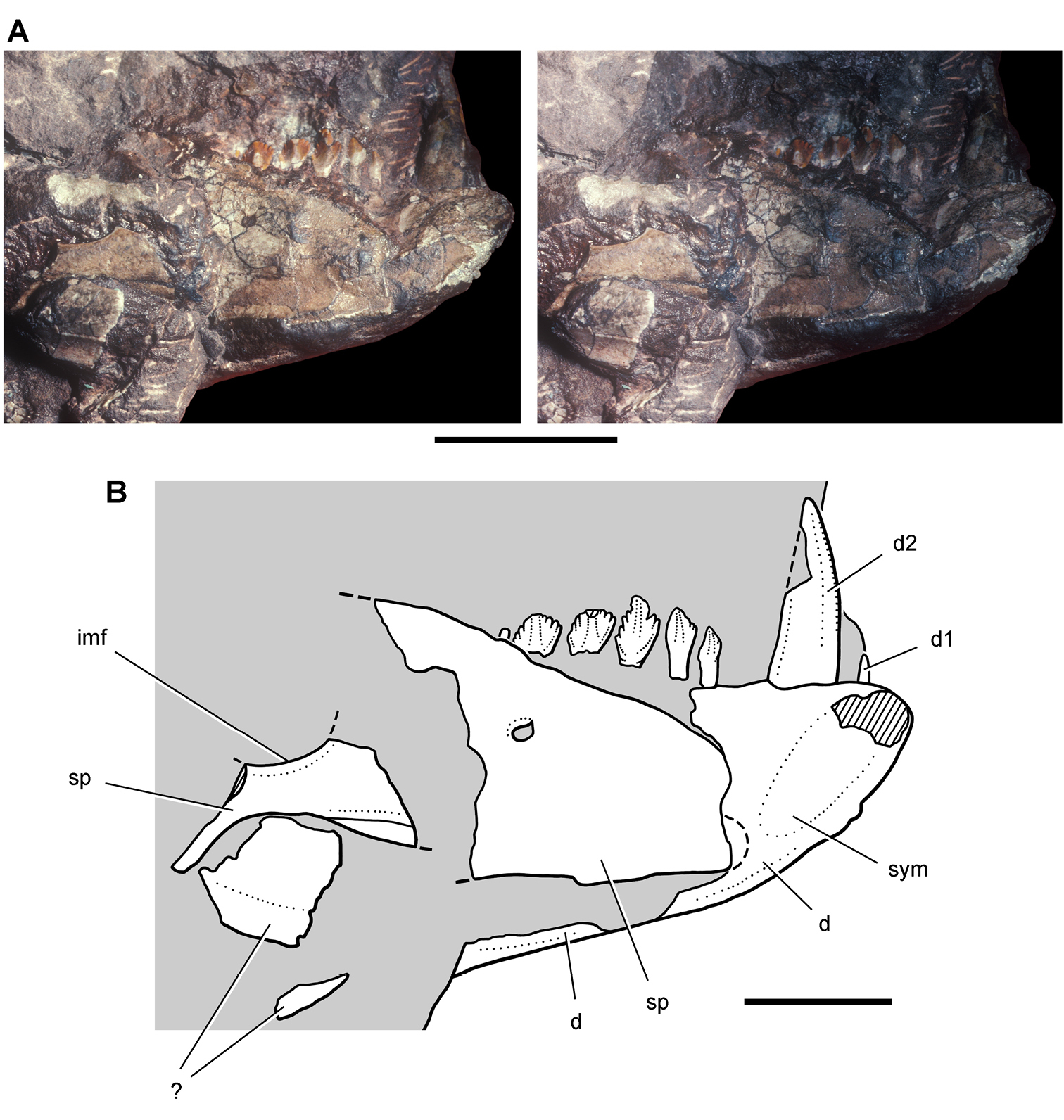
Lower jaw of the heterodontosaurid Lycorhinus angustidens from the Lower Jurassic Upper Elliot Formation of South Africa. Anterior end of the lower jaw of an adult individual (NHMUK RU A100). Stereopair (A) and line drawing (B) of the anterior end of the left lower jaw in medial view. Hatching indicates broken bone; dashed lines indicate estimated edges; tone indicates matrix. Scale bars equal 2 cm in A and 1 cm in B. Abbreviations: d dentary d1, 2 dentary tooth 1, 2 imf internal mandibular fenestra sp splenial sym symphysis.
Lower jaw of the heterodontosaurid Lycorhinus angustidens from the Lower Jurassic Upper Elliot Formation of South Africa. Posterior end of the lower jaw of an adult individual (NHMUK RU A100). Stereopair (A) and line drawing (B) of the posterior end of the right lower jaw in lateral view. Dashed lines indicate estimated edges; tone indicates matrix. Scale bars equal 2 cm. Abbreviations: a angular asaf anterior surangular foramen cp coronoid process d dentary emfo external mandibular fossa sa surangular sp splenial.
Skull of the heterodontosaurid Lycorhinus angustidens from the Lower Jurassic Upper Elliot Formation of South Africa. Skull reconstruction based on the holotypic dentary (SAM-PK-K3606) and referred specimens (NHMUK RU A100, BP/1/4244). Dashed lines indicate estimated edges. Abbreviations: adi arched diastema antfo antorbital fossa be buccal emargination d dentary d1-3, 15 dentary teeth 1–3, 15 fo foramen j jugal m maxilla m1, 14, 15 maxillary tooth 1, 14, 15 nf narial fossa pd predentary pm premaxilla pm1, 3 premaxilla tooth 1, 3.
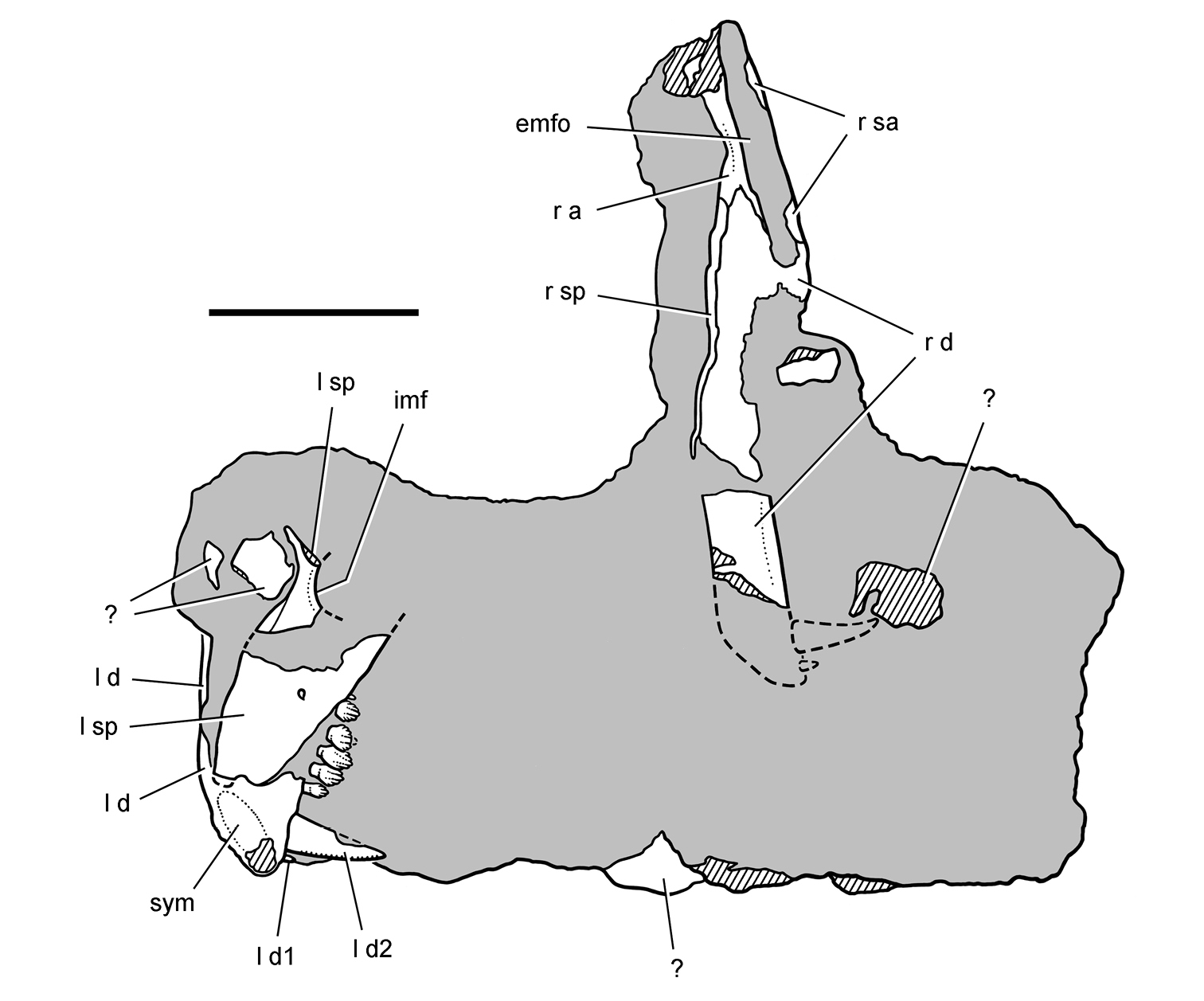
Partial skull of Manidens condorensis from the Middle Jurassic Cañadón Asfalto Formation of Argentina. Skull reconstructions in lateral view A Reversed from Pol et al. (2011) B This study. Dashed lines indicate estimated edges. Abbreviations: a angular antfo antorbital fossa asaf anterior surangular foramen be buccal emargination bo basioccipital bt basal tubera d dentary d1, 2, 11 dentary tooth 1, 2, 11 emfo external mandibular fossa f frontal gl glenoid gr groove j jugal jfl jugal flange jh jugal horn m maxilla m1, 11 maxillary tooth 1, 11 n nasal pd predentary pm premaxilla po postorbital pof postorbital fossa popr paroccipital process q quadrate qj quadratojugal ri ridge sa surangular sq squamosal.
Partial skull of the heterodontosaurid Pegomastax africanus gen. n. sp. n. from the Lower Jurassic Upper Elliot Formation of South Africa. Partial skull (SAM-PK-K10488). Photograph (A) and line drawing (B) of the partial skull preserving the postorbital and anterior portion lower jaws. Hatching indicates broken bone; dashed lines indicate estimated edges; tone indicates matrix. Scale bar equals 2 cm. Abbreviations: adf anterior dentary foramen apd articular surface for the predentary cp coronoid process d dentary d1, 2, 8, 11 dentary tooth 1, 2, 8, 11 di diastema l left Mc Meckel’s canal pd predentary po postorbital pr posterior ramus pra prearticular r right rt replacement tooth sym symphysis t tooth vr ventral ramus.
Lower jaws of the heterodontosaurid Pegomastax africanus gen. n. sp. n. from the Lower Jurassic Upper Elliot Formation of South Africa. Anterior portion of the lower jaws (SAM-PK-K10488). Stereopair of dentaries and predentary. Scale bar equals 2 cm.
Lower jaw of the heterodontosaurid Pegomastax africanus gen. n. sp. n. from the Lower Jurassic Upper Elliot Formation of South Africa. Predentary and dentaries (SAM-PK-K10488). Stereopair (A) and line drawing (B) of the predentary and anterior portion of the dentaries in ventrolateral view. Hatching indicates broken bone; dashed lines indicate estimated edges; grey tone indicates matrix. Scale bars equal 1 cm. Abbreviations: adf anterior dentary foramen apd articular surface for the predentary d dentary d1 dentary tooth 1 l left pd predentary r right sym symphysis t tooth vf vascular foramen.
Dentary teeth of the heterodontosaurid Pegomastax africanus gen. n. sp. n. from the Lower Jurassic Upper Elliot Formation of South Africa. Dentary tooth row (SAM-PK-K10488). Stereopair (A) and line drawing (B) of left dentary teeth 1-8 in lateral view. Hatching indicates broken bone; dashed lines indicate estimated edges; grey tone indicates matrix; pink tone indicates wear facets. Scale bars equal 5 mm. Abbreviations: be buccal emargination d1, 2, 5–8 dentary teeth 1, 2 5–8 wf wear facets.
Dentary teeth of the heterodontosaurid Pegomastax africanus gen. n. sp. n. from the Lower Jurassic Upper Elliot Formation of South Africa. Posterior dentary teeth (SAM-PK-K10488). Stereopair (A) and line drawing (B) of left dentary teeth 7-11 in medial view. Hatching indicates broken bone; tone indicates matrix. Scale bars equal 5 mm. Abbreviations: be buccal emargination d dentary d7, 11 dentary tooth 7, 11 l left pri primary ridge rt replacement tooth.
Lower jaw of the heterodontosaurid Pegomastax africanus gen. n. sp. n. from the Lower Jurassic Upper Elliot Formation of South Africa. Reconstruction of the predentary and dentary in lateral view (based on SAM-PK-K10488). Dashed lines indicate estimated edges. Abbreviations: adf anterior dentary foramen apd articular surface for the predentary be buccal emargination d dentary d1, 2, 11 dentary teeth 1, 2, 11 emf external mandibular fenestra pd predentary pri primary ridge.

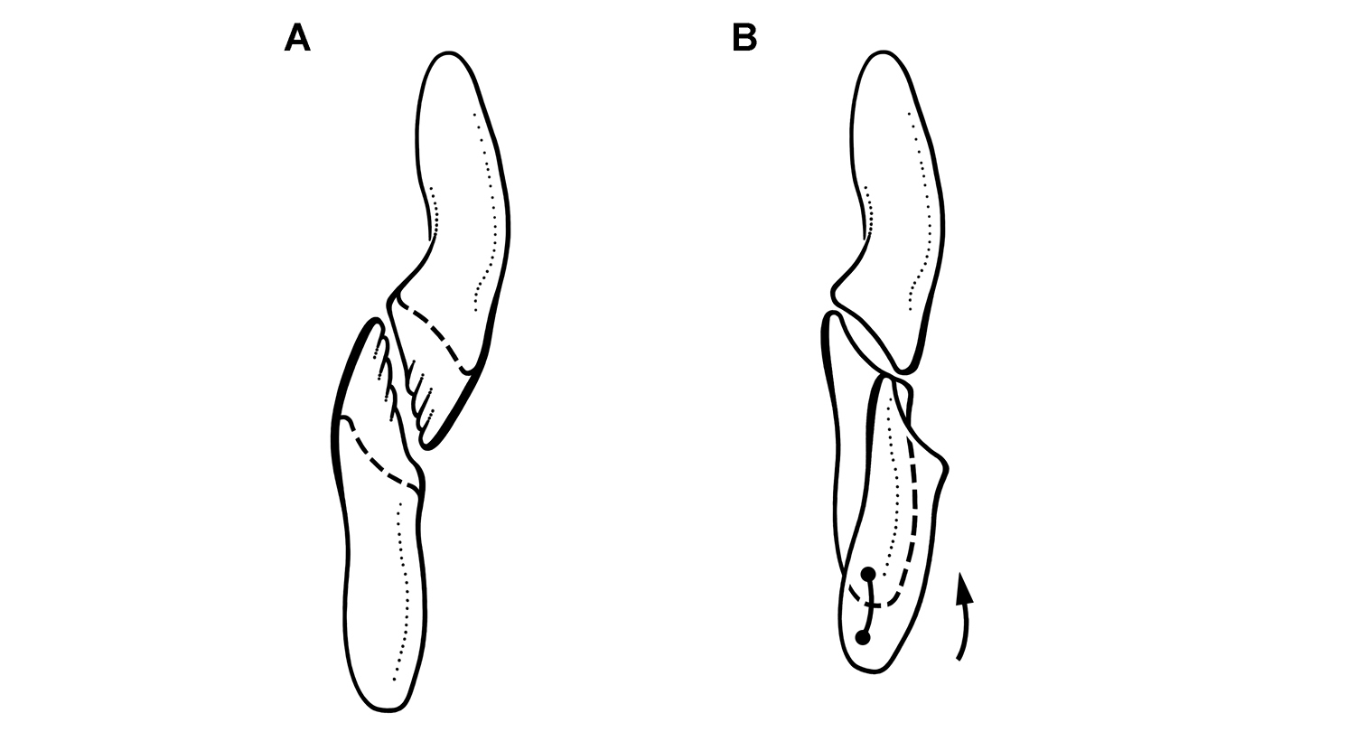
Hypotheses for jaw movement during tooth-to-tooth occlusion in Heterodontosaurus tucki. Jaw movements (arrows) to account for shearing wear facets during isognathous occlusion (from Crompton and Attridge 1986) A Dorsomedial movement of the lower jaws against stable maxillary tooth rows B Dorsal movement of the lower jaws with lateral flaring of the maxillary tooth rows C Medial rotation of the dentaries pivoting anteriorly at the predentary-dentary joint.
Occlusion and wear between maxillary and dentary teeth in Heterodontosaurus tucki. Reconstruction of left maxillary and dentary teeth in anterior view at different stages of wear A Dentition in a subadult with unworn crowns and near vertical occlusal plane with dashed lines indicating the occlusal surface of a worn crown (based on AMNH 24000) B Worn crowns from the middle of the tooth rows showing labiolingually concave wear facets, which are more concave in dentary than maxillary crowns (based on SAM-PK-K1332). The dentary tooth is shown at the initiation and conclusion of a masticatory stroke, with a dashed line showing the tooth margin hidden from view and registration dots and an arrow indicating tooth movement during occlusion.
Snout and upper dentition in Heterodontosaurus tucki. Cast of snout and upper dentition (UCRC PVC11) from an early mold of adult skull SAM-PK-K1332 A Cast of snout and upper dentition in left lateral view B Stereopair of anterior portion of the left antorbital fossa in posterolateral view. Scale bar equals 2 cm in A and 1 cm in B. Abbreviations: aantfe accessory antorbital fenestra eantfe external antorbital fenestra iantfe internal antorbital fenestra j jugal l lacrimal or left m maxilla m2, 10, maxillary tooth 2, 10 n nasal nf narial fossa pm premaxilla pm2, 3 premaxillary tooth 2, 3 pmfo promaxillary fossa r right.
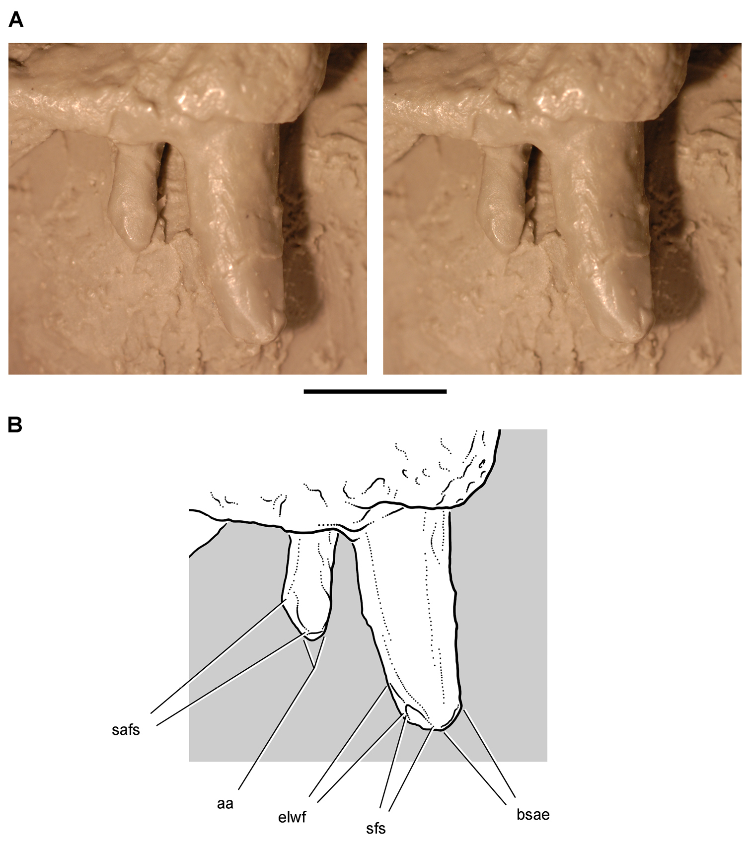
Premaxillary dentition in Heterodontosaurus tucki. Cast of premaxillary tooth 2 and 3 in left lateral view (UCRC PVC11) from an early mold of adult skull SAM-PK-K1332. Stereopair (A) and line drawing (B) showing spalling, abrasion, and the mesial and distal edges of ligual tooth-to-bill wear facets. Scale bar equals 5 mm. Abbreviations: aa apical abrasion bsae breakage surface with abraded edges elwf edge of lingual wear facet sfs spalled fracture surface safs spalled abraded fracture surface.
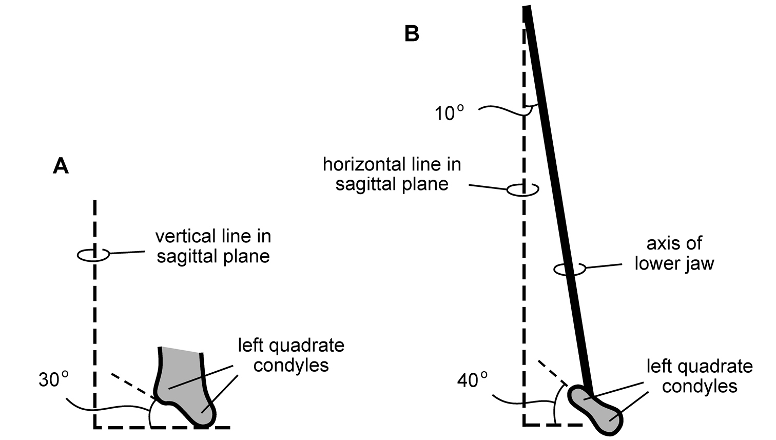
Configuration of jaw articulation and long axis of the lower jaw in Heterodontosaurus tucki from the Lower Jurassic Upper Elliot and Clarens formations of South Africa. Diagrammatic depiction of jaw configuration based on subadult skull AMNH 24000 and adult skull SAM-PK-K1332 A Left quadrate condyles in anterior view showing 30° angle from the horizontal B Left quadrate condyles and long axis of the lower jaw in ventral view showing 40° angle from a transverse axis and 10° divergence from the midline, respectively.
Relative position of the lower jaw in Heterodontosaurus tucki from the Lower Jurassic Upper Elliot and Clarens formations of South Africa. Position of the lower jaw during occlusion (A) and moderate gape (B) as seen in left lateral view of a cast of the cranium and lower jaws of adult specimen SAM-PK-K1332. Scale bar equals 2 cm.
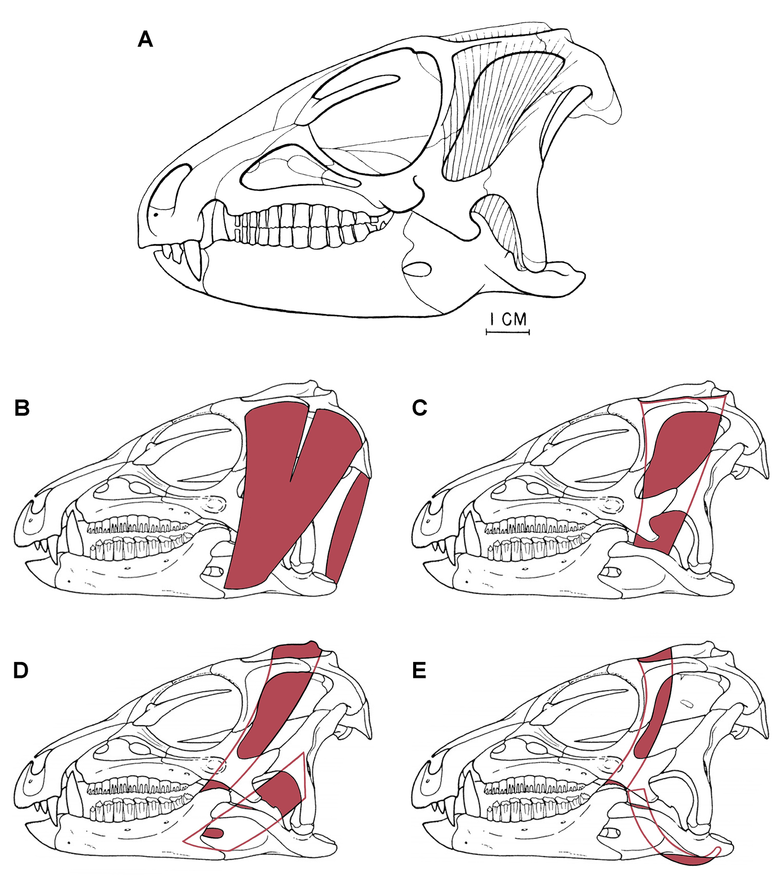
Previous jaw muscle reconstructions in Heterodontosaurus tucki from the Lower Jurassic Upper Elliot and Clarens formations of South Africa A Initial adductor muscle reconstruction (from Crompton and Attridge 1986) B Reconstruction of the adductor mandibulae externus superficialis (left) and the depressor mandibulae (right) (adapted from figures in Norman et al. 2011) C Reconstruction of the adductor mandibulae externus medialis (adapted from figures in Norman et al. 2011) D Reconstruction of the adductor mandibulae externus profundus (left) (misidentified as “adductor mandibulae externus posterior” in legend for Fig. 35, Norman et al. 2011) and the adductor mandibulae posterior (right) (adapted from figures in Norman et al. 2011) E Reconstruction of the pseudotemporalis (left) and the pterygoideus posterior (right) (adapted from figures in Norman et al. 2011).
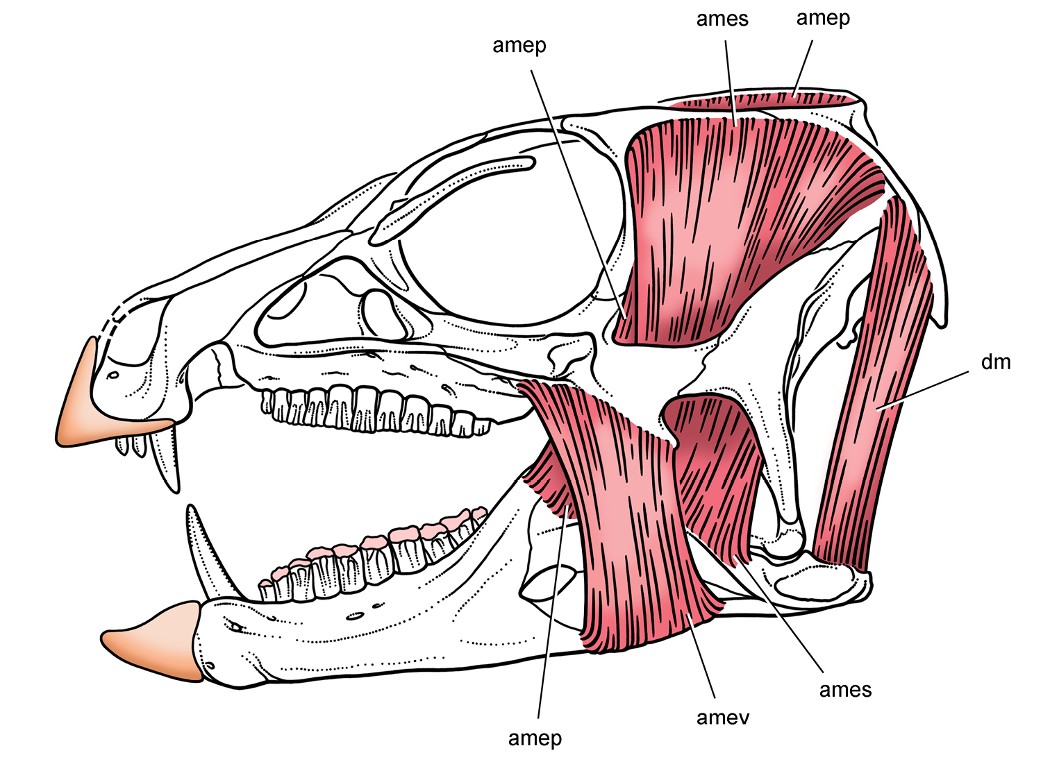
Jaw musculature and keratin sheathing in Heterodontosaurus from the Lower Jurassic Upper Elliot and Clarens formations of South Africa. Reconstruction of select jaw muscles and keratin sheathing of upper and lower bills supported in this study. Pink tone indicates wear facets. Abbreviations: amep adductor mandibulae externus profundus ames adductor mandibulae externus superficialis amev adductor mandibulae externus ventralis dm depressor mandibulae.
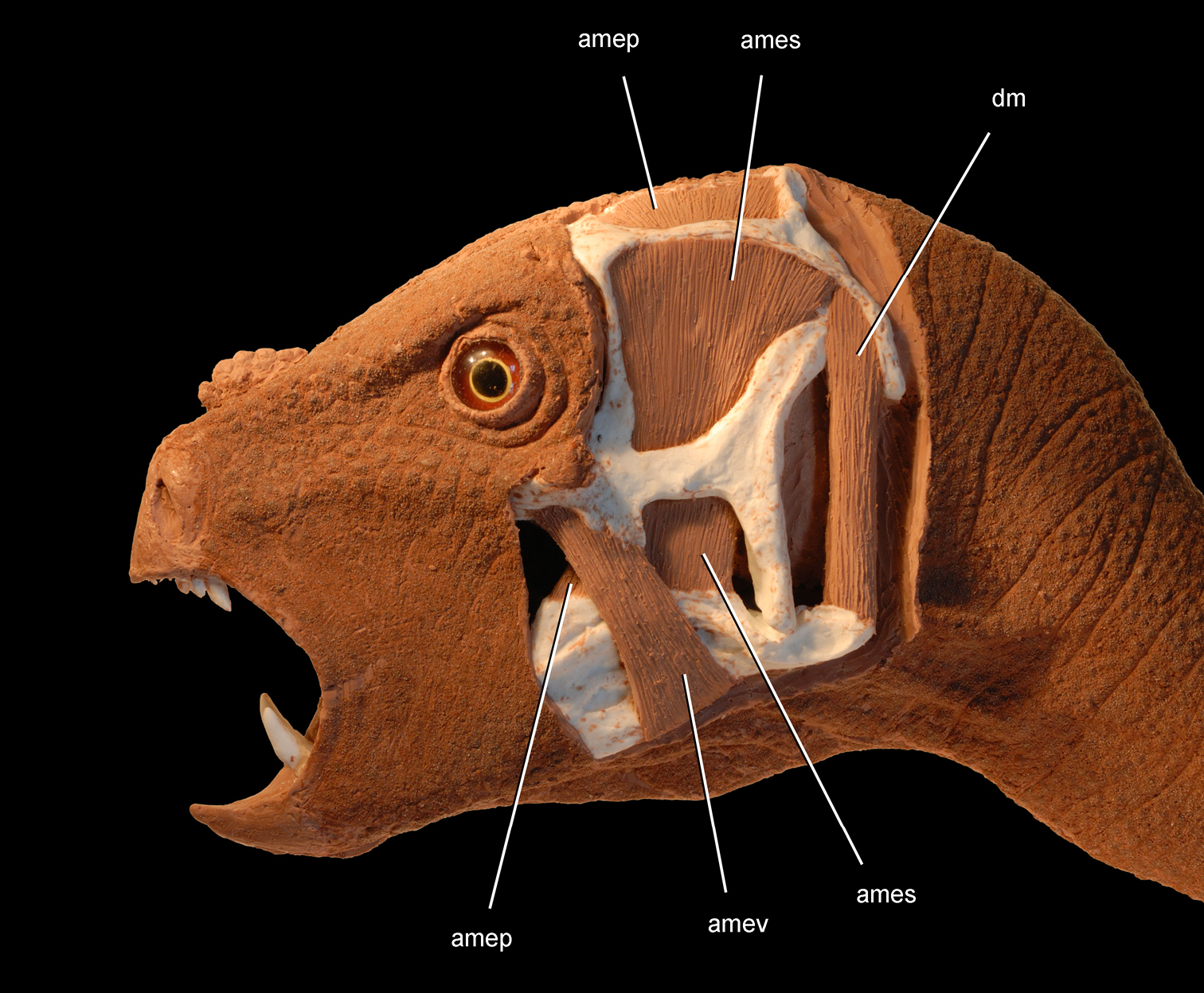
Jaw adductor musculature in Heterodontosaurus tucki from the Lower Jurassic Upper Elliot and Clarens formations of South Africa. Flesh head sculpting over a cast of an adult skull (SAM-PK-K1332) showing surface tissues (anterior half) and select jaw muscles (posterior half). Abbreviations: amep adductor mandibulae externus profundus ames adductor mandibulae externus superficialis amev adductor mandibulae externus ventralis dm depressor mandibulae.
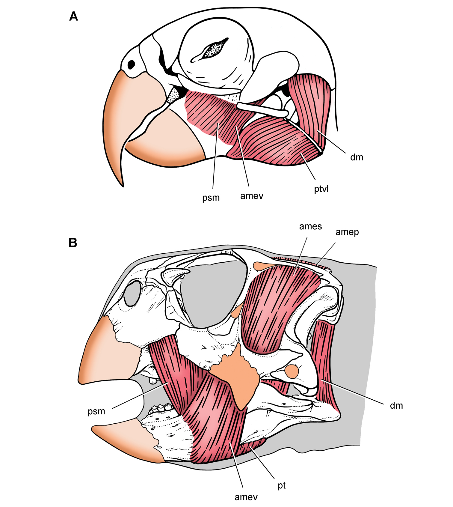
Jaw adductor musculature and keratin sheathing in a psittaciform bird and parrot-beaked dinosaur. Differentiation of facial components of the adductor muscle mass to increase bite force A Pionites leucogaster (white-bellied parrot) (from Sereno et al. 2010) B Psittacosaurus gobiensis with inferred jaw muscles and keratin sheathing (from Sereno et al. 2010). Abbreviations: amep adductor mandibulae externus profundus ames adductor mandibulae externus superficialis amev adductor mandibulae externus ventralis dm depressor mandibulae psm pseudomasseter pt pterygoideus ptvl pterygoideus ventralis lateralis.
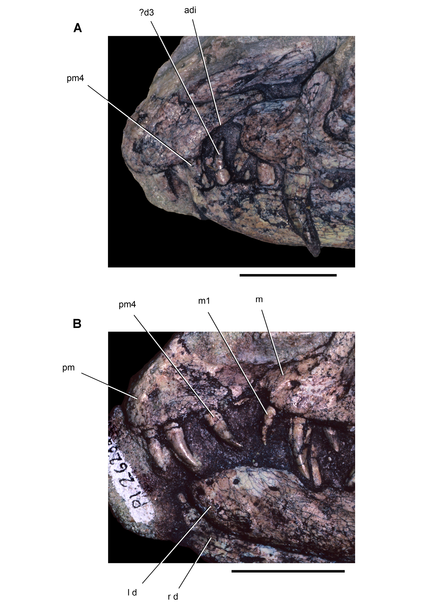
Snout and anterior dentition of dinosaurian carnivores. Snout and anterior dentition in left lateral view of coelophysoid theropods A Coelophysis bauri (MCZ 4327) from the Late Triassic B “Syntarsus” kayentakatae (MNA V2623) from the Early Jurassic. Scale bars equal 2 cm. Abbreviations: adi arched diastema d dentary d3 dentary tooth 3 l left m maxilla m1 maxillary tooth 1 pm premaxilla pm4 maxillary tooth 4 r right.
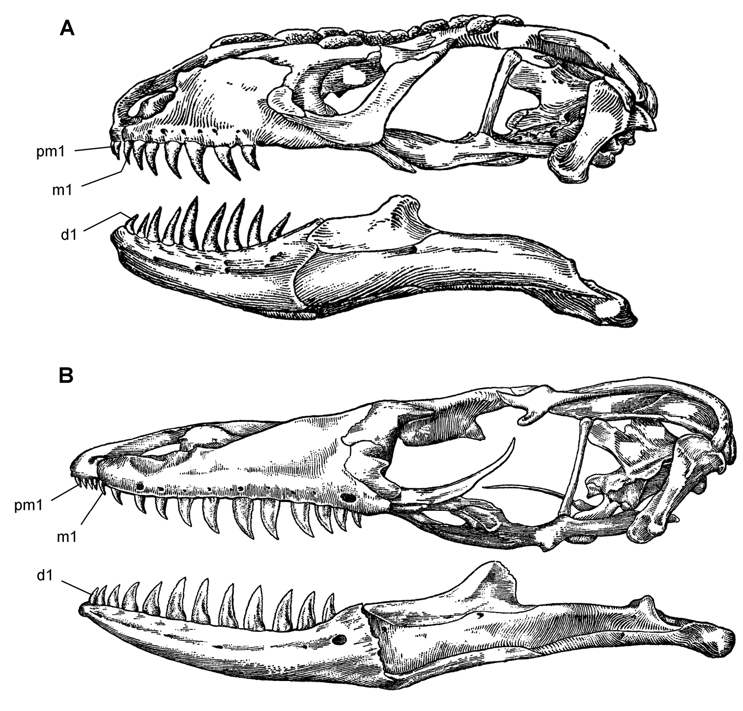
Skulls of extant lepidosaurian carnivores. Skulls in left lateral view (from McDowell and Bogert 1954) A Heloderma horridum (beaded lizard) B Varanus varius (lace monitor). Abbreviations: d1 dentary tooth 1 m1 maxillary tooth 1 pm1 premaxillary tooth 1.
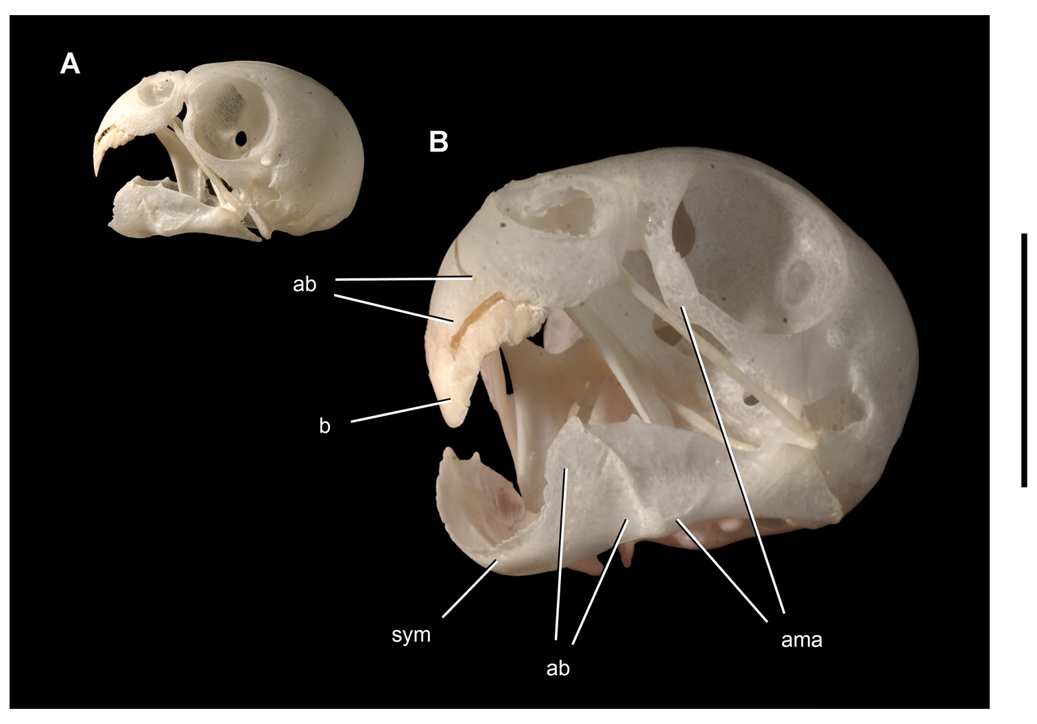
Bone and keratinous sheath in the upper bill of a parrot. Skull of the budgerigar Melopsittacus undulatus (UCRC R1) with keratinous sheath of upper bill A Lateral view B Magnified anterolateral view. Scale bar equals 1cm in B. Abbreviations: ab attachment platform for bill ama area of muscle attachment b bill sym symphysis.
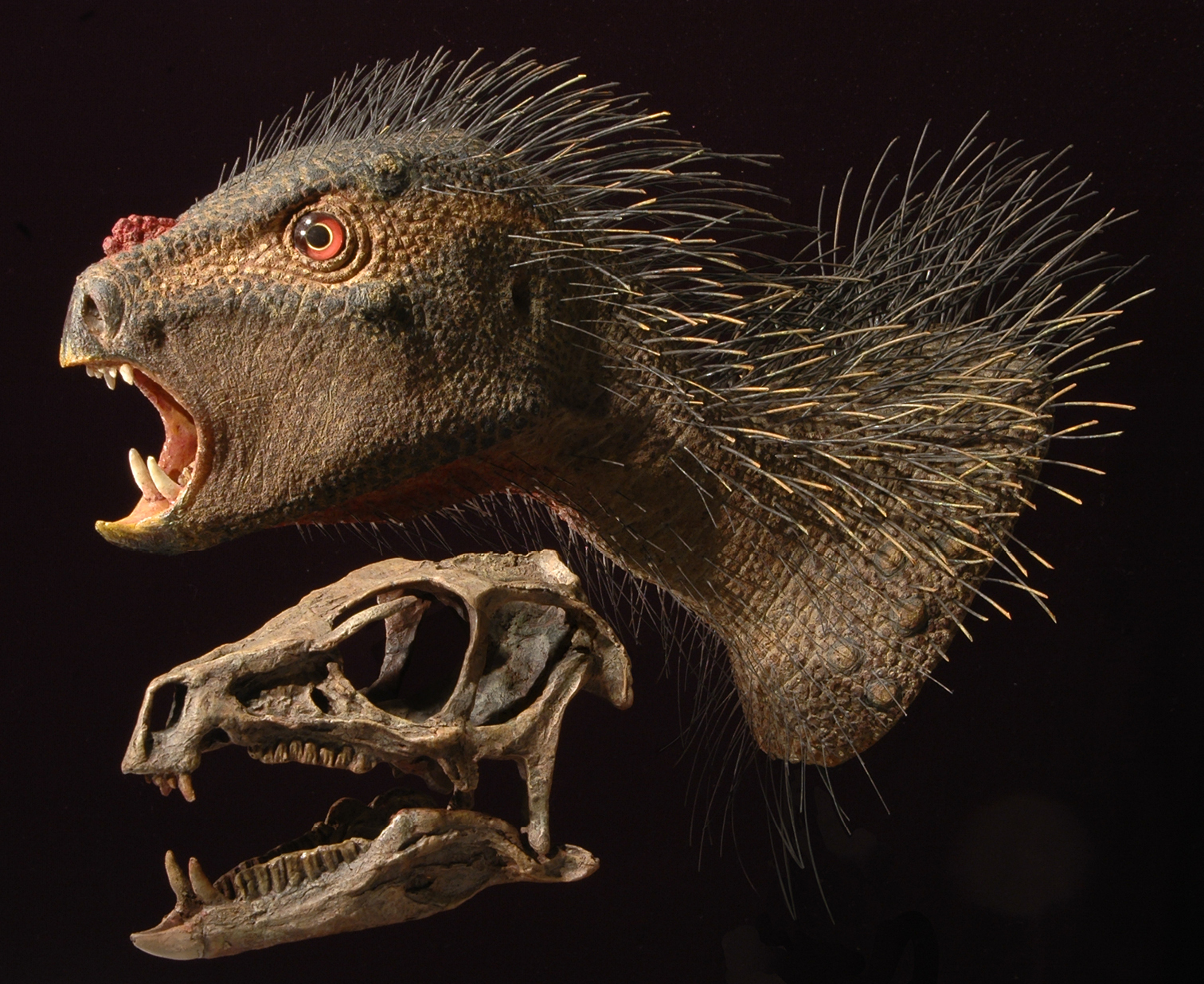
Flesh reconstruction of the head and neck of Heterodontosaurus tucki from the Lower Jurassic Upper Elliot and Clarens formations of South Africa. Flesh head and neck and skull reconstructions with similar gape. The flesh reconstruction was modeled over a skull cast with skin texture based on bone surface details in the skull of Heterodontosaurus tucki (SAM-PK-K1332) and bristles based on those preserved in Tianyulong confuciusi (STMN 26-3). Reconstructions by Tyler Keillor.

Initial phylogenetic hypothesis for heterodontosaurid relationships. Qualitative parsimony analysis by Sereno (1986) with Heterodontosauridae as the sister taxon to Euornithopoda (= Ornithopoda) and with Abrictosaurus basal to Lycorhinus and Heterodontosaurus among heterodontosaurids. Some terminal taxa in the original analysis are collapsed into suprageneric taxa; red text indicates heterodontosaurid taxa.
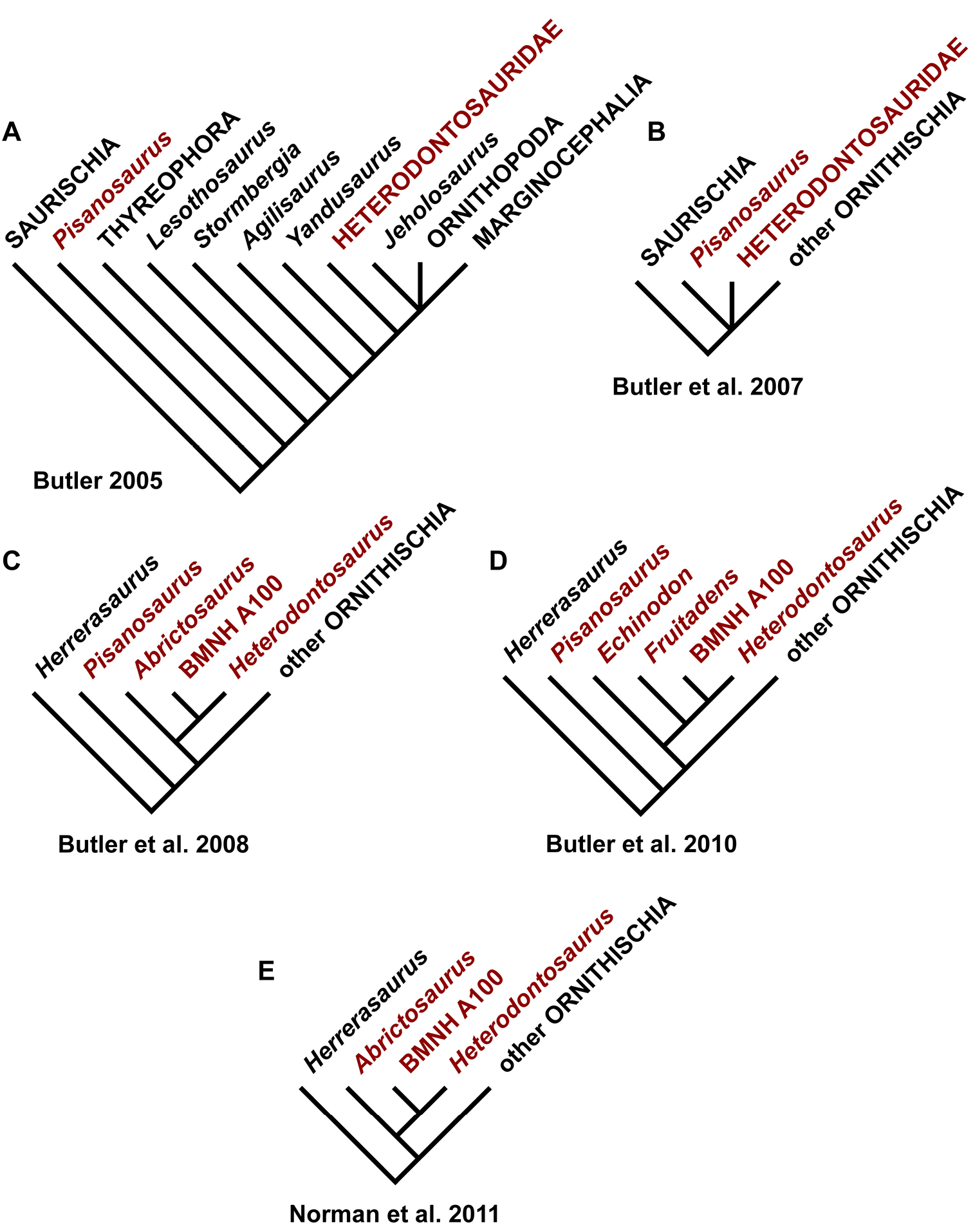
Phylogenetic hypotheses for heterodontosaurids by Butler and colleagues. A Heterodontosaurids with the exception of Pisanosaurus allied with euornithopods (after Butler 2005) B Heterodontosaurids and Pisanosaurus as basal ornithischians (after Butler et al. 2007) C Heterodontosaurids and Pisanosaurus as basal ornithischians with Lycorhinus (NHMUK RU A100, formerly BMNH A100) closer to Heterodontosaurus than Abrictosaurus (after Butler et al. 2008) D Heterodontosaurid relationships including Echinodon and Fruitadens as basal taxa (after Butler et al. 2009) E Heterodontosaurid relationships as in Butler et al. (2008) (after Norman et al. 2011). Some terminal taxa in the original analyses are collapsed into suprageneric taxa; red text indicates heterodontosaurid taxa; “BMNH A100” is now catalogued as NHMUK RU A100.
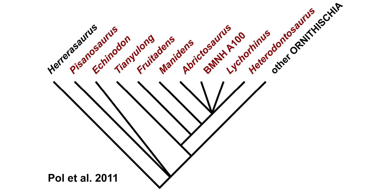
More inclusive phylogenetic hypothesis for heterodontosaurids. Parsimony analysis of Pol et al. (2011) based on character data modified from Butler et al. (2009). The analysis includes the South American genus Manidens and the Asian genus Tianyulong. Some terminal taxa in the original analysis are collapsed into suprageneric taxa; red text indicates heterodontosaurid taxa; “BMNH A100” is now catalogued as NHMUK RU A100.
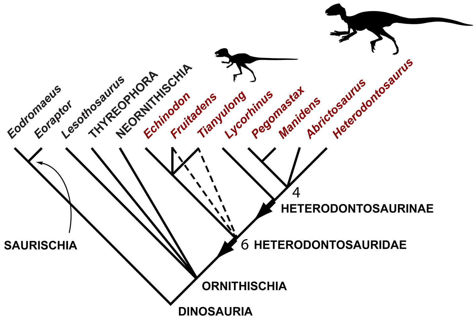
Phylogenetic hypothesis for heterodontosaurids based on character data assembled in this study. Consensus tree summarizing 6 minimum-length trees (38 steps) based on maximum parsimony analysis of 30 characters in 8 heterodontosaurid genera (consistency index = 0.79; retention index = 0.88; Appendix I). Outgroups (constrained as shown) include basal saurischians (Eoraptor, Eodromaeus) and representative ornithischians (Lesothosaurus, Thyreophora, Neornithischia). Dashed lines indicate loss of resolution with an increase of one step in tree length; numbers at nodes indicate the decay index when four poorly known heterodontosaurids (Echinodon, Fruitadens, Lycorhinus, Pegomastax) are removed from the analysis. Red text highlights heterodontosaurid genera; arrows indicate stem-based definitions for Heterodontosauridae and Heterodontosaurinae; body icons of Tianyulong and Heterodontosaurus are shown at appropriate relative size (based on Figures 30 and 72).
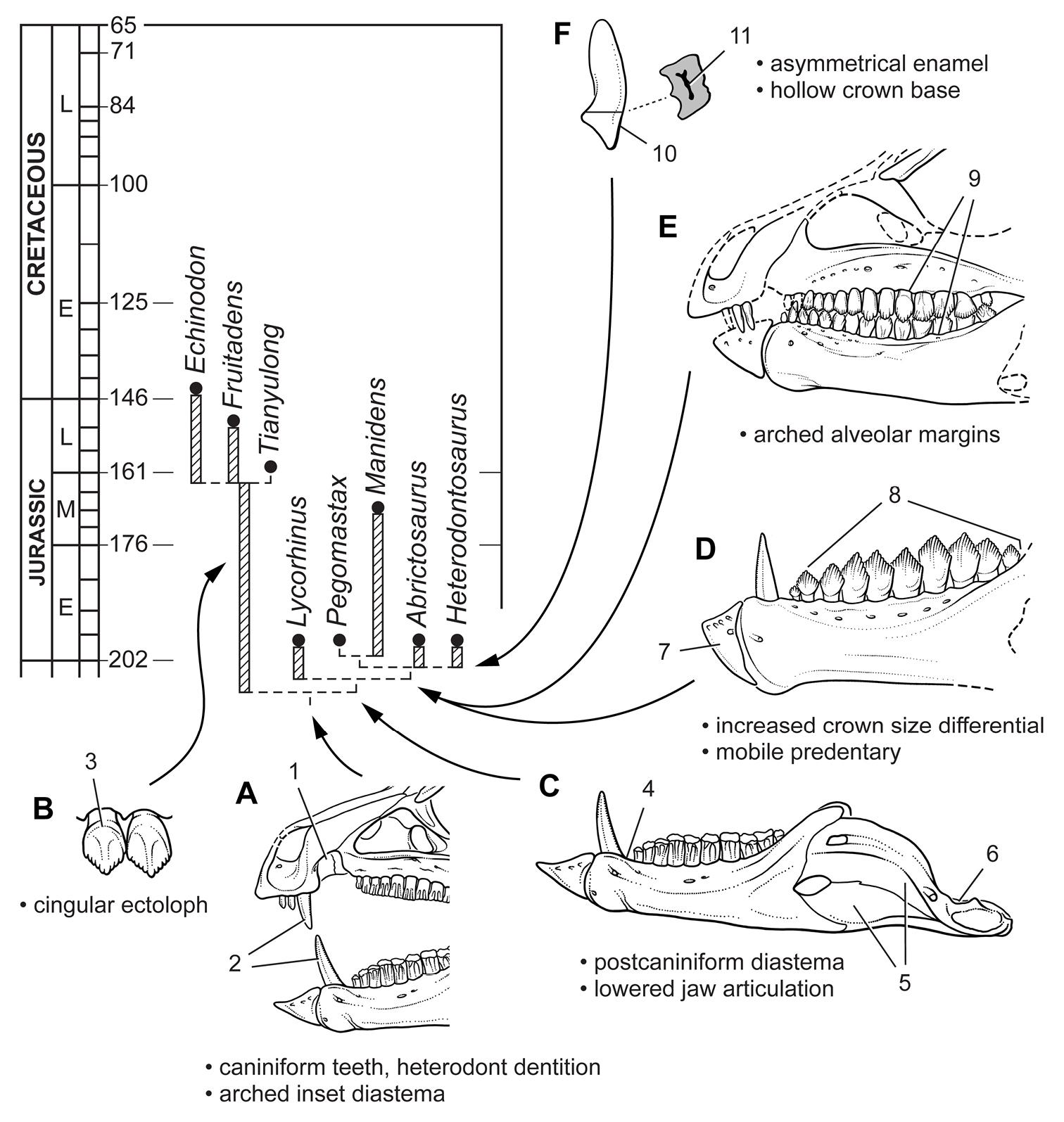
Evolution of key masticatory specializations among heterodontosaurids. A Heterodontosauridae B Simple-crowned heterodontosaurids C Heterodontosaurinae D Advanced heterodontosaurines E Abrictosaurus and Heterodontosaurus F Heterodontosaurus. Abbreviations: 1 arched inset diastema 2 heterodont dentition with caniniform teeth 3 cingular ectoloph 4 postcaniniform diastema 5 external mandibular fossa 6 lowered jaw articulation 7 mobile predentary articulating against saddle-shaped dentary articular surface 8 increased differential in crown size 9 arched alveolar margins in cheek dentition 10 asymmetrical enamel 11 hollow crown base.
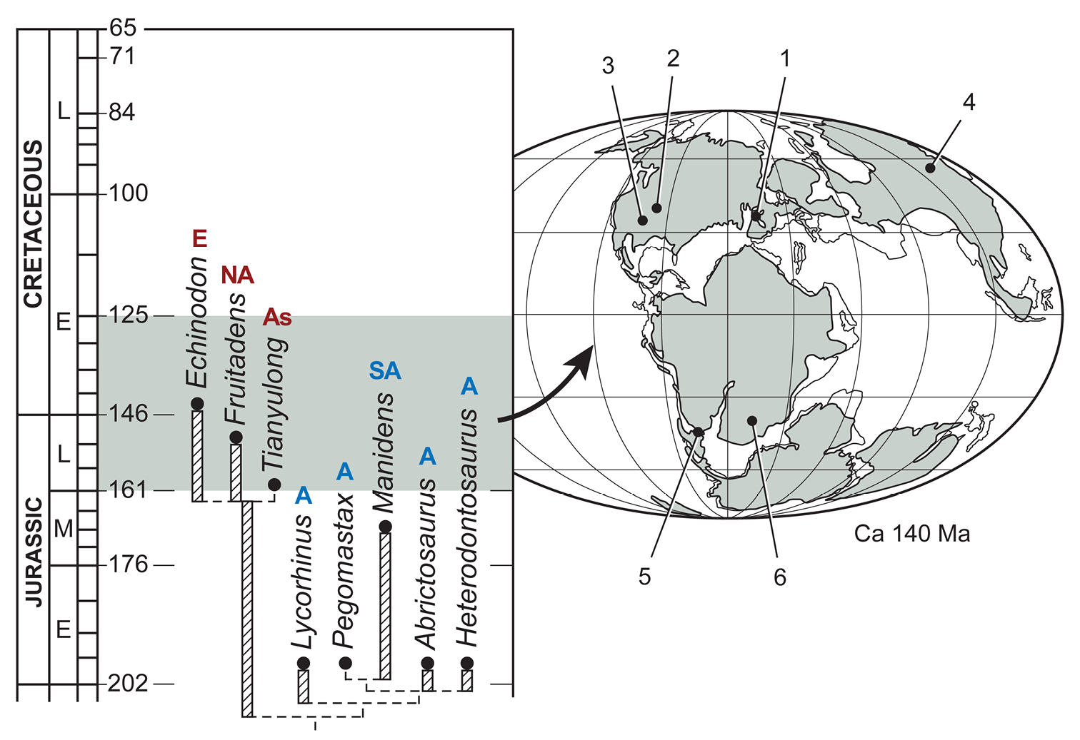
Biogeographic distribution in time for heterodontosaurids included in the phylogenetic analysis. Localities for included taxa are plotted on an Early Cretaceous (Berriasian-Valanginian) paleogeographic map (Smith et al. 1994) and recent time scale (Gradstein and Ogg 2009). Abbreviations for taxon localities and biogeographic areas: 1 Echinodon 2 Fruitadens 3 Kayenta heterodontosaurid 4 Tianyulong 5 Manidens 6 Abrictosaurus, Lycorhinus, Heterodontosaurus, Pegomastax A Africa As Asia E Europe NA North America SA South America.