Related articles by
Research Article
Other versions:


- ContentsContents
- Article InfoArticle Info
- CiteCite
- MetricsMetrics
- CommentComment
- RelatedRelated
- FigsFigs
- TabsTabs
- MapMap
- TaxaTaxa
- RefsRefs
- CitedCited
- Inst. CodeInst. Code
- NanopubsNanopubs
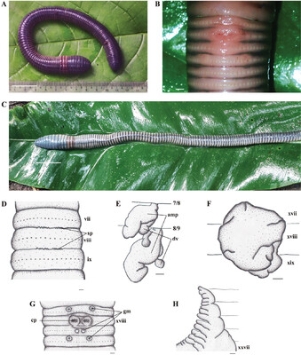
Figure 4.
Metaphire rusydii sp. n., holotype. A Living specimen (dorsal view); B Living specimen (ventral view of male pore region); C Preserved specimen (dorsal view); D Spermathecal pore; E Spermathecae, right side on intrasegmental 7/8/9 (am = ampulla; dv = diverticulum); F Prostate gland; G Male pore region (cp = opening copulatory pouch); H Intestinal caecum. Scale bars: 50 mm (A), 1 mm (C–H).
Subscribe to email alerts for current Article's categories