Monograph |
|
Corresponding author: Maria Cristina Díaz ( taxochica@gmail.com ) Academic editor: Pavel Stoev
© 2023 Maria Cristina Díaz, Marissa Nuttall, Shirley A. Pomponi, Klaus Rützler, Sarah Klontz, Christi Adams, Emma L. Hickerson, G. P. Schmahl.
This is an open access article distributed under the terms of the Creative Commons Attribution License (CC BY 4.0), which permits unrestricted use, distribution, and reproduction in any medium, provided the original author and source are credited.
Citation:
Díaz MC, Nuttall M, Pomponi SA, Rützler K, Klontz S, Adams C, Hickerson EL, Schmahl GP (2023) An annotated and illustrated identification guide to common mesophotic reef sponges (Porifera, Demospongiae, Hexactinellida, and Homoscleromorpha) inhabiting Flower Garden Banks National Marine Sanctuary and vicinities. ZooKeys 1161: 1-68. https://doi.org/10.3897/zookeys.1161.93754
|
Abstract
Sponges are recognized as a diverse and abundant component of mesophotic and deep-sea ecosystems worldwide. In Flower Garden Banks National Marine Sanctuary region within the northwestern Gulf of Mexico, sponges thrive among diverse biological and geological habitats between 16–200+ m deep (i.e., coral reefs and communities, algal nodules, and coralline algae reefs, mesophotic reefs, patch reefs, scarps, ridges, soft substrate, and rocky outcrops). A synoptic guide is presented, developed by studying common sponge species in the region, through direct sampling and in-situ photographic records. A total of 64 species is included: 60 are Demospongiae (14 orders), two are Hexactinellida (one order), and two are Homoscleromorpha (one order). Thirty-four taxa are identified to species and 13 were identified to have affinity with, but were not identical to, a known species. Fifteen taxa could only be identified to genus level, and the species remain as uncertain (incerta sedis), with the potential to represent new species or variants of known species. One specimen received only a family assignation. This study extends geographic or mesophotic occurrence data for eleven known species and includes several potentially new species. This work improves our knowledge of Gulf of Mexico sponge biodiversity and highlights the importance of the region for scientists and resource managers.
Keywords
Algal reefs, biodiversity, Gulf of Mexico, mesophotic reefs, Porifera, sponges
Introduction
Flower Garden Banks National Marine Sanctuary consists of portions of 17 topographic features in the northwestern Gulf of Mexico. The reefs and banks occur along the continental shelf, from 70–120 miles off the coast of Texas and Louisiana (Fig.

Sponges are recognized as a diverse and abundant component of mesophotic and deep-sea ecosystems (
The major goal of this study was to update current knowledge of Porifera biodiversity from mesophotic depths at the sanctuary region and to promote this knowledge among major stakeholders. We have developed a synoptic identification guide that can be used by a wide range of end-users (i.e., marine scientists and students, conservationists, environmental managers, naturalists, recreational divers, etc.). This identification guide summarizes the current taxonomic status and distinct features for 63 species distinguished of the common sponge species encountered in the region. This work will improve our knowledge of sponge biodiversity in the Gulf of Mexico and enhance studies of sponges from mesophotic and deep-water ecosystems in the region. Furthermore, this first illustrated guide to species at mesophotic depths in the northwestern Gulf of Mexico will facilitate the comparisons with recently studied mesophotic sponge fauna from other deep mesophotic habitats occurring at Pulley Ridge in the southeast Gulf of Mexico (MCD unpublished data), Cuba (
Materials and methods
Area of study
This study focused on topographic features in the northwestern Gulf of Mexico in and around Flower Garden Banks National Marine Sanctuary located on the continental shelf edge in the northwestern Gulf of Mexico in the USA (Fig.
Collection methods and data
Collections were made using one of three remotely operated vehicles (ROV), including Phantom S2, owned and operated by
University of North Carolina at Wilmington (

Species data and morphological characterization
Each species within this guide is represented by an in-situ image, the lowest available scientific name, species author/date, higher taxonomy, depth, and sample number (indicated in the figure legend). The species data are divided in six sections: “Diagnostic features” describes distinctive morphologic features; Similar species with which it might be confused; “Distribution and abundance” refers to overall regional distribution from the World Porifera Database and other recent references (
Fifty-two of the 64 species included on this study were identified by the analysis of one or more samples. Therefore, the majority (~ 84%) of the identifications were confirmed by evaluation of skeletal morphology (skeleton type, size, and architecture) as well as features of the external morphology. Skeleton analysis was carried out using methods described in
We use the same criteria to describe the external morphology as in the recent guide to sponges from deep marine protected areas from the southeastern USA (
Species checklist terms and abbreviations
aff. affinis; the species might appear similar but is not that species. Implies a higher degree of uncertainty compared to cf. (
cf. confer; to be compared with. Indicates that most of the diagnostic characters correspond to a given species, but some characters are unclear. The identification is provisional but is likely to be definitive after comparing with reference material or consulting a specialist of the taxon (
FGBNMS Flower Garden Banks National Marine Sanctuary;
GOM Gulf of Mexico;
sp. nov. new species to science. Specimen has unique characters that can support our interpretation about its distinct and unique identity;
spp. species in plural. Refers to multiple species from a particular genus.
Results
Taxonomic scope
Sixty-four species were identified from a collection of 54 samples with photographs and ten photographs (without samples) from inside and around Flower Garden Banks National Marine Sanctuary (Suppl. material
Geographic scope
Eleven species included in this study are either first reports for the occurrence of that species at mesophotic depths, or first occurrences in the Gulf of Mexico or specifically in the northwestern Gulf of Mexico. Biemna cribaria, Placospherastra antillensis, Batzella rubra, and Erylus trisphaerus are here reported at mesophotic depths (> 50 m deep) for the first time. First reports in the Gulf of Mexico include Stellettinopsis megastylifera, Erylus alleni, and Erylus goffrilleri. First reports in the northwestern area of the Gulf of Mexico include Agelas dilatata, Neophrissospongia cf. nolitangere, Erylus trisphaerus, and Ircinia campana.
The occurrence and qualitative abundance estimate for most of the species in this study, along 17 banks or features in Flower Garden Banks National Marine Sanctuary and vicinity are summarized in the Suppl. material
Taxonomic accounts
Phylum Porifera
Class Demospongiae
Subclass Heteroscleromorpha
Order Agelasida
Family Agelasidae
Agelas cf. citrina
Diagnostic features

Similar species
Agelas clathrodes has key-holed and round oscula. Agelas cerebriformis has a convoluted surface but it is brown and tubular.
Distribution and abundance
Agelas citrina occurs on shallow coral reefs throughout the Caribbean and the Florida Keys. Found on mesophotic reefs at FGBNMS, Cuba (50–61 m deep), and South Carolina MPA (52 m depth). At FGBNMS, rare to moderate in abundance and a widespread distribution occurring at 11 sites.
Ecology
Lower mesophotic reefs and heavily silted reefs in FGBNMS region. A strong, particular garlic-like odor is associated with all regional variants. This morphotype is referred to as cf. since it does not have the typical flabellate shape nor the orangish color.
Identification
KR, SK, CA, MCD.
References
Agelas clathrodes
Diagnostic features

Similar species
Agelas citrina flabellate specimens are similar but lack key holed oscula and usually have a paler pinkish or yellowish color.
Distribution and abundance
Common in shallow and mesophotic reefs in North and South Carolina, eastern Florida, throughout the Caribbean, the Guyana shelf, and Brazil.
Ecology
Coral reefs, coral communities, and coralline algae reefs in FGBNMS region.
Identification
MCD, MFN.
References
Agelas dilatata
Diagnostic features
Flabellate to fan- and cup-shaped, < 3 cm thick, sometimes pedunculated. Brown in color. The surface is smooth with abundant and homogeneously arranged round oscula (4–10 mm) on the upper side, and small unevenly dispersed ostia (1–2 mm wide) on the underside.

Similar species
Agelas dispar, a fan-shaped brown species, which is thicker and possesses mostly key-holed oscula.
Distribution and abundance
Previously considered restricted to the Bahamian-Greater Antilles shallow coral reefs (18–30 m deep) and Cuba (90–115 m). This is the first report for the NW GOM, where it is rare at Sonnier Bank.
Ecology
Coralline algae reefs. Specimen is overgrown by a film of green algae. A unique alkaloid isolated from a Yucatan specimen is bioactive against a multidrug-resistant pathogen Pseudomonas aeruginosa (
Identification
MCD.
References
Order Axinellida
Family Axinellidae
Ptilocaulis cf. walpersii
Diagnostic features
Flagelliform branching; single or multiple branches (ca. 1–2 cm wide, ≤ 50 cm height). Red to orange in color. Branches have different lengths, and they can be straight, bent, or laterally fused forming flabellate bodies. Surface rugose and porous, with flattened or rounded processes. Oscula are sparse along the side of branches, hardly visible. Branches are compressible and firm. The identification given to this specimen is based on the external morphology and observations of the live photo.

Similar species
Ptilocaulis marquezi (with oxeas and styles) and Higginsia coralloides (with acanthose micro-oxeas added to large oxeas and styles). Ptilocaulis walpersii has only styles as spicules. The cf. is placed since the spicules could not be corroborated. Higginsia coralloides consists of shorter (≤ 10 cm height) and thicker branches (3–5 cm wide).
Distribution and abundance
Ptilocaulis walpersii is widely distributed on shallow coral reefs throughout the Caribbean, Florida, and Bermuda (0.5–35 m); recently reported at the southern GOM, 4–20 m deep (
Ecology
Coralline algae reefs.
Identification
MCD, CA.
References
Order Axinellida
Family Heteroxyidae
Myrmekioderma gyroderma
Diagnostic features
Massive amorphous to thick encrusting (3.5 cm thick). Brown reddish to orange in color externally, orange internally. Surface highly ornamented by plates and shallow grooves. Oscula in low abundance and irregularly arranged.

Similar species
Very similar to Didiscus oxeatus, and Myrmekioderma rea; this last species presents a smoother appearance and few thinner grooves. Only a spicule analysis allows to distinguish between them. Didiscus spp. have discorhabds as microscleres, Myrmekioderma rea has only straight trichodragmata, while Myrmekioderma gyroderma has twisted long trichodragmata.
Distribution and abundance
Shallow and mesophotic reefs, throughout the Caribbean and Gulf of Mexico. At the FGBNMS the species presents low to medium abundance (2–100 individuals) at nine sites.
Ecology
Coralline algae reefs, algal nodules, lower mesophotic reefs.
Identification
KR, SK, CA, MCD.
References
Order Axinellida
Family Raspailiidae
Ectyoplasia ferox
Diagnostic features

Similar species
This species is quite variable in color and level of rugosity. Massive and smooth forms of Cliona varians can be confused with it. Spicule composition allows a definitive diagnosis.
Distribution and abundance
Throughout the Caribbean, Gulf of Mexico, and SE Brazil, very common in shallow coral reefs. Mesophotic reefs in Cuba. At FGBNMS the species is rare to low in abundance (1–10) at five sites.
Ecology
Coralline algae reefs, coral communities, algal nodules, lower mesophotic reefs.
Identification
KR, SK, CA, MCD.
References
Didiscus oxeatus
Diagnostic features

Similar species
Myrmekioderma gyroderma and Myrmekioderma rea are very similar externally; the distinction of their microscleres allows their differentiation. Didiscus spp. have discorhabds and Myrmekioderma spp. have trichodragmata (see
Distribution and abundance
Throughout the Caribbean, SE Brazil, and northern GOM on shallow reefs. Mesophotic reefs at FGBNMS, Lesser and Greater Antilles, Florida, Bahamas, and Brazil (
Ecology
Coralline algae reefs, algal nodules.
Identification
KR, SK, CA, MCD.
Reference
Order Axinellida
Family Stelligeridae
Higginsia coralloides
Diagnostic features
Bushy with several digitate branches diverging from a thicker peduncle. Vermillion red alive. The surface is composed of irregular tubercules, corrugations, or conules with projecting spicules that trap sediment; similar to a cauliflower surface, with interstitial areas where inconspicuous ostia and oscula can be found. Consistency is spongy but firm.

Similar species
Younger specimens of Ptilocaulis marquezi (with oxeas and styles) and Ptilocaulis walpersii (with styles) might be confused with Higginsia coralloides (with acanthose micro-oxeas added to large oxeas and styles).
Distribution and abundance
Shallow coral reef and hard substrate at Guyana Shelf, Grenada, Bahamas, Florida, Nicaragua, Yucatan, North Carolina, possibly Brazil (
Ecology
Lower mesophotic reefs, heavily silted reefs, coralline algae reefs.
Identification
KR, SK, CA, MCD.
Reference
Order Biemnida
Family Biemnidae
Biemna cribaria
Diagnostic features
Massive sub-spherical barrel growth form, with a top central dip. Color dark brown externally, tan internally. Multiple digitate projections on the surface on the top and side areas of the sponge. Oscula are aggregated on the concave upper depression.

Similar species
The overall shape is reminiscent of other barrel sponges such as Ircinia strobilina or Spheciospongia vesparium, but the digitate projections of Biemna cribaria, and the skeleton allow their differentiation.
Distribution and abundance
The sponge is rare in occurrence but reported at 20 m from Cuban and Jamaican reefs (
Ecology
Coral communities, coralline algae reefs.
Identification
MCD.
Reference
Neofibularia nolitangere
Diagnostic features
Massive base with thick lobes (10–30 cm high x 10–15 cm wide). Brown to yellowish in color externally, tan internally. The surface is irregularly corrugated to velvety smooth. The oscula are on top of lobes with a thin paler membrane. This species can grow as a thick barrel or massive crusts. The sponge is soft and friable in consistency. It is well known by its tendency to cause skin irritation.

Similar species
The massive size and reddish-brown external color reminiscent of some Neopetrosia spp. Spicules allow clear differentiation.
Distribution and abundance
Coral reefs or rock pavements in shallow depths in southwestern Caribbean (Colombia and Panama), Florida and North Carolina. At FGBNMS it is low to high (2–100+) in abundance at five sites. Thousands of polychaete worms swarmed from the inside of the sponge when it was collected.
Ecology
Coralline algae reefs, algal nodules, and lower mesophotic reefs.
Identification
KR, SK, CA, MCD.
Reference
Order Bubarida
Family Dictyonellidae
Acanthella cubensis
Diagnostic features

Similar species
Ptilocaulis walpersii (with only oxeas), Ptilocaulis marquezi (with oxeas and styles), while Acanthella cubensis has styles in a wide size range, and sinuous strongyles.
Distribution and abundance
Acanthella cubensis occurs on shallow coral reefs in Cuba, south Caribbean, Florida, and North Carolina. At FGBNMS the species is from rare to medium (1–100) in abundance at 12 sites. The species occurs also at mesophotic rock pavements in South Carolina, inside proposed Charleston shelf MPA at 54 m (
Ecology
Coralline algae reefs, algal nodules, lower mesophotic reefs.
Identification
CA, MCD.
Reference
Acanthella cf. mastophora
Diagnostic features
Globular, slightly flattened (4 cm in diameter and 2–4 cm in height). Pale yellow. The surface has ‘woolly’-warty appearance due to roundish papillae. Between the papillae there are furrows (1–3 mm deep). The surface is very hairy due to abundant fibrous dense filaments of unknown origin, and projecting spicule brushes. Firm and compressible, with flexible surface projections. The cf. notation is due to predominance of oxea instead of styles. Otherwise, it is very similar to Acanthella mastophora in color, surface, and reticulate spicule arrangement.

Similar species
Small specimens of Cinachyrella alloclada may have similar appearance. Spicules would allow clear distinction.
Distribution and abundance
Acanthella mastophora is found in south Florida, North Carolina, Azores and Eastern Atlantic (76–394 m deep). Widespread distribution at FGBNMS with rare to low (1–10) abundances at nine localities.
Ecology
Coralline algae reefs, algal nodules, lower mesophotic reefs.
Identification
MCD, CA.
Reference
Auletta tuberosa
Diagnostic features
Clusters of tubes (0.25 –1 cm diameter), arborescent, with short and narrow peduncle; tubes anastomose and are crooked, uneven, and bumpy. Orange to yellowish tan in color. The surface is felt-like, smooth visually. Oscula or vents, on top of the tubes (2–4 mm diameter). Soft and compressible in consistency.

Similar species
the protuberances of the tubes and ramose thick branches make this a unique species. The spicules include slender oxeas, styles, and wavy strongyles allowing distinction from Auletta syncinularia, which contains only styles and wavy strongyles.
Distribution and abundance
Auletta tuberosa is reported from 60–80 m depth at Guyana, southern Caribbean, eastern Antilles, Florida, Bahamas, and southeast GOM (off Cape Sable) where it was originally described. At FGBNMS it has a wide-spread distribution, occurring at 12 sites, with abundance ranging from rare to common (1–100 individuals).
Ecology
Coralline algae reefs, algal nodules, lower mesophotic reefs.
Identification
MCD, CA.
Reference
Auletta syncinularia
Diagnostic features
(young specimen). Single white tube (1–3 mm wide, 1.6 cm high). Surface rugose. One oscule on top of the tube. As adults this species grows as a cluster of smooth slender tubes (3–10 cm long, 1 cm wide), with a peduncle (2–3 cm long). Spicules are highly conserved (sinuous strongyles and styles present in two or three size classes.

Similar species
The young specimens of Auletta sp. nov. 1 (described below).
Distribution and abundance
Auletta syncinularia is reported from the Gulf of Mexico, Florida, Barbados, and Azores (70–159 m deep); elsewhere, down to 200 m depth (
Ecology
This species was found associated with a rock where a large hexactinellid was growing (DFH3-23).
Identification
MCD.
Reference
Auletta
Diagnostic features
Single or double slender tubes (0.5–1 cm in diameter), drab orange in color. Adult specimens are 8–13 cm long, and < 5 mm wide (DFH8-15A). A young specimen (GFOE 3-8H) was 2 cm high and 2–4 mm wide. Surface is smooth, microscopically hispid, and porous. Oscula on top of each tube are 4 mm wide. A very thin white membrane surrounds each oscule. Tubes are compressible, but they become harder and thinner towards the base.

Similar species
The surface and slender tube shape of Auletta syncinularia is similar to this undescribed species of Auletta. It differs from Auletta syncinularia in having oxeas and anisoxeas (straight and sinuous) and lacking the size categories of styles.
Distribution and abundance
Found at mesophotic reefs at FGBNMS. Wide-spread distribution in the FGBNMS with medium abundances, from low to common (2–100 individuals) at 13 sites.
Ecology
Coralline algae reefs, algal nodules, lower mesophotic reefs.
Identification
MCD, CA.
Reference
Order Scopalinida
Family Scopalinidae
Scopalina ruetzleri
Diagnostic features
Thick encrusting, occasionally lobate, 1–2 cm thick. The color in life is bright orange to pinkish orange. The surface is conulose (1–2 mm high and 1–4 mm apart) with many large contractile ostia (500 µm wide). The oscula are 1–3 mm in diameter and have a delicate, transparent membrane. The consistency is very soft, delicate, limp, and easily tom.

Similar species
Thicker specimens of Prosuberites laughlini (with tylostyles) may be confused in the field with Scopalina ruetzleri (with styles). Spicule analysis allows differentiation.
Distribution and abundance
Common and widespread distribution in shallow water coral reefs and mangroves in the Caribbean, Bermuda, Brazil, and GOM. At FGBNMS it had low abundance (2–10) at two sites.
Ecology
Coralline algae reefs, algal nodules.
Identification
MCD.
Reference
Order Clionaida
Family Clionaidae
Placospherastra antillensis
Diagnostic features
Thick encrusting (1–5 mm thick). Color in life orange, dark orange, brown-orange, or yellowish. The surface consists of elongated plates, separated by meandering ridges and grooves with pores. The system of plates and ridges is irregular in shape. Consistency hard, rough to the touch.

Similar species
The plates and canals on the surface are similar to Placospongia spp. surface. The intense orange color of Placospherastra antillensis, and the spicules allow their differentiation.
Distribution and abundance
Usually under coral rubble and in reef caves, 20–23 m depth in Bonaire and Belize. First report at mesophotic depths. At FGBNMS the species has a widespread distribution occurring at 11 sites with rare to medium abundance (1–100).
Ecology
Coralline algae reefs, algal nodules, lower mesophotic reefs.
Identification
KR, SK, CA.
References
van Soest, 2009;
Order Haplosclerida
Family Chalinidae
Haliclona
Diagnostic features

Similar species
This species can be confused with smooth specimens of Pseudaxinella belindae, which is more red-orange in color and has styles for spicules instead of small oxea.
Distribution and abundance
Mesophotic reefs in northwestern GOM at FGBNMS, and east GOM at Pulley Ridge (MCD, unpublished data). Rare to low abundance at two sites.
Ecology
Coralline algae reefs, algal nodules, lower mesophotic reefs. This is probably an undescribed species of Haliclona.
Identification
CA, MCD.
Reference
Order Haplosclerida
Family Callyspongiidae
Callyspongia (Cladochalina) cf.armigera
Diagnostic features
Short repent branch, gray to cream in color, with thorny conules and a porous surface. Few oscula visible (3–4 mm wide). This species commonly grows as erect branches, although repent specimens are reported in the literature.

Similar species
The abundant thorny conules and the stiff consistency of this species allows its morphological diferentiation, Pleraplysilla sp. 2 (Fig.
Distribution and abundance
An occasional species in shallow coral reefs throughout the Caribbean, south GOM, and Florida. Found at mesophotic reefs in Cuba (52 m) and in northwestern GOM at FGBNMS, occurring in low abundance (2-10) at one site.
Ecology
Coralline algae reefs.
Identification
KR, SK, CA, MCD.
Reference
Order Haplosclerida
Family Petrosiidae
Neopetrosia proxima
Diagnostic features
Thickly encrusting to massive lobate (2–9 cm in thickness). Pinkish to brown externally, tan internally. The surface is smooth but feels like sandpaper. Abundant oscula, 2–3 mm in diameter, and 1–3 cm apart. Oscula have a thin white membrane that contrasts with the darker surface color. The sponge is very firm to hard.

Similar species
This species is similar to other species of Neopetrosia described by
Distribution and abundance
A common species on shallow rocky shores and reefs to deeper reef habitats with a variety of wave exposures (
Ecology
Coralline algae reefs, silted lower mesophotic reefs.
Identification
KR, SK, CA, MCD.
Reference
Petrosia
Diagnostic features
Round to flattened branching (branches 1–2 cm wide), occasionally anastomosing, with roundish tips. Branches are erect, horizontal, or creeping along the substrate. Red-brown to purple externally and tan internally in live. The tips are paler in color. The surface is very smooth. White rimed oscula, 1–2 mm wide, separated by several cm. The sponge is compressible but firm.

Similar species
The growth form of this Petrosia (Petrosia) species in unique.
Distribution and abundance
Mesophotic reefs and rocky pavement in the northwestern GOM at FGBNMS, east GOM at Pulley Ridge, and in South Carolina (52–72 m) (
Ecology
Coralline algae reefs, algal nodules, lower mesophotic reefs. The purple color probably originates from endosymbiotic cyanobacteria Synechococcus spongiarium.
Identification
MCD.
Reference
Petrosia weinbergi
Diagnostic features
Thick crusts (1–2 cm in thickness) to plate-shaped. Dark green to brown in color externally and tan internally, with oscula contrasting by a wide white rim. The surface is smooth to slightly undulating. The oscula are slightly raised from the surface and white, 1–2 mm wide and 2–5 cm apart. Usually, this species forms small patches, and the specimen in Fig.

Similar species
The ear-shaped specimens of Petrosia weinbergi are similar to Petrosia pellasarca. The former is greenish in color and lacks the small toxa. Similar species: include crustose forms of Cliona varians and Cliona aprica may look similar to crustose forms of Petrosia weinbergi.
Distribution and abundance
This species is rare in shallow reefs throughout the Caribbean and at mesophotic reefs in the Greater Antilles, Guyana, Brazil and in the northwestern GOM at FGBNMS. At FGBNMS the species is found in rare to low (1–10) abundance at two localities. Depth ranges from 8–500 m.
Ecology
Coralline algae reef, algal nodule, lower mesophotic reef.
Identification
KR, SK, CA, MCD.
References
Xestospongia muta
Diagnostic features
Barrel-shaped. with a wide apical vent surrounded by a 2–5-cm wall which thickens towards the sponge base. Smaller specimens may present as a cone-shaped form. Red-brown externally, tan internally. Surface ranges from smooth to irregularly ridged or pitted. Few small openings (2–3 mm in diameter) may be oscula. Inner wall without detachable dermis, rough. The detachable dermis ends on the inside rim of the vent. The atrial cavity extends to ca. half the cup height. Consistency is brittle, easily crumbled.

Similar species
Xestospongia sp. nov. 1, described below, is shorter, with a flat top, thicker rimmed walls, and much smaller atrium than X. muta.
Distribution and abundance
An iconic species from shallow reefs in Florida, throughout the Caribbean, to southeastern Brazil, southern GOM, and northwestern GOM at FGBNMS. Mesophotic reefs in Cuba, south Florida, northwestern GOM at FGBNMS, and east GOM at Pulley Ridge.
Ecology
Coral reefs, coral communities, coralline algae reefs, algal nodules.
Identification
John Reed, MCD.
Reference
Xestospongia
Diagnostic features
Massive thick barrel sponge, with rounded edges and a small apical oscule or pseudo-oscule (3 cm in diameter). The sponge top is flattened. The color is pink to dark reddish, with whitish spots, tan internally. The surface is smooth to spikey and microscopically porous. A very thin transparent membrane can be distinguished on the oscule rim, and branching thin spicule tracts can be distinguished at high magnification.

Similar species
This species is similar to Xestospongia muta but it is fat, shorter, with a flat top, thicker walls, and a smaller “atrium” than Xestospongia muta.
Distribution and abundance
Mesophotic reefs in Cuba, south Florida, northwestern GOM at FGBNMS region, and east GOM at Pulley Ridge. This was the most abundant species at the mesophotic reefs in Cuba, and it is currently being described by a Cuba-USA team. At FGBNMS, it has been recognized once at Geyer Bank; probably confused with Xestospongia muta previously.
Ecology
Algal nodules.
Identification
KR, SK, CA, MCD.
Reference
Order Haplosclerida
Family Niphatiidae
Niphates erecta
Diagnostic features
Single erect branch to multiple branches or arborescent. Pink to gray in color. The surface is porous, microhispid, and rough to the touch. Many oscula dispersed along the branch with a slight elevation compared to the surface. Many oscula had a barnacle inside. The sponge is firm, slightly compressible.

Similar species
Niphates amorpha with erect branches and Niphates erecta can be confused. The possible conspecificity of these two forms remains to be clarified.
Distribution and abundance
Very common species throughout the Caribbean, Bermuda, Florida, and Brazil at shallow (< 50 m) and mesophotic depths (50–100 m). At FGBNMS the species is reported with rare to high abundance (1–100+) at seven localities.
Ecology
Coralline algae reef, algal nodule, lower mesophotic reef.
Identification
KR, SK, CA, MCD.
References
Order Haplosclerida
Family Phloeodictyidae
Siphonodictyon
Diagnostic features
Large massive sponge with abundant long yellow brown oscular tubes (3–12 cm long and 1.5–2 cm wide) that project between shorter, amorphous to digitate drab yellow fistules (1–4 cm high and < 10 cm long). Only a soft and smooth oscular tube was collected and had only oxeas as spicules. 18S sequences (738 bp) show that this species is separated phylogenetically from a clade formed by sequences of Siphonodictyon coralliophagum and Siphonodictyon brevitubulatum available on GenBank (Diaz, Segura, and Pomponi, unpublished data).

Similar species
Oceanapia spp. may have similar oscular tubes and fistules (
Distribution and abundance
The species was seen once at Geyer Bank at FGBNMS.
Ecology
Found in algal nodule beds. This species is a bio-eroding sponge.
Identification
Iris Segura, KR, MCD.
Reference
Ruetzler 1971; Ruetzler et al. 2014.
Siphonodictyon brevitubulatum
Diagnostic features

Similar species
Siphonodictyon coralliophagum has much larger and thicker bright yellow oscular tubes and fistules.
Distribution and abundance
The species is reported from Jamaica, Costa Rica, Colombia, and Martinique, and northwestern GOM in the FGBNMS. At the FGBNMS it was observed once at Geyer Bank, while analyzing the photograph of Syphonodictyon sp. nov. 1 (Fig.
Ecology
Algal nodules. This species is a bio-eroding sponge.
Identification
MCD.
Reference
Order Poecilosclerida
Family Chondropsidae
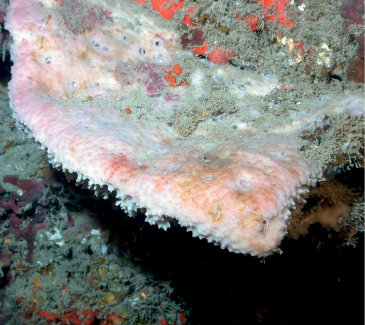
Batzella rubra
Diagnostic features
Thinly encrusting sponge, growing over dead coral or other sponges. Deep orange to bright red color in live. The surface is smooth and ornamented by paler colored dermal canals that branch away from the oscula, wide close to the oscula and thinner away from it. The consistency is compressible where the sponge is thicker.

Similar species
The sponge can be confused with other red encrusting species, but the particular ‘dripping’ morphology of the dermal canals makes them easy to distinguish.
Distribution and abundance
This species is reported from shallow reefs in Cuba and Bahamas. This is the first report for the species at mesophotic reefs. At FGBNMS the specimen on the photograph was found at east Flower Garden Bank and the species has rare to moderate abundance at ten other sites.
Ecology
Coralline algae reefs, algal nodules, lower mesophotic reefs.
Identification
MCD.
Reference
Batzella cf. rubra
Diagnostic features
Thinly encrusting sponge (1–10 mm thick) growing over dead coral. Orange to red color in life, black to purple in alcohol. The surface is smooth to bumpy with whitish branching dermal canals, and roundish papillae with two or three clumps of ostia. The cf. is due to the rounded papillae only described for Batzella mollis, a species found at the “Juan Fernandez and Desventuradas islands” off the Chilean Pacific coast.

Similar species
Distribution and abundance
This is the first record from mesophotic reefs. At FGBNMS the species has been recorded with rare to low abundance (1–10) in three localities.
Ecology
Coralline algae reefs, algal nodules, lower mesophotic reefs.
Identification
KR, SK, CA, MCD.
References
Order Poecilosclerida
Family Microcionidae
Clathria
Diagnostic features
Massive with short, protruding, flattened, digitate branches, forming a roundish bush. Colored red in life. Surface minutely porous and with a translucent veil (dermis). Oscula not visible. This species belongs to the genus Clathria; however, species identifications require spicule analysis.

Similar species
Several branching bushy Clathria species are described by
Distribution and abundance
Arborescent Clathria species are diverse and well-known from the Gulf of Mexico (
Ecology
Sandy substrates.
Identification
KR, SK, CA, MCD.
Reference
Order Suberitida
Family Halichondriidae
Halichondria
Diagnostic features
Massive to thick encrusting, or globular to lobate with lobes ≤ 15 cm high. Yellow orange sponge alive, tan pinkish in alcohol. The surface is rugose to verrucose, with deep grooves and holes < 0.5 mm wide; the deep grooves, where thin ectosome is absent, have a feathery appearance. Oscula 0.5–2 cm wide, with a yellow membranous collar that is 5–8 mm high when the oscula are fully open. Compressible in consistency.

Similar species
Myrmekioderma rea when it grows as a thick massive crust, and massive lobate Axynissa ambrosia can be easily confused with this species. Its spicules (oxea in a wide size range) and surface features are unique among the Halichondriidae.
Distribution and abundance
The sponge is common at FGBNMS on mesophotic habitats between 55–73 m. This is an undescribed species. The species occurs at four sites with rare to medium abundance (1–100).
Ecology
Coralline algae reefs, algal nodules, lower mesophotic reefs.
Identification
CA, SWK, MCD.
References
Topsentia bahamensis
Diagnostic features
Massive, columnar shape with round or blunt tip (10 cm high, 2–4 cm in diameter). The sponge is red-brown externally and tan internally in living sponges. The surface is smooth visually with a sandpaper feel. Five dispersed oscula (1–3 mm in diameter). Very firm in consistency, but crumbly.

Similar species
Topsentia ophirhaphidites, which has deformed small oxea added to the larger oxea. Petrosiids in general by their reddish brown color and hard brittle consistency. Spicule study needed to distinguish it.
Distribution and abundance
Currently reported from shallow reefs in southern GOM in northern Yucatan and Belize, and at mesophotic depths in the Bahamas and northwestern GOM at the FGBNMS.
Ecology
Coralline algae reefs, algal nodules.
Identification
CA, SWK, MCD.
Reference
Order Suberitida
Family Suberitidae
Rhizaxinella clava
Diagnostic features
The “corn dog sponge”. A pale brown, clavate, stipitate sponge (15 cm in total length) with a long thin peduncle (2 mm in diameter at attachment area) and an upper globose soft body (1 cm in diameter at its thickest part). The surface is very smooth and velvety. The apical oscule is slit-shaped and has a collared membrane visible in the in-situ photograph. The sponge is firm but slightly compressible.

Similar species
The “corn dog sponge” is similar externally to the “lollipop sponge”, Stylocordyla chupachups and to other members of the family Stylocordylidae, which are mostly present in cold deep waters.
Distribution and abundance
Currently reported at mesophotic depths in the Florida Keys, Guyana, Surinam, and northwestern GOM at the FGBNMS. At FGBNMS the species is widespread, occurring at 15 sites with abundance from rare to very common (1–100+).
Ecology
Coralline algae reefs, silted lower mesophotic reefs.
Identification
KR, CA, SWK, MCD.
Reference
Order Tetractinellida
Sub-order Astrophorina
Family Corallistidae
Corallistes typus
Diagnostic features
Small cups or plates, with undulating rims, walls 1–3 cm thick, usually with a thick peduncle. Brown with faintly pink tinges. The surface is smooth, with rims sometimes covered by sediment. Oscula not visible.

Similar species
An integrative study of 247 “lithistid” samples from the tropical western Atlantic (
Distribution and abundance
Southern, eastern, and northern Caribbean, Florida, and Bahamas 61–914 m deep (
Ecology
Coralline algae reefs, lower mesophotic reefs.
Ideentification
KR, CA, SWK.
References
Neophrissospongia cf. nolitangere
Diagnostic features
Plate or ear-shaped sponges with 1–2 cm thick walls with rounded margins, and 8–12 cm across. Tan-brown, plate. The cf. denomination was given since minute oscula on inner surface (0.5–1 mm wide) and a pedicel described for Neophrissospongia nolitangere were not seen in the image or during voucher analysis.

Similar species
Corallistes typus and other species from the same genus. Neophrissospongia differs from Corallistes by the spiny or tuberculate nature of the dichotriaene top in the former, and the smooth nature on the later.
Distribution and abundance
Neophrissospongia nolitangere is common at deep waters from the eastern Atlantic, Azores and Mediterranean.
Ecology
Algal nodules.
Identification
KR, CA, SWK, MCD.
References
Pissera and Levi 2002;
Order Tetractinellida
Sub-order Astrophorina
Family Ancorinidae
Stellettinopsis cf. megastylifera
Diagnostic features
Round to massive sponge. Brownish gray to dirty white in color. The surface is prickly, hispid, feels like sandpaper; numerous holes ≤ 3–5 mm in diameter in sponge body. Few larger oscula 1–4 cm wide. Hard and dense in consistency. Species identity requires further analysis and comparative work (

Distribution and abundance
The species is reported from shallow depths growing on coral reefs, rocks, sand, or mangroves (3–25 m deep) in the Colombian Caribbean, Curacao, Panama, Belize, southern GOM, and Dominican Republic. Rare species. This is the first report of this species for the north GOM mesophotic. At FGBNMS, moderate abundance at three sites.
Ecology
Coralline algae reefs, algal nodules.
Identification
SWK, CA, KR.
References
Order Tetractinellida
Sub-order Astrophorina
Family Geodiidae
Erylus alleni
Diagnostic features
Stalks with heart-shaped tops (3–7 cm in height, 1–3 cm wide). Dark brown in color externally, tan internally. Very smooth surface. One or two oscula per stalk located at the tips (1–5 mm wide). The oscula continued by an atrium 1–2 cm deep. Dense in consistency.

Similar species
The stalked growth form and certain spicule details (elliptical aspidasters, two categories of oxyasters) allow its differentiation with co-occurring similar species such as Erylus goffrilleri and Erylus trisphaerus.
Distribution and abundance
Mesophotic depths in Puerto Rico and Brazil. Rare species. This is the first report of this species for the GOM, at FGBNMS found with rare to low abundance (1–10) at 7 sites.
Ecology
Coralline algae reefs, algal nodules.
Identification
KR, CS, SWK, MCD.
Reference
Erylus goffrilleri
Diagnostic features
Massive amorphous to lobate. Dark brown color that lightens towards the base of the lobes. Smooth surface, slight wrinkles when out of water. One apical oscule, on top of each lobe (4–8 mm wide), with a paler colored membrane. Compressible but dense in consistency, easily breakable.

Similar species
Several species of Erylus from the tropical western Atlantic are very similar in external appearance. The calthrop-like triaenes and the tylasters allows its distinction from co-occurring species.
Distribution and abundance
Reported in shallow and mesophotic reefs in the Bahamas and Jamaica. First report of this species for the GOM at FGBNMS region, found at low abundance (1–10), at Geyer Bank.
Ecology
Algal nodules.
Identification
KR, CS, SWK, MCD.
Reference
Erylus trisphaerus
Diagnostic features

Similar species
The peanut-shaped aspidasters, with two to three swollen areas, allows its distinction from co-occurring species of the genus.
Distribution and abundance
This is a rare species originally described from Apalachee Bay in north Florida at 13 m deep. Since then, it has been reported from shallow reefs in Alacranes Reef (Southern GOM), Cuba, and Curacao. This is the first report from mesophotic reefs and first report from northwestern GOM at FGBNMS where it occurs with rare to low abundance (1–10) at six sites.
Ecology
Coralline algal reefs, algal nodules, lower mesophotic reefs.
Identification
KR, CA, SWK, MCD.
Reference
Geodia cf. curacaoensis
Diagnostic features
Spherical, approx. 7 cm in diameter, with a roundish black apical plate (2 cm wide). The sponge color is pale brown with reddish tinges. The surface is mostly smooth, with patches with sediments or turf around the apical plate, and occasionally on body side. Many oscula (2 mm wide) concentrated on the apical plate. Hard as a rock. The cf. is assigned due to the black color of the sieve plate, larger oscula, and the because size of the large category of oxea of Geodia curacaoensis is twice the size of oxea from the FGBNMS specimen.

Similar species
This specimen is very similar to Geodia curacaoensis, in overall external morphology, and spicule composition.
Distribution and abundance
Geodia curacaoensis was described from mesophotic depths in Curacao and was recorded in shallow reefs at Alacranes Reef, south GOM, and at mesophotic depths in Cuba. This morphotype was found at FGBNMS in low (2–10) abundance at six sites.
Ecology
Coralline algae reefs, algal nodules, lower mesophotic reefs.
Identification
KR, CA, SWK, MCD.
Reference
Order Tetractinellida
Sub-order Astrophorina
Family Theonellidae
Discodermia
Diagnostic features

Similar species
The image of a Discodermia dissoluta specimen (
Distribution and abundance
The genus Discodermia is common in deep waters at the tropical western Atlantic. Discodermia dissoluta is the most widespread distributed species of the genus in the region from the GOM to Brazil. At FGBNMS, rare to low (1–10) abundance at two sites.
Ecology
Lower mesophotic reefs.
Identification
KR, CA, SWK, MCD.
Reference
Order Tetractinellida
Sub-order Astrophorina
Family Thrombidae
Yucatania sphaeroidocladus
Diagnostic features
Encrusting to massive sponge (1–9 cm in thickness). Ochre brown externally in life. The sponge has a vermetid gastropod growing within its body. Surface microhispid, rough to the touch, oscula numerous and scattered, rounded to oval, 1–4.5 mm wide. Some of the openings at the surface correspond to the vermetids.

Similar species
Any massive-amorphous to subglobular species that accumulate debris can be confused with this species. Spicule composition is essential for its identification.
Distribution and abundance
Mesophotic depths from eastern Brazil, Guiana, Trinidad, and continental platform of the Yucatan Peninsula. Widely distributed at the mesophotic depths in FGBNMS (12 sites), with rare or medium abundance (1–100).
Ecology
Coralline algae reefs, algal nodules, lower mesophotic reefs.
Identification
KR, CA, SWK, MCD.
References
Order Tetractinellida
Sub-order Spirophorina
Family Tetillidae
Cinachyrella
Diagnostic features
Globular sponge (12 cm wide). Neon yellow in and out, covered by sediment obscuring the sponge color. The surface appears smooth, rough to the touch; microhispid microscopically. Few apical oscula (6–8 mm wide). Dense in consistency. This specimen was initially identified as Tetilla sp. However, an 18S barcoding study shows this species is 99.9% Cinachyrella sp. (Iris Segura, pers. comm., 2022). The specimen studied lacks protrienes and anatrianes found in all Cinachyrella species from the region. This specimen has long oxea: 2500–3000 × 10–50 µm, small oxea: 130–170 × 5 µm, and sigma (c-, and s-shaped), 20–30 µm long × < 1 µm wide. The determination of this species requires further comparative work and the analyses of other genetic markers.

Similar species
Globular yellowish species of the genera Cinachyrella, Cinachyra, or Tetilla.
Distribution and abundance
At FGBNMS this species is rare, appearing once at one site.
Ecology
Coralline algal reefs.
Identification
Iris Segura, MCD.
Reference
Order Chondrosida
Family Chondrosiidae
Chondrosia collectrix
Diagnostic features
Thick encrusting (1–3 cm in thickness) to lobate, brown, black to tan in color externally with darker spotted areas, tan internally. Smooth surface, and round oscula, with elevated membranes. This species has very cartilaginous consistency.

Similar species
Chondrilla caribensis is very similar in growth form and color but it lacks oscula with a collared membrane and possesses typical and abundant spheraster spicules.
Distribution and abundance
The species is distributed at coral reefs, seagrasses, and/or mangroves in Florida, Bermuda, throughout the Caribbean, Brazil, and southern Gulf of Mexico. At FGBNMS is widely distributed with low to high abundance (10–100+) occurring at 12 sites.
Ecology
Inhabits lower mesophotic reef and heavily silted reef, coralline algae reef, algal nodules.
Identification
KR, CA, SWK, MCD.
Reference
Order Verongiida
Family Aplysinidae
Aiolochroia crassa
Diagnostic features
Massive/lobate to amorphous. Variable color from yellow to pink and purple. The surface has roundish conules. The oscula are on top of lobe(s), have small flat oscular membranes the same color than the sponge. Compressible and dense in consistency.

Similar species
Some specimens of Verongula rigida, with few ridges at the surface, can be confused with this species.
Distribution and abundance
This species is very common at shallow coral reefs in Florida, Bermuda, Bahamas, throughout the Caribbean, Brazil, and Gulf of Mexico. At mesophotic depths in FGBNMS it is widely distributed, occurring at ten sites.
Ecology
Coral communities.
Identification
KR, CA, SWK, MCD.
Reference
Aplysina
Diagnostic features

Similar species
The overall growth form of this species is similar to that of Aplysina archeri. However, Aplysina archeri tubes are usually much larger, and invariably a pink to violet color at all its depth range of distribution. A similar whitish Aplysina with larger dimensions was observed in Cuba at 58 and 81 m deep.
Distribution and abundance
This is a unique and rare species of Aplysina, found in mesophotic reefs at FGBNMS and Pulley Ridge. At FGBNMS it was found once at the east Flower Gardens bank.
Ecology
Found at coralline algal reef, algal nodule, lower mesophotic reef.
Identification
KR, CA, SWK, MCD.
References
Aplysina cf. archeri
Diagnostic features
Clusters of short tubes that spread laterally. SP04 was 10–20 cm high, and 3–5 cm in diameter, and SP25 was 3–10 cm high 3–6 cm wide, pink to deep purple in color. Fistulose rods sporadically grow out from the tubes. Surface rugose and microconulose. Roundish tube tops, more pronounced on SP25. The specimens are tentatively identified from the photographs as Aplysina cf. archeri due to the predominance of short roundish tubes, and lateral growth; no samples were available for analysis.

Similar species
At least three species of Aplysina can be a cluster of short tubes, viz. A. fistularis, A. insularis, and A. muricyana but their color is mostly yellow. Collection and genetic data would be very helpful to discern these species.
Distribution and abundance
Aplysina archeri is common at shallow coral reefs in Florida (Dry Tortugas) and throughout the Caribbean. This species might be a morphological variant of the species or a closely related species which occurs as a single or clumps of elongated purplish tubes.
Ecology
Coral communities, sandy substrates.
Identification
MCD.
References
Verongula rigida
Diagnostic features
Massive lobate to tubular species. This sample had multiple tubes of different lengths (4–20 cm high, 2–4 cm wide). Reddish yellow in color when alive, purple as dry or in alcohol. The surface is rugose to verrucose, ribbed, but smooth to the touch, not like sandpaper. One oscule (0.8–1 cm) on top of each tube, the opening extending the length of sponge. Oscula with a flat diaphragm-like contractile membrane darker in color. The consistency is firm but compressible, fibrous, and tough.

Similar species
Specimens of this species with slightly ribbed surface can be confused with Aiolochroia crassa. Some morphotypes of Smenospongia aurea can also look like short tubes of Verongula rigida.
Distribution and abundance
This species is common at shallow coral reefs throughout the Caribbean. At FGBNMS, rare, found only at three sites.
Ecology
Heavily silted lower mesophotic reefs.
Identification
KR.
Reference
Verongula reiswigi
Diagnostic features
Large tube or vase, wider at the base or at the mid body. The color is yellow with green or pinkish tinges alive, purple when dry or in alcohol. The outer surface is ribbed, forming a regular pattern that covers the whole sponge surface. One large opening (oscule or pseudo-oscule) at top of each “vase” (> 5 cm), with a very thin membrane (1–2 mm) that surrounds the whole rim. The consistency is firm but compressible.

Similar species
The overall shape, “oscule” size, and surface of this species makes it unique.
Distribution and abundance
This species is rare in occurrence at shallow and mesophotic coral reefs in Florida, Bahamas, Cuba, and eastern Caribbean. Rare at FGBNMS, seen only once at one site,
Ecology
Heavily silted lower mesophotic reef.
Identification
MCD.
References
Order Verongiida
Family Ianthellidae
Ianthellide
Diagnostic features
Thin encrusting sponge, < 1 mm thick, bright yellow in life, purple in alcohol (Fig.

A In-situ photograph of a thin yellow Verongid growing on an hexactinellid skeletal framework highlighted by the red arrow, 147 m deep. Sample GFOE3-23A B fragment of sponge observed under light microscope 100X; arrows show large dark cells potential spherulous cells (SC), and smaller dark cells, potentially granular cells (GC) C smear of a sample fragment observed with a light microscope at 400× magnification. Note the large ovoid choanocyte chambers (red arrow).
Similar species
Two Ianthellidae genera without fibers have been described, Hexadella and Vansoestia, which have species with thin bodies, and yellowish color. However, those species have a more detachable leathery body, with surfaces ornamented by dermal canals, and prominent oscula.
Distribution and abundance
At FGBNMS the species was collected once at Elvers Bank.
Ecology
Coralline algae reefs, lower mesophotic reefs, algal nodules.
Identification
MCD.
Reference
Díaz et al. 2015.
Order Dendroceratida
Family Darwinellidae
Aplysilla aff. sulfurea
Diagnostic features
Thin encrusting sponge, 1–2 mm when preserved in alcohol. Pale yellow/orange in color alive. In alcohol it turns dark purple. In life the surface bears low conules, and oscula a few mm wide.
Similar species
The overall growth form, color, and color change in alcohol is similar to those in Aplysilla sulfurea.
Distribution and abundance
Aplysilla sulfurea is the type species for the genus and was described from the Adriatic and Mediterranean seas and eastern Atlantic. The reports from Bermuda, Florida, and southern Africa probably represent different species of similar habit and color. At FGBNMS the species is widely distributed at 14 sites with a range of abundance from rare to medium (2–100).

Ecology
Coralline algae reefs, lower mesophotic reefs, algal nodules.
Identification
CA, MCD.
References
Order Dictyoceratida
Family Dysideidae
Dysidea
Diagnostic features

Similar species
This species is similar to the Mediterranean species Dysidea fragilis. There is an inaccurate citation of Dysidea fragilis by
Distribution and abundance
At FGBNMS the species is widely distributed at ten sites with a range of abundance from rare (1 per site) to common (11–100).
Ecology
Coralline algae reef, lower mesophotic reef, and algal nodules.
Identification
KR, MCD.
Reference
Pleraplysilla
Diagnostic features
Very thin crust, pale pink color. The surface is smooth with irregularly distributed small conules (< 1 mm high). Oscula with a collar membrane and thick canals that run towards the oscula. The conules are produced by single or branching fibers that depart from a spongin basal plate.

Similar species
Very similar in external appearance to Vansoestia caribensis, a skeleton-less sponge of the family Ianthellidae, Order Verongiida. This species has abundant single or dendritic fibers, dark in color with an apparent pith, and some foreign spicules inside. This sponge is very similar to the species of Pleraplysilla depicted by
Distribution and abundance
Reported by
Ecology
Coralline algae reefs, lower mesophotic reefs.
Identification
KR, SK, CA, MCD.
Reference
Pleraplysilla
Diagnostic features
Massive bushy, 5 cm wide, 7 cm high. Tan in color alive. Sharp conules, 2–3 mm high, separated by 3–5 mm. The sponge is compressible but firm. The sponge has dendritic fibers, pale in color, which incorporate broken spicules. This sponge is currently identified as an undescribed species of Pleraplysilla. 28S analysis of this specimen clearly places it within the Order Dictyoceratida, but not within the Family Dysideidae. 18S analysis places it as 99.5% similar to Pleraplysilla spinifera, the type of the genus; however, that is an eastern Atlantic and Mediterranean species. This result supports the generic assignation of this new species from GOM (Diaz, Segura, and Pomponi, unpublished data).

Similar species
Its massive habit is similar to that of the shallow Caribbean mangrove species Pleraplysilla stocki, and to Pleraplysilla spinifera from the Mediterranean.
Distribution and abundance
The species has been collected once from FGBNMS, and once from Pulley Ridge, at the southeast GOM (MCD).
Ecology
Coralline algal reef.
Identification
Iris Segura, MCD.
Reference
Order Dictyoceratida
Family Irciniidae
Ircinia campana
Diagnostic features

Similar species
This might be a different species from the shallow reef species, but close genetic and morphological comparison must be carried out to distinguish Ircinia species (
Distribution and abundance
Widespread throughout the Caribbean at shallow coral reefs and seagrass meadows. This is the first report of the species for the northwestern GOM and at mesophotic depths in the GOM. At FGBNMS, low (1–10) in abundance and only documented at Stetson Bank.
Ecology
Lower mesophotic reefs, coralline algae reefs.
Identification
MCD.
Reference
Ircinia cf. campana
Diagnostic features
Flabellate to fan. Brown, gray, to pinkish in color. The surface is regularly conulose. Abundant round oscula (2–3 mm) on the upper surface, sometimes clumped. The cf. is to highlight the uncommon plate-shape habitus for the species, indicating that this morphotype could represent either a different species or a variant of Ircinia campana. Further genetic and morphologic comparisons are required.
Distribution and abundance
Widespread throughout the Caribbean on shallow coral reefs and seagrass meadows. This is the first report of the species for the northwestern GOM and at mesophotic depths. Single specimen found at one locality.
Ecology
Coralline algal reefs.
Identification
MCD.
References
Ircinia strobilina
Diagnostic features
The sponge is sub-globular to massive and cake-shaped, gray to black color in life. Large specimens show an upper depression where oscula abound. The surface has characteristic large conules (2–15 mm high, 5–15 mm apart). Oscula 4–10 mm in diameter, either single or in groups, with a thin membrane. The specimens are tough in consistency.

Distribution and abundance
Widespread throughout the Caribbean, Bermuda, GOM, and Brazil. The species has been previously reported in the northern and southern GOM (
Ecology
Coral communities, coralline algae reefs, lower mesophotic reefs.
Id
MCD.
Reference
Ircinia
Diagnostic features
Single, two, or three tubes connected at the base. Tubes taper towards the tip. Pink to white in life. The tubes in Fig.

Similar species
This is a very unique Ircinia species, probably undescribed.
Distribution and abundance
This is an undescribed species of Ircinia. At FGBNMS the species was seen once at two localities. MCD has seen this species once in the Bahamas.
Ecology
Silted coralline algae reefs, silted lower mesophotic reefs.
Identification
CA, KR, SK, MCD.
Reference
Ircinia
Diagnostic features
Cushion shape to massive (5 cm thick). The surface is finely conulose (< 1 mm high, and 1–2 mm apart). Color alive is pink to reddish brown externally, tan internally. Small oscula 2–4 mm in diameter) with a thin white membrane around their rim, sparsely distributed on the sponge. The sponge is compressible but tough to cut.

Similar species
The species appears similar to Neopetrosia proxima and its closest species. Neopetrosia proxima lacks the conules, has a harder consistency, and a spicular skeleton, while Ircinia possess a skeleton of spongin fibers and filaments that may incorporate foreign particles (sand, and broken spicules).
Distribution and abundance
At FGBNMS the species was found once at one site.
Ecology
Coralline algal reefs.
Identification
CA, KR, SK, MCD.
Reference
Order Dictyoceratida
Family Thorectidae
Smenospongia cf. echina
Diagnostic features
Globular to cushion shape, dirty yellow to grayish in life, brownish purple in alcohol. The surface has shallow roundish warts (≤1 cm wide) but feels smooth to the touch. Oscula from 2 mm to > 1 cm wide, with a slightly elevated membrane.

Similar species
Similar to verongiid species, Smenospongia spp. tends to turn purplish after collection.
Distribution and abundance
Smenospongia echina occurs in low abundance at shallow and mesophotic reefs in Puerto Rico (60–72 m), Belize, Florida (Dry Tortugas), Cayman Islands, and Cuba. At FGBNMS the species occurs in rare to low abundance (1–10) at four sites.
Ecology
Lower mesophotic reefs, coralline algae reefs.
Ideentification
CA, KR, SK, MCD.
Reference
Class Homoscleromorpha
Order Homosclerophorida
Family Plakinidae
Plakortis cf. zyggompha
Diagnostic features
Thick encrusting (5–10 mm thick). Pinkish brown in life. The surface is very smooth, velvety to the touch. Dense consistency. Sponge was overgrowing the base of an albino Aplysina spp. (DFH9-2B). Spicules larger than those of Plakortis zyggompha (de Laubenfels, 1934).

Similar species
Plakortis angulospiculatus and Plakortis halichondroides, with the same dark brown color and thick crustose shape; Plakortis zyggompha is always much thinner (< 5 mm) and oscula are much smaller. A genetic comparison would clarify the taxonomic status of the FGBNMS material.
Distribution and abundance
The species is originally described from mesophotic depths (84–165 m), and it is also reported from Florida (Dry Tortugas), Belize (cryptic habitats), and Jamaica (mangroves). At FGBNMS the species was rare and found at only three sites.
Ecology
Algal reef, algal nodule, lower mesophotic reef.
Identification
SK, KR, CA.
References
Class Hexactinellida
Order Hexasterosphora
Family Dactylocalycidae
Dactylocalyx pumiceus
Diagnostic features
Basal funnel expanded distally forming a plate, or a cup, with a wavy rim. Inner cup surface has elongated pits or grooves, several cm long, < 1 cm wide. This sample has not yet been examined but the overall growth form points to this species.

Distribution and abundance
This is a species with great latitudinal distribution from Florida and Gulf of Mexico to Brazil, distributed along the western coast of the Atlantic Ocean between 300N and S, at depth of 91–1996 m. The species is also reported off the coast of Portugal. At FGBNMS the species was collected at Elvers Bank where it was found in low (1–10) abundance.
Ecology
Lower mesophotic reef.
Identification
MCD.
Reference
Iphiteon panicea
Diagnostic features
Massive, flabellate, white, glass sponge, attached to a rock. The white elongated zoanthid, Vitrumanthus schrieri

Similar species
When zoanthids are extended the species can look like the hexactinellid Verrucocoeloidea liberatorii Reiswig & Dohrmann, 2014.
Distribution and abundance
This species has a northwestern Caribbean distribution (88–1957 m deep). At FGBNMS the species was collected once at Elvers Bank.
Ecology
Lower mesophotic reefs, sandy bottoms. The zoanthid Vitrumanthus schrieri (Parazoanthidae) was originally described in association with the glass sponge Verrucocoeloidea liberatorii. In this sample, the identity of the zoanthid was obtained by barcoding data (28S gene) (Iris Segura, pers. comm., 2022).
Identification
MCD.
References
Discussion and conclusions
This checklist of 64 sponge species represents only a portion of the sponge fauna inhabiting mesophotic depths in the Flower Garden Banks National Marine Sanctuary region. Caribbean coral reefs that have been studied for years, including surveys of both open and sciophilous (shaded) habitats, such as the Belizean barrier reef (
Our most recent collection conducted off the Sanctuary boundaries in 2019 contributed specimens of seven potentially new species of the genera Auletta, Petrosia, Xestospongia, Cinachyrella, Siphonodictyon, and Pleraplysilla, and one specimen from the family Ianthellidae. These include species with significant biomass representation and widespread occurrence such as Petrosia sp. nov. 1 and Xestospongia sp. nov. 1, or species with novel or important ecological features such as the skeleton-less Ianthellidae sp. 1 overgrowing an Hexactinellid, and the bioeroding species Siphonodictyon sp. nov. 1. Molecular analyses (using 28S and 18S genes) are in progress to complement the morphological characteristics to refine and, in some cases, confirm the identification of these potentially new mesophotic sponge species. Part of the data from this concurrent study was essential for the generic determination of three of the species included in this paper, Cinachyrella sp. 1, Pleraplysilla sp. nov. 2, and Siphonodictyon sp. nov. 1.
The biological role of sponges in coral ecosystems should ignite interest to continue studying this fauna from less studied mesophotic habitats. Sponges are known for their high diversity and high biomass in shallow and mesophotic coral reefs (
Acknowledgements
We would like to recognize the extensive contribution of the people who collected the specimens used in this manuscript including the crew of the R/V ‘Manta’, University of North Carolina at Wilmington Undersea Vehicle Program ROV pilots (Eric Glidden, Lance Horn, Glen Taylor, and Jason White), and the Global Foundation for Ocean Exploration ROV team (Joshua Carlson, Karl McLetchie, Jeff Laning, Todd Gregory, Roland Brian). The scientific results and conclusions, as well as any views or opinions expressed herein, are those of the author(s) and do not necessarily reflect the views of NOAA or the Department of Commerce. We also thank John Reed (Harbor Branch Oceanographic Institute – FAU) for providing photographs from the 2022 NOAA OER expedition to the FGBNMS region), and Dr. Iris Segura (Harbor Branch Oceanographic Institute – FAU) for contributing barcoding data that allowed the generic designation of three sponge and one zoanthid species. Funding for this project was provided in part by Flower Garden Banks National Marine Sanctuary, the NOAA Deep-Sea Coral Program, and the National Marine Sanctuary Foundation.
References
- Aerts LAM (1998) Sponge/coral interactions in Caribbean reefs: Analysis of overgrowth patterns in relation to species identity and cover. Marine Ecology Progress Series 175: 241–249. https://doi.org/10.3354/meps175241
- Alcolado PM (1984) Nuevas especies de esponjas encontradas en Cuba. Poeyana 271: 1–22. [New species of sponges from Cuba]
- Alvarez B, van Soest RWM, Rützler K (1998) A Revision of Axinellidae (Porifera: Demospongiae) in the Central West Atlantic Region. Smithsonian Contributions to Zoology 598(598): 1–47. https://doi.org/10.5479/si.00810282.598
- Bell JJ (2008) The functional roles of marine sponges. Estuarine, Coastal and Shelf Science 79(3): 341–353. https://doi.org/10.1016/j.ecss.2008.05.002
- Boury-Esnault N, Rützler K (1997) Thesaurus of Sponge Morphology. Smithsonian Contributions to Zoology 596(596): 1–55. https://doi.org/10.5479/si.00810282.596
- de Laubenfels MW (1934) New sponges from the Puerto Rican deep. Smithsonian Miscellaneous Collections 91(17): 1–28.
- De Laubenfels MW (1936) A Discussion of the Sponge Fauna of the Dry Tortugas in Particular and the West Indies in General, with Material for a Revision of the Families and Orders of the Porifera. Carnegie Institute of Washington Publication 467. Tortugas Laboratory Paper (30): 1–225. [pls 1–22]
- de Laubenfels MW (1950) The porifera of the Bermuda archipelago. Transactions of the Zoological Society of London 27(1): 1–154. https://doi.org/10.1111/j.1096-3642.1950.tb00227.x
- de Laubenfels MW (1953) Sponges from the Gulf of Mexico. Bulletin of Marine Science of the Gulf and Caribbean 2(3): 511–557.
- de Weerdt WH (2000) A monograph of the shallow-water Chalinidae (Porifera, Haplosclerida) of the Caribbean. Beaufortia 50(1): 1–67.
- de Voogd NJ, Alvarez B, Boury-Esnault N, Carballo JL, Cárdenas P, Díaz MC, Dohrmann M, Downey R, Goodwin C, Hajdu E, Hooper JNA, Kelly M, Klautau M, Lim SC, Manconi R, Morrow C, Pinheiro U, Pisera AB, Ríos P, Rützler K, Schönberg C, Vacelet J, van Soest RWM, Xavier J (2023) World Porifera Database. [Accessed on December 20 2022] https://doi.org/10.14284/359
- Díaz MC, Pomponi SA (2018) New Poecilosclerida from mesophotic coral reefs and the deep-sea escarpment in the Pulley Ridge region, eastern Gulf of Mexico: Discorhabdella ruetzleri n.sp. (Crambeidae) and Hymedesmia (Hymedesmia) vaceleti n.sp. (Hymedesmiidae). In: Klautau M, Pérez T, Cárdenas P, de Voogd N (Eds) Deep sea and cave sponges. Zootaxa 4466(1), 229–237. https://doi.org/10.11646/zootaxa.4466.1.17
- Díaz MC, Rützler K (2001) Sponges: An essential. Component of Caribbean Coral Reefs. Bulletin of Marine Science 69(2): 535–546.
- Díaz MC, Pomponi SA, van Soest RWM (1993) A systematic revision of the central West Atlantic Halichondrida (Demospongiae, Porifera). Part III: Description of valid species. In: Uriz MJ, Rützler K (Eds) Recent advances in ecology and systematics of sponges. Scientia Marina 57(4), 283–306.
- Díaz MC, Busutil L, García-Hernández MR, Pomponi SA (2019) Cuba’s Mesophotic Coral Reefs- Sponge Photo Identification Guide. In: Reed JK, Farrington F (Eds) Cooperative Institute for Ocean Exploration, Research, and Technology (CIOERT) at Harbor Branch Oceanographic Institute, Florida Atlantic University (HBOI-FAU). Harbor Branch Oceanographic Institute Contribution Number 2256. http://www.cioert.org/wp-content/uploads/2019/06/D%C3%ADaz-et-al-Cubas-Mesophotic-Coral-Reefs-Sponge-Photo-Identification-Guide-Edition–1 [Accessed 10 June 2022]
- Díaz MC, Pomponi SA, Farrington S, Reed JK (2021) Photo Identification Guide of the Sponges inhabiting the Shelf-edge Marine Protected Areas and Deep-water Reefs of the Southeastern USA (1st Edn). Harbor Branch Oceanographic Institute Contribution Number, 2294. http://www.cioert.org/expeditions/mesophotic-reef-ecosystems
- Esteves EL, de Paula TS, Lerner C, Lôbo-Hajdu G, Hajdu E (2018) Morphological and molecular systematics of the ‘Monanchora arbuscula complex’ (Poecilosclerida : Crambeidae), with the description of five new species and a biogeographic discussion of the genus in the Tropical Western Atlantic. Invertebrate Systematics 32(2): 457–503. https://doi.org/10.1071/IS16088
- Fiore CL, Baker DM, Lesser MP (2013) Nitrogen Biogeochemistry in the Caribbean Sponge, Xestospongia muta: A Source or Sink of Dissolved Inorganic Nitrogen? PLoS ONE 8(8): e72961. https://doi.org/10.1371/journal.pone.0072961
- Gómez P (2006) Yucatania clavus, new genus and species of the family Thrombidae (Porifera: Demospongiae: Astrophorida) from the continental shelf off Yucatan, Mexico. Proceedings of the Biological Society of Washington 119(3): 339–345. https://doi.org/10.2988/0006-324X(2006)119[339:YCNGAS]2.0.CO;2
- Gómez P (2007) Inventario de las esponjas del Parque Nacional Sistema Arrecifal Veracruzano, con nuevos Registros de Especies (Porifera: Demospongiae). In: Jiménez-Hernández MJ, Granados-Barba A, Ortiz-Lozano L (Eds) Investigaciones Científicas en el Sistema Arrecifal Veracruzano. Universidad Autónoma de Campeche, Campeche, 51–72.
- Gómez P (2014) The genus Clathria from the Gulf of Mexico and Mexican Caribbean, with redescription and resurrection of Clathria carteri (Poecilosclerida: Microcionidae). Zootaxa 3790(1): 51–085. https://doi.org/10.11646/zootaxa.3790.1.3
- Green G, Fuentes L, Gómez P (1986) Nuevos registros de Porifera del arrecife La Blanquilla, Veracruz, México. Anales del Centro de Ciencias del Mar y Limnología 13(3): 127–146.
- Hartman WD, Hubbard R (1999) A new species of Thrombus (Porifera: Demospongiae: Astrophorida) from Trinidad, West Indies. Bulletin of Marine Science 64(1): 1–8.
- Hickerson EL, Schmahl GP (2012) Sponges of Deepwater Communities in the Northwestern Gulf of Mexico. Developed by Flower Garden Banks National Marine Sanctuary. https://flowergarden.noaa.gov/doc/posters/spongesdeepwaternwgom.pdf
- Kealoha AK, Doyle SM, Shamberger KEF, Sylvan JB, Hetland RD, DiMarco SF (2020) Localized hypoxia may have caused coral reef mortality at the Flower Garden Banks. Coral Reefs 39(1): 119–132. https://doi.org/10.1007/s00338-019-01883-9
- Kelly JB, Thacker RW (2021) New shallow water species of Caribbean Ircinia Nardo, 1833 (Porifera: Irciniidae). Zootaxa 5072(4): 301–323. https://doi.org/10.11646/zootaxa.5072.4.1
- Kise H, Montenegro J, Santos MEA, Hoeksema BW, Ekins M, Ise Y, Higashiji T, Fernandez-Silva I, Reimer JD (2022) Evolution and phylogeny of glass-sponge-associated zoantharians, with a description of two new genera and three new species. Zoological Journal of the Linnean Society 194(1): 323–347. https://doi.org/10.1093/zoolinnean/zlab068
- Lehnert H, van Soest RWM (1998) Shallow water sponges of Jamaica. Beaufortia 48(5): 71–103.
- Mah CL (2020) New species, occurrence records and observations of predation by deep-sea Asteroidea (Echinodermata) from the North Atlantic by NOAA ship Okeanos Explorer. Zootaxa 4766(2): 201–260. https://doi.org/10.11646/zootaxa.4766.2.1
- Meesters E, Knijn R, Willemsen P, Pennartz R, Roebers G, van Soest RWM (1991) Sub-rubble communities of Curaçao and Bonaire coral reefs. Coral Reefs 10(4): 189–197. https://doi.org/10.1007/BF00336773
- Mothes B, Lerner CB, Silva CMM (1999) Revision of Brazilian Erylus (Porifera: Astrophorida: Demospongiae) with description of a new species. In: Hooper JNA (Ed.) Origin and Outlook. Memoirs of the Queensland Museum 44: 369–380.
- Mullins DA (2021) NudiNotes Column. Dive Log Australasia Magazine 387: 34–35.
- Nuttall MF, Hickerson EL, Blakeway RD, Schmahl GP, Sammarco PW (2022) Do oil and gas lease stipulations in the Northwestern gulf of Mexico need expansion to better protect vulnerable coral communities? How low relief habitats support high coral biodiversity. Frontiers in Marine Science 8: e780248. https://doi.org/10.3389/fmars.2021.780248
- Office of National Marine Sanctuaries (2016) Flower Garden Banks National Marine Sanctuary Expansion Draft Environmental Impact Statement. U.S. Department of Commerce, National Oceanic and Atmospheric Administration, Office of National Marine Sanctuaries, Silver Spring, MD, 156 pp.
- Office of National Marine Sanctuaries (2020) Flower Garden Banks National Marine Sanctuary Expansion Final Environmental Impact Statement. U.S. Department of Commerce, National Oceanic and Atmospheric Administration, Office of National Marine Sanctuaries, Silver Spring, MD, 146 pp.
- Pang RK (1973) The systematics of some Jamaican excavating sponges (Porifera). Postilla 161: 1–75. https://doi.org/10.5962/bhl.part.24559
- Parra-Velandia FJ, Zea S, van Soest RWM (2014) Reef sponges of the genus Agelas (Porifera: Demospongiae) from the Greater Caribbean. Zootaxa 3794(3): 301–344. https://doi.org/10.11646/zootaxa.3794.3.1
- Pawlik JR (2011) The chemical ecology of sponges on Caribbean reefs: Natural products shape natural systems. Bioscience 61(11): 888–898. https://doi.org/10.1525/bio.2011.61.11.8
- Pech-Puch D, Pérez-Povedano M, Martínez-Guitián M, Lasarte C, Vázquez Ucha J, Bou G, Rodriguez J, Beceiro A, Jiménez C (2020) In Vitro and In Vivo Assessment of the Efficacy of Bromoageliferin, an Alkaloid Isolated from the Sponge Agelas dilatata, against Pseudomonas aeruginosa. Marine Drugs 18(6): e326. https://doi.org/10.3390/md18060326
- Perez T, Díaz MC, Ruiz C, Condor-Lujan B, Klautau M, Hajdu E, Lobo-Hajdu G, Zea S, Pomponi SA, Thacker RW, Carteron S, Tollu G, Pouget-Cuvelier A, Thlamon P, Marechal J-P, Thomas OP, Ereskovsky AV, Vacelet J, Boury-Esnault N (2017) How a collaborative integrated taxonomic effort has trained new spongiologists and improved knowledge of Martinique Island (French Antilles, eastern Caribbean Sea) marine biodiversity. PLoS ONE 12(3): e0173859. https://doi.org/10.1371/journal.pone.0173859
- Pinheiro US, Hajdu E, Custódio MR (2007) Aplysina Nardo (Porifera, Verongida, Aplysinidae) from the Brazilian coast with description of eight new species. Zootaxa 1609(1): 1–51. https://doi.org/10.11646/zootaxa.1610.1.zootaxa.1609.1.1
- Pomponi SA, Kelly M, Reed JK, Wright AE (2001) Diversity and bathymetric distribution of lithistid sponges in the tropical western Atlantic. Bulletin of the Biological Society of Washington 10: 344–353.
- Pomponi SA, Díaz MC, van Soest RWM, Bell LJ, Busutil L, Gochfeld DJ, Kelly M, Slattery M (2019) Sponges. In: Loya Y, Puglise KA, Bridge T (Eds) Mesophotic Coral Ecosystems of the World. Springer, New York, 563–588. https://doi.org/10.1007/978-3-319-92735-0_32
- Reed J (2022) Dive notes from Mohawk ROV dives on Flower Garden Banks, Wright NOAA OE cruise, May 2022.
- Reed JK, González-Díaz P, Busutil López L, Farrington S, Martínez-Daranas B, Cobián Rojas D, Voss J, Díaz MC, David A, Hanisak MD, González Mendez J, García Rodríguez A, González-Sánchez PM, Viamontes Fernández J, Estrada Pérez D, Studivan M, Drummond F, Pomponi SA (2018) Cuba’s mesophotic reefs and associated fish communities. Revista de Investigaciones Marinas 38(1): 56–125. [ISSN: 1991-6086] [Harbor Branch Oceanographic Institute Contribution Number 2151] http://www.cioert.org/wp-content/uploads/2018/09/2018-Reed-et-al-Cubas-Mesophotic-Coral-Reefs-and-Associated-Fish-Communities-RIM-pub.pdf]
- Reed JK, Farrington S, Díaz MC, Pomponi SA, Hanisak D (2021) Photo Identification Guide of the Benthic Taxa Inhabiting the Mesophotic Reefs of the Florida Keys National Marine Sanctuary. Harbor Branch Oceanographic Technical Report Number 197, 220 pp. http://www.cioert.org/wp-content/uploads/2021/09/2021-Reed-et-al-FKNMS-Mesophotic-Reefs-Photo-Guide.pdf [accessed 10 June 2022]
- Reiswig HM (2002) Family Dactylocalycidae Gray, 1867. In: Hooper JNA, van Soest RWM (Eds) Systema Porifera. A guide to the classification of sponges (2 volumes). Kluwer Academic/Plenum, NY, 1293–1300. [1708 + XLVIII] [ISBN 978-1-4615-0747-5] https://doi.org/10.1007/978-1-4615-0747-5_134
- Reiswig HM, Dohrmann M (2014) Three new species of glass sponges (Porifera: Hexactinellida) from the West Indies, and molecular phylogenetics of Euretidae and Auloplacidae (Sceptrulophora). Zoological Journal of the Linnean Society 171(2): 233–253. https://doi.org/10.1111/zoj.12138
- Rützler K, Piantoni C, van Soest RWM, Díaz MC (2014) Diversity of sponges (Porifera) from cryptic habitats on the Belize barrier reef near Carrie Bow Cay. Zootaxa 3805(1): 1–129. https://doi.org/10.11646/zootaxa.3805.1.1
- Sandes J, Lira J, Pinheiro U, Muricy G (2020) Taxonomy of Melophlus Thiele, 1899 and Stellettinopsis Carter, 1879, with description of two new species from Brazil (Demospongiae: Astrophorina). Marine Biodiversity 50(2): 1–24. https://doi.org/10.1007/s12526-019-01037-8
- Santos Neto C, Nascimento E, Cavalcanti T, Pinheiro U (2018) Taxonomy of Oceanapia Norman, 1869 (Demospongiae: Haplosclerida: Phloeodictyidae) from the Brazilian coast. Zootaxa 4455(2): e363. https://doi.org/10.11646/zootaxa.4455.2.6
- Schmahl GP, Hickerson EL, Precht WF (2008) Biology and Ecology of Coral Reefs and Coral Communities in the Flower Garden Banks Region, Northwestern Gulf of Mexico. In: Riegl BM, Dodge RE (Eds) Coral Reefs of the USA. Springer Netherlands, 221–261. https://doi.org/10.1007/978-1-4020-6847-8_6
- Schmahl GP, Hickerson EL, Nuttall MF (2012) Science-based design of coral protected areas in the Gulf of Mexico. Proceedings of the 12th International Coral Reef Symposium, Cairns, Australia, 9–13 July 2012.
- Schuster A, Pomponi SA, Pisera A, Cárdenas P, Kelly M, Wörheide G, Erpenbeck D (2021) Systematics of ‘lithistid’ tetractinellid demosponges from the Tropical Western Atlantic-implications for phylodiversity and bathymetric distribution. PeerJ 9: e10775. https://doi.org/10.7717/peerj.10775
- Semmler R, Hoot WC, Reaka ML (2016) Are mesophotic coral ecosystems distinct communities and can they serve as refugia for shallow reefs? Coral Reefs 36(2): 433–444. https://doi.org/10.1007/s00338-016-1530-0
- Sigovini M, Keppel E, Tagliapietra D (2016) Open Nomenclature in the biodiversity era. Methods in Ecology and Evolution 7(10): 1217–1225. https://doi.org/10.1111/2041-210X.12594
- Slattery M, Lesser MP, Gochfeld DJ et al. (2017) Biogeographic connectivity of Caribbean mesophotic sponge communities. In: Gochfeld DJ, Wright CA (Eds) Proceedings of the AAUS 36th Scientific Symposium. American Academy of Underwater Sciences, Dauphin Island, 67–70.
- Ugalde D, Gomez P, Simoes N (2015) Marine sponges (Porifera: Demospongiae) from the Gulf of México, new records and redescription of Erylus trisphaerus (de Laubenfels, 1953). Zootaxa 3911(2): 151–183. https://doi.org/10.11646/zootaxa.3911.2.1
- Ugalde D, Fernandez JCC, Gómez P, Lôbo-Hajdu G, Simões N (2021) An update on the diversity of marine sponges in the southern Gulf of Mexico coral reefs. Zootaxa 5031(1): 001–112. https://doi.org/10.11646/zootaxa.5031.1.1
- van Soest RWM (1978) Marine sponges from Curaçao and other Caribbean localities. Part I. Keratosa. In: Hummelinck PW, Van der Steen LJ (Eds) Uitgaven van de Natuurwetenschappelijke Studiekring voor Suriname en de Nederlandse Antillen. No. 94. Studies on the Fauna of Curaçao and other Caribbean Islands 56(179), 1–94.
- van Soest RWM (1980) Marine sponges from Curaçao and other Caribbean localities. Part II. Haplosclerida. In: Hummelinck PW, Van der Steen LJ (Eds) Uitgaven van de Natuurwetenschappelijke Studiekring voor Suriname en de Nederlandse Antillen. No.104. Studies on the Fauna of Curaçao and other Caribbean Islands 62(191), 1–173.
- van Soest RWM (1984) Marine sponges from Curaçao and other Caribbean localities. Part III. Poecilosclerida. In: Hummelinck PW, Van der Steen LJ (Eds) Uitgaven van de Natuurwetenschappelijke Studiekring voor Suriname en de Nederlandse Antillen. No. 112. Studies on the Fauna of Curaçao and other Caribbean Islands 66(199), 1–167.
- van Soest RWM (2017) Sponges of the Guyana Shelf. Zootaxa 4217(1): 1–225. https://doi.org/10.11646/zootaxa.4217.1.1
- van Soest RWM, Rützler K (2002) Family Tetillidae Sollas, 1888. In: Hooper JNA, an Soest vRWM (Eds) Systema Porifera: a guide to the classification of sponges. Kluwer/Plenum, New York, 85–98. [xlviii + 1704 pp.] https://doi.org/10.1007/978-1-4615-0747-5_8
- van Soest RWM, Stentoft N (1988) Barbados Deep-Water Sponges. In: Hummelinck PW, Van der Steen LJ (Eds) Uitgaven van de Natuurwetenschappelijke Studiekring voor Suriname en de Nederlandse Antillen. No. 122. Studies on the Fauna of Curaçao and other Caribbean Island 70(215), 1–175.
- Van Soest RWM, Meesters EH, Becking LE (2014) Deep-water sponges (Porifera) from Bonaire and Klein Curaçao, Southern Caribbean. Zootaxa 3878(5): 401–443. https://doi.org/10.11646/zootaxa.3878.5.1
- Vicente J, Ríos JA, Zea S, Toonen RJ (2019) Molecular and morphological congruence of three new cryptic Neopetrosia spp in the Caribbean. Peer J 7: e6371. https://doi.org/10.7717/peerj.6371
- Villamizar E, Laughlin RA (1991) Fauna Associated with the Sponges Aplysina archeri and Aplysina lacunosa in a Coral Reef of the Archipiélago de Los Roques, National Park, Venezuela. In: Reitner J, Keupp H (Eds) Fossil and Recent Sponges. Springer, Berlin, Heidelberg. https://doi.org/10.1007/978-3-642-75656-6_44
- Wagner D, Etnoyer PJ, Schull J, David AW, Nizinski MS, Hickerson EL, Battista TA, Netburn AN, Harter SL, Schmahl GP, Coleman HM, Hourigan TF (2017) Science Plan for the Southeast Deep Coral Initiative (SEDCI): 2016–2019. [NOAA Technical Memorandum NOS NCCOS 230, NOAA National Ocean Service, Charleston, SC, 96 pp.]
- Wiedenmayer F (1977) Shallow-water sponges of the Western Bahamas. Experientia Supplementum 28: 1–287. [pls 1–43] https://doi.org/10.1007/978-3-0348-5797-0
- Wintermann-Kilian G, Kilian EF (1984) Marine Sponges of the Region of Santa Marta (Colombia). Part II. Homosclerophorida, Choristida, Spirophorida, Hadromerida, Axinellida, Halichondrida, Poecilosclerida. Studies on Neotropical Fauna and Environment 19(3): 121–135. https://doi.org/10.1080/01650528409360650
- Wulff JL (1994) Sponge feeding by Caribbean angelfishes, trunkfishes, and filefishes. In: van Soest RWM, van Kempen TMG, Braekman JC (Eds) Sponges Time Space. Rotterdam, Balkema, 265–271.
- Wulff JL, Buss LW (1979) Do sponges help hold coral reefs together? Nature 281(5731): 474–475. https://doi.org/10.1038/281474a0
- Zea S, Henkel TP, Pawlik JR (2014) The Sponge Guide: a picture guide to Caribbean sponges (3rd Edn.). www.spongeguide.org
Supplementary material
Table of localities for mesophotic sponge species in the North Western Gulf of Mexico
Data type: occurrences, geographic coordinates, habitat type, and localities
Explanation note: The table summarizes distribution data for sponge species collected and observed at 17 localities at the Flower Garden Banks National Marine Sanctuary Region.