


Citation: Jin X-X, Li C-D (2014) A new species of Ptilomymar (Hymenoptera, Mymaridae) and a key to the described species. ZooKeys 439: 127–134. doi: 10.3897/zookeys.439.8304
Ptilomymar dianensis sp. n. (Hymenoptera, Mymaridae) from southwest China is described and illustrated. A key to the six described species is given. The type specimens are deposited in the insect collections of Northeast Forestry University, China.
Chalcidoidea, Mymaridae, Ptilomymar dianensis, taxonomy, new species, China
Ptilomymar was established by
Specimens were collected from Yunnan Province (southwest China) using yellow pan traps. Specimens were dissected and mounted dorsally or laterally in Canada balsam on slides following the method described by
NEFU Northeast Forestry University, Harbin, China.
Morphological terminology and abbreviations are those of
OD Mid ocellar diameter
OOL Ocular-ocellar length
LOL Least ocellar length
POL Postocellar length
Fln Flagellar segment
Gtn Gastral tergum
(Note: females are not known for orientalis; males are not known for dictyon and rete)
| 1 | ♀: flagellum clavate, funicle 8-segmented and clava 1-segmented | 2 |
| − | ♂: flagellum filiform, 11-segmented | 6 |
| 2 | Scape distinctly enlarged ventrally in apical half (Fig. 1) | 3 |
| − | Scape not distinctly enlarged ventrally in apical half | 4 |
| 3 | Pedicel about 1.6× as long as fl1; fl1 distinctly longer than wide (Fig. 1); fore wing about 3.6× as long as wide, with a triangular dark brown marking behind marginal vein (Fig. 4); metanotum about 0.25× as long as scutellum | Ptilomymar dianensis sp. n. |
| − | Pedicel about 5.0× as long as fl1; fl1 as long as or at most slightly longer than wide; fore wing about 5.4× as long as wide, without a broad dark band behind marginal vein; metanotum slightly less than 0.5× as long as scutellum | Ptilomymar magnificum |
| 4 | Propodeum with strong reticulations lateral to the translucent carinae; petiole not much longer than wide; gt1 with small translucent carinae | Ptilomymar rete |
| − | Propodeum almost smooth lateral to the translucent carinae; petiole at least 2× as long as wide; gt1 with large translucent carinae | 5 |
| 5 | Fl7 and fl8 each distinctly shorter than fl3–6 individually; gt1 with a pair of scale-like setae on each side; ovipositor not exserted | Ptilomymar dictyon |
| − | Fl3–8 almost subequal in length; gt1 without scale-like setae; ovipositor distinctly exserted | Ptilomymar besucheti |
| 6 | Propodeum with unbranched spiracular setae | Ptilomymar orientalis |
| − | Propodeum with branched spiracular setae | 7 |
| 7 | Scape distinctly enlarged ventrally in apical half (Fig. 10) | 8 |
| − | Scape not distinctly enlarged ventrally in apical half | Ptilomymar besucheti |
| 8 | Pedicel about 1.3× as long as fl1; fl1 distinctly longer than wide; fore wing with a triangular dark brown marking behind marginal vein (Fig. 11); metanotum 0.25× as long as scutellum | Ptilomymar dianensis sp. n. |
| − | Pedicel about 3.0× as long as fl1; fl1 as long as or at most slightly longer than wide; fore wing without a broad dark band behind marginal vein; metanotum slightly less than 0.5× as long as scutellum | Ptilomymar magnificum |
http://zoobank.org/457CE7F5-F306-410B-BE28-E46C5D092CCB
♀ (NEFU), China, Yunnan Province, Mengla County, Menglun Town, Mannanxing, 11–13.I. 2013, Hui-Lin Han, Ye Chen.
Two males. CHINA. Yunnan. Same data as holotype (1♂, NEFU); Jinghong City, Yexianggu, 17–18.I. 2013, Hui-Lin Han, Ye Chen (1♂, NEFU).
Scape distinctly enlarged ventrally in apical half; pedicel about 1.6× as long as fl1; fl1 distinctly longer than wide; fore wing 3.62× as long as wide, with a triangular dark brown marking behind marginal vein, and a narrow brown strip just beyond venation; gt1 with large translucent carinae; ovipositor distinctly exserted.
Ptilomymar dianensis is distinguished from most other species except Ptilomymar magnificum by the shape of the scape that is distinctly enlarged ventrally in apical half (the scape not distinctly enlarged ventrally in apical half in the remaining species), Ptilomymar dianensis differs from Ptilomymar magnificum by its longer fl1 (shorter in Ptilomymar magnificum), wider fore wing (narrower in Ptilomymar magnificum), and shorter metanotum, 0.25× as long as scutellum (longer metanotum, slightly less than 0.5× as long as scutellum in Ptilomymar magnificum). Ptilomymar dianensis differs from Ptilomymar rete by its larger translucent carinae (smaller in Ptilomymar rete) and distinctly exserted ovipositor (not distinctly exserted in Ptilomymar rete). Ptilomymar dianensis differs from Ptilomymar orientalis by its branched spiracular setae on propodeum (unbranched spiracular setae in Ptilomymar orientalis), wider fore wing (narrower in Ptilomymar orientalis), and larger facets (smaller in Ptilomymar orientalis). Ptilomymar dianensis differs from Ptilomymar besucheti and Ptilomymar dictyon by its longer fl1 (shorter in the latter two), wider fore wing (narrower in the latter two), distinctly exserted ovipositor (not exserted in Ptilomymar dictyon), fl3–8 almost subequal in length (fl7 and fl8 each distinctly shorter than fl3–6 individually in Ptilomymar dictyon).
Description. Female. Head dark brown with ocelli black. Antenna brown with fl1 slightly lighter, scape and pedicel yellowish-brown. Mesosoma dark brown with pronotum and petiole brown. Fore wing hyaline, with a triangular dark brown marking behind marginal vein, and a narrow brown strip just beyond venation. Venation brown with stigmal vein dark brown. Legs yellowish-brown with last tarsal segments brown. Metasoma dark brown with ovipositor brown.
Head. Eye about 1.5× as long as wide; facets large, each nearly the size of an ocellus. Vertex 0.82× as long as wide, with strong reticulate sculpture; POL about 6.5× as long as OOL. Antenna (Fig. 1). Scape 5.45× as long as wide, longitudinally striate, distinctly enlarged ventrally in apical half; pedicel almost smooth, 1.31× as long as wide, and 1.55× as long as fl1; fl1 distinctly longer than wide; fl2 slightly longer than pedicel, 1.64× as long as fl1; clava 2.48× as long as wide.
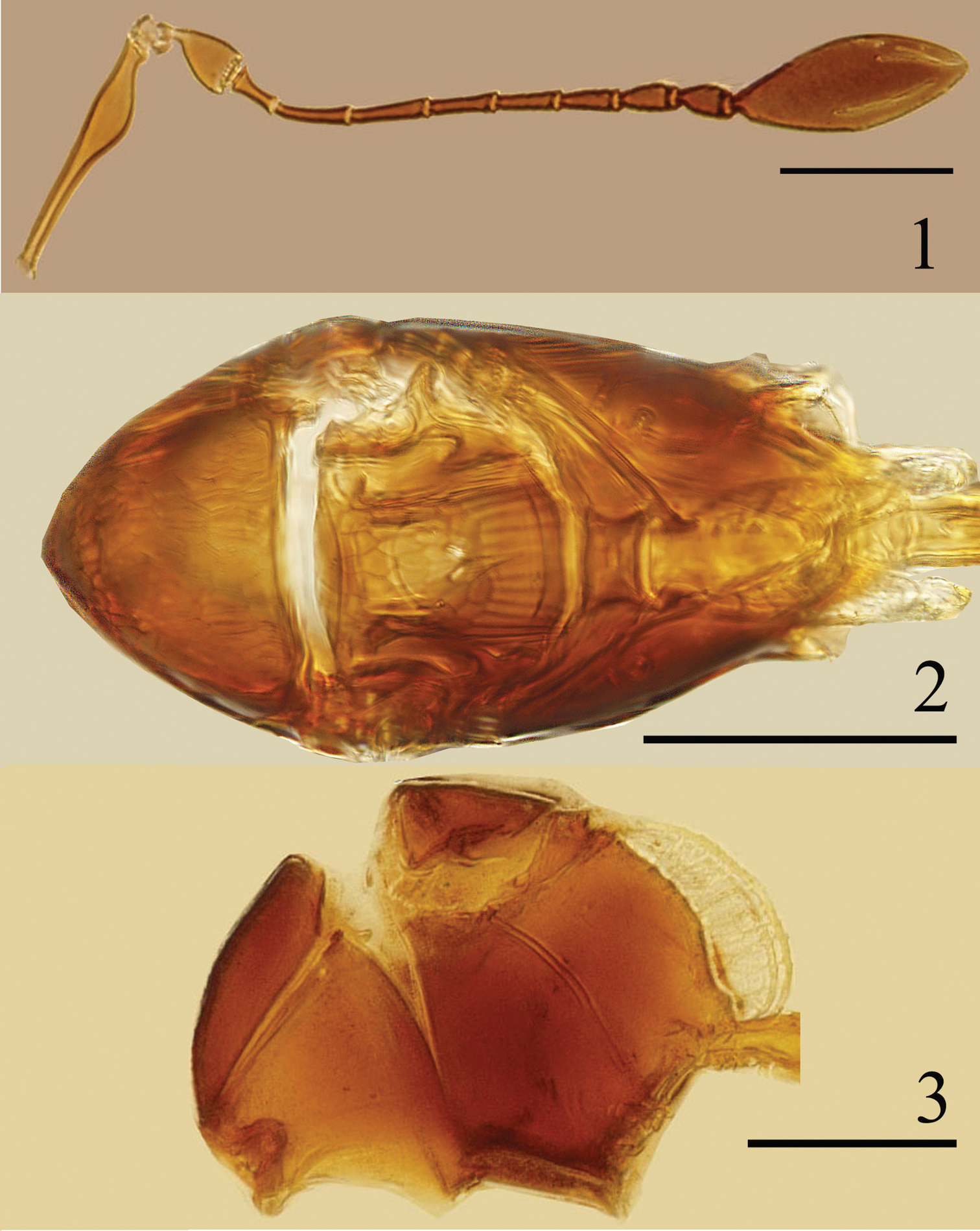
Ptilomymar dianensis sp. n., holotype female: 1 antenna 2 mesosoma, dorsal 3 mesosoma, lateral. Scale bars=100 μm.
Mesosoma (Fig. 2) 1.95× as long as wide. Mesoscutum 0.58× as long as wide, with strong reticulation. Scutellum with strong reticulation on anterior scutellum and longitudinal striate on posterior scutellum; with a pair of campaniform sensilla nearer posterior margin than anterior margin. Metanotum 0.25× as long as scutellum. Mid panel of metanotum subrectangle, with longitudinal striate. Propodeum slightly shorter than mesoscutum, without reticulate sculpture, with 2 large subparallel translucent carinae (Figs 2, 3, 6, 7) and 2 branched setae, each on lateral to spiracle.
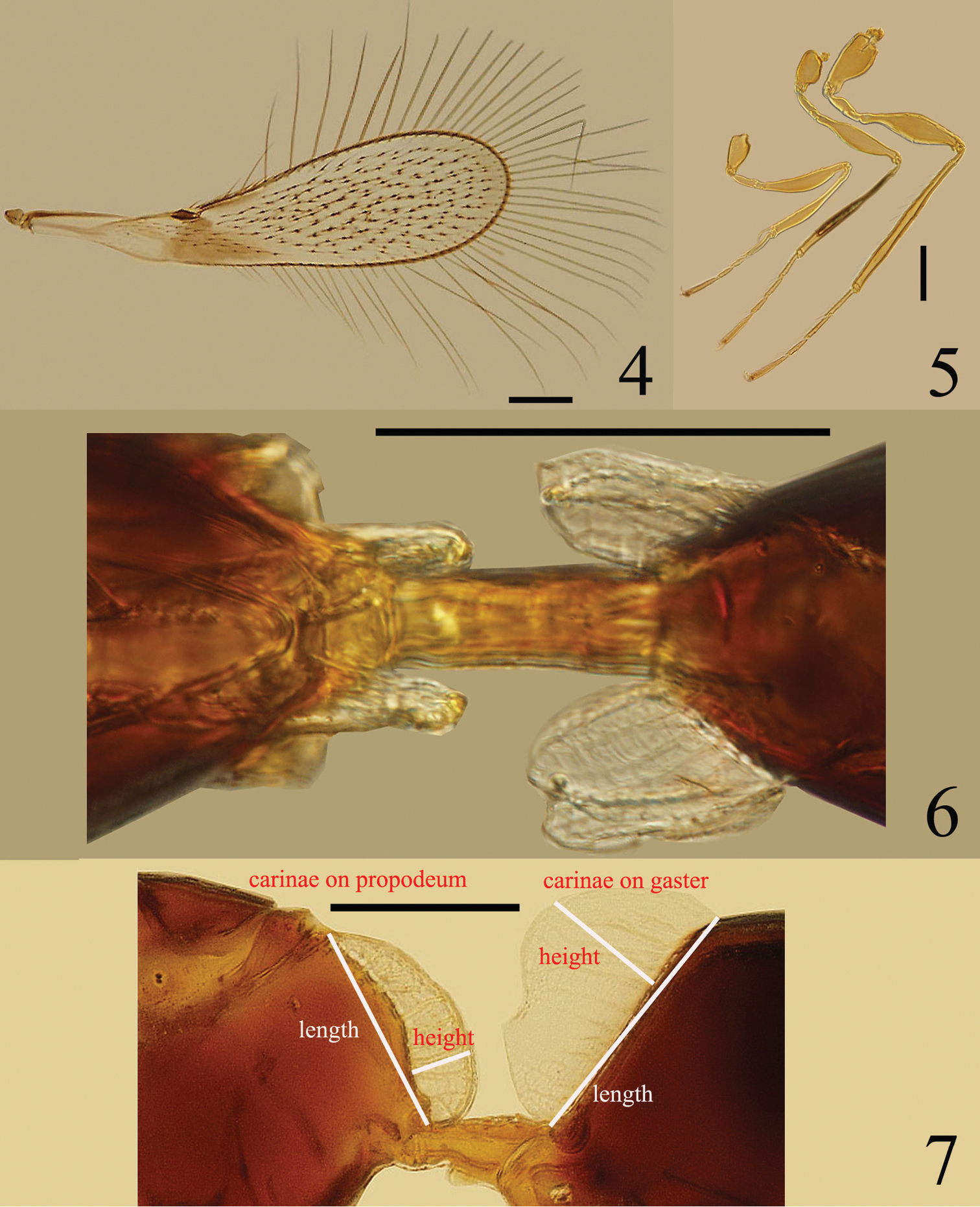
Ptilomymar dianensis sp. n., holotype female: 4 fore wing 5 legs 6 carinae on mesosoma and metasoma, dorsal 7 carinae on mesosoma and metasoma, lateral. Scale bars=100 μm.
Fore wing (Fig. 4) 3.62× as long as wide, longest marginal setae 1.38× as long as greatest wing width. Stigmal vein with 4 campaniform sensilla apically.
Legs (Fig. 5) with femora, especially metafemur, swollen medially. Mesocoxa without teeth-like structures on the posterior surface.
Metasoma. Petiole (Fig. 6) about 2.8× as long as wide. Gaster (Fig. 8) oblong, Gt1 (Fig. 7) with 2 large translucent carinae and 1 smaller carinae and a pair of scale-like setae on each side; ovipositor distinctly exserted, about 0.7× as long as mesotibia.
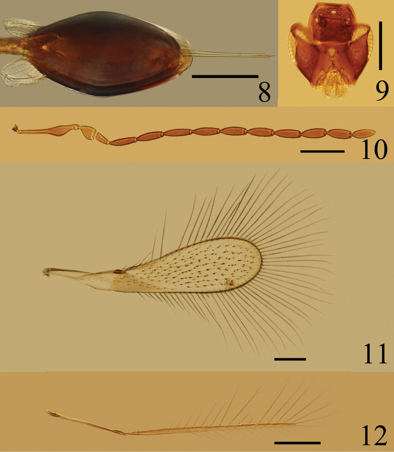
Ptilomymar dianensis sp. n., holotype female: 8 gaster. Paratype male: 9 head, dorsal 10 antenna 11 fore wing 12 hind wing. Scale bars=100 μm.
Measurements (length/width, μm): Body length: 500. OD 9.6, OOL 9.6, LOL 33.6, POL 62.4. Antenna: scape 144.0/ 26.4, pedicel 40.8/ 31.2, fl1 26.4, fl2 43.2, fl3 45.6, fl4 38.4, fl5 36.0, fl6 33.6, fl7 33.6, fl8 31.2, clava 136.8/ 55.2. Fore wing 752.4/ 207.9, longest marginal setae 287.1. Propodeum with carinae length 115.2, height 33.6 (measured in lateral view – Fig. 3); gaster with dorsolateral carina length 144, height 67.2 (measured in lateral view – Fig. 7), and ventromedian carina length 120, height 33.6. Ovipositor 201.6.
Similar to female except as follows. Antenna (Fig. 10) with all the flagellar segments longer than wide. Fore wing (Fig. 11) 3.89–4.06× as long as wide. Hind wing (Fig. 12) 0.76–0.78× as long as fore wing, disc with only one row of setae.
Measurements (length/width, μm): Body length 550–580. Antenna: scape 139.2–144.0/ 21.6–26.4, pedicel 43.2/ 28.8–31.2, fl1 33.6, fl2 64.8, fl3 67.2, fl4 38.4, fl5 64.8, fl6 62.4, fl7 62.4, fl8 62.4, fl9 60.0, fl10 60.0, fl11 57.6. Fore wing 643.5–693.0/ 158.4–178.2, hind wing 504.9–524.7.
Unknown.
Chinese: dian=Yunnan Province, and refers to the distribution of the species in the Yunnan Province of China.
We are grateful to Dr. Hui-Lin Han and Mr. Ye Chen, Northeast Forestry University, Harbin, China, for specimen collection, Dr. Anis, Department of Zoology, Aligarh Muslim University, Aligarh, Inida, for providing Hayat & Anis reference, Prof. Hong-Ying Hu, Xinjiang University, Urumqi, Xinjiang, China, for providing Yoshimoto reference, and Dr. D. Chesters, Institute of Zoology, Chinese Academy of Sciences, Beijing, P. R. China, for correction of English.