


Citation: Petrulevičius JF, Popov YA (2014) First fossil record of Discocephalinae (Insecta, Pentatomidae): a new genus from the middle Eocene of Río Pichileufú, Patagonia, Argentina. ZooKeys 422: 23–33. doi: 10.3897/zookeys.422.6750
A new genus and species of Discocephalini, Acanthocephalonotum martinsnetoi gen. n. et sp. n. is described from Río Pichileufú, middle Eocene of Patagonia, Argentina at palaeolatitude ~ 46°S. The new species is the first fossil representative of the Discocephalinae. This taxon is extant in equatorial to subtropical America, and some species reach warm temperate latitudes (Buenos Aires province). The new genus is distinguished from the other genera of Discocephalini by the combination of these characters: interocular width greater than head length; head massive and quadrangular with the anterior margin almost straight; juga touching each other; labrum thick and curved; triangular ante-ocular process extending beyond the eye; broad spine-like antero-lateral process of the pronotum; pronotum explanate and bean shaped; scutellum triangular with a circular tongue reaching the anterior side of abdominal segment 7; and wings well developed with membrane just surpassing end of abdomen.
Acanthocephalonotum gen. n. , Heteroptera, Discocephalini
Pentatomidae is a diverse and globally distributed family of insects with nearly 900 described genera and 4722 living species (
The single specimen comes from the Patagonian locality of Río Pichileufú, Río Negro, Argentina (
The fossil is housed at the Museo Asociación Paleontológica Bariloche (repository prefix MAPBAR), San Carlos de Bariloche, Río Negro, Argentina. Recent specimens of Discocephalinae are housed in the Entomological collection (Box 1895) of the Museo de La Plata (MLP), La Plata, Argentina. The holotype of Glyphuchus sculpturatus Stål, 1858 is housed in the Naturhistoriska riksmuseet, Stockholm, Sweden.
The fossil and recent specimens from Argentina were photographed with a Nikon D5000 digital camera. The new species was drawn with a camera lucida attached to a Wild M8 stereomicroscope.
Acanthocephalonotum martinsnetoi sp. n.
Pronotum with the humeral and posterior angles developed; origin of the labium caudad of the anterior limit of the eyes; head wider than long, anterior margin of head almost straight; labrum thick and curved; juga touching each other before clypeus; interocular width greater than head length (1.16 ×); triangular ante-ocular process extending beyond the eye and perpendicular to the sagittal plane; pronotum with an antero-lateral process (broad spine-like), parallel to the sagittal plane; scutellum triangular with a developed and circular tongue; wings well developed with membrane just surpassing end of abdomen; costal margin bending acutely before end of basal half (boomerang shaped); apex of the scutellum not reaching the apex of corium.
Type species: Acanthocephalonotum martinsnetoi sp. n.
From the Latin acanthus, meaning spiny, the Greek κεφαλή, meaning head and the Greek νώτος, meaning dorsal and signalling dorsal part of prothorax. “After the head and pronotum with broad spine-like processes”.
http://zoobank.org/53C87349-C765-463A-B96D-EA1E5A9896C4
Figs 1, 2Same as for the genus, by monotypy.
The specimen is mainly complete and articulated in dorsal position with a composite view of dorsal and ventral structures.
Body: 4.7 mm long and 3.6 mm wide at pronotum; width (at the base of the hemelytra) / specimen length ratio, 0.78; antennae and legs not visible; head broad, almost rectangular with numerous punctures, wider than long; anterior margin of head almost straight in almost all its width; head 1.15 mm wide in its anterior margin, 0.8 mm long; eyes, 0.24 mm wide, 0.11 mm long; anteocular length 0.36 mm; inter-ocular width 0.95 mm; inter-ocular width / head length ratio, 0.84; distance between ocelli 0.48 mm; distance between eyes and ocellus 0.2 mm; juga (= mandibular plates) touching each other before clypeus; apex of juga contiguous about 0.11 mm; lateral margins of juga deeply concave; clypeus bullet shaped; ante-ocular process extending beyond the eye and perpendicular to the sagittal plane, subtriangular shaped, 0.23 mm long, with its anterior margin convex and posterior margin concave and beside the eye; labrum thick and curved (ventral structure); origin of the labium caudad of the anterior limit of the eyes (ventral structure); pronotum with a broad spine-like antero-lateral process, stout and acute, parallel to the sagittal plane, 0.2 mm long; head length / pronotal width ratio, 0.87; pronotum with numerous punctures, strongly explanate and bean-shaped, 3.6 mm wide, 1 mm long; lateral margins rounded and irregular; scutellum triangular with a developed and circular tongue; scutellum about 2.8 mm wide at base, 1.9 mm long; tongue, 1.2 mm wide and 0.75 mm long; apex of tongue surpassing the corium; apex of scutellum reaching the anterior side of abdominal segment 7; posterior margin of abdominal segment 7 with three straight sides; gonocoxites 8 (ventral structure) with sub-triangular truncate shape, outer lateral margins obliques, posterior ones straights; laterotergites 8 large, sub-triangular, truncate in inner lateral margins.
Wings: well developed membrane just surpassing end of abdomen; corium with punctures; costal margin bending acutely before end of basal half (boomerang shaped); costal angles of corium above ante-penultimate tergum; R slightly curved and followed by punctures by both sides; M slightly zigzagged; CuA almost straight and followed by punctures by both sides; venation not visible in membrane.
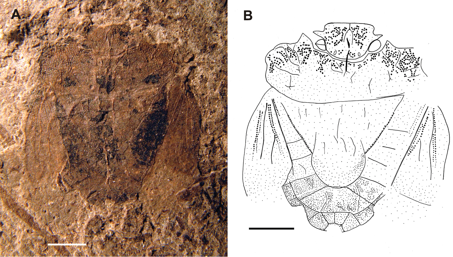
Habitus of Acanthocephalonotum martinsnetoi gen. n. et sp. n. Holotype specimen MAPBAR 4137 A Photograph B line drawing. Scale bars represent 1 mm.
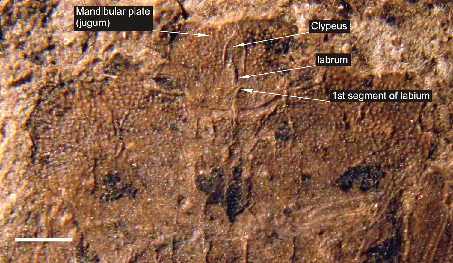
Photograph of detail of head and thorax of Acanthocephalonotum martinsnetoi gen. n. et sp. n. Holotype specimen MAPBAR 4137. Scale bar represents 1 mm.
holotype specimen MAPBAR 4137.
Volcanic caldera-lake beds, Río Pichileufú, quarry RP3 (
Dedicated to the memory of Rafael Gioia Martins-Neto, outstanding palaeoentomologist and “irmão de alma”, who unexpectedly and prematurely passed away in 2010 at age 56.
The specimen is a female in dorsal position albeit some ventral structures of head and genitalia are visible resulting in a composite view. Females of Discocephalinae are recognized by having external genital structures as laterotergites and gonocoxites (Fig. 3A, C). Laterotergites 8 are joined by a transverse band visible from the dorsal side (Fig. 3A), but this structure is not visible in the fossil specimen (Figs 1, 2). Males of recent representatives of the group have a pygophore that is easily lost, leaving the free posterior face of the seventh segment with three sides (Fig. 3B, D).
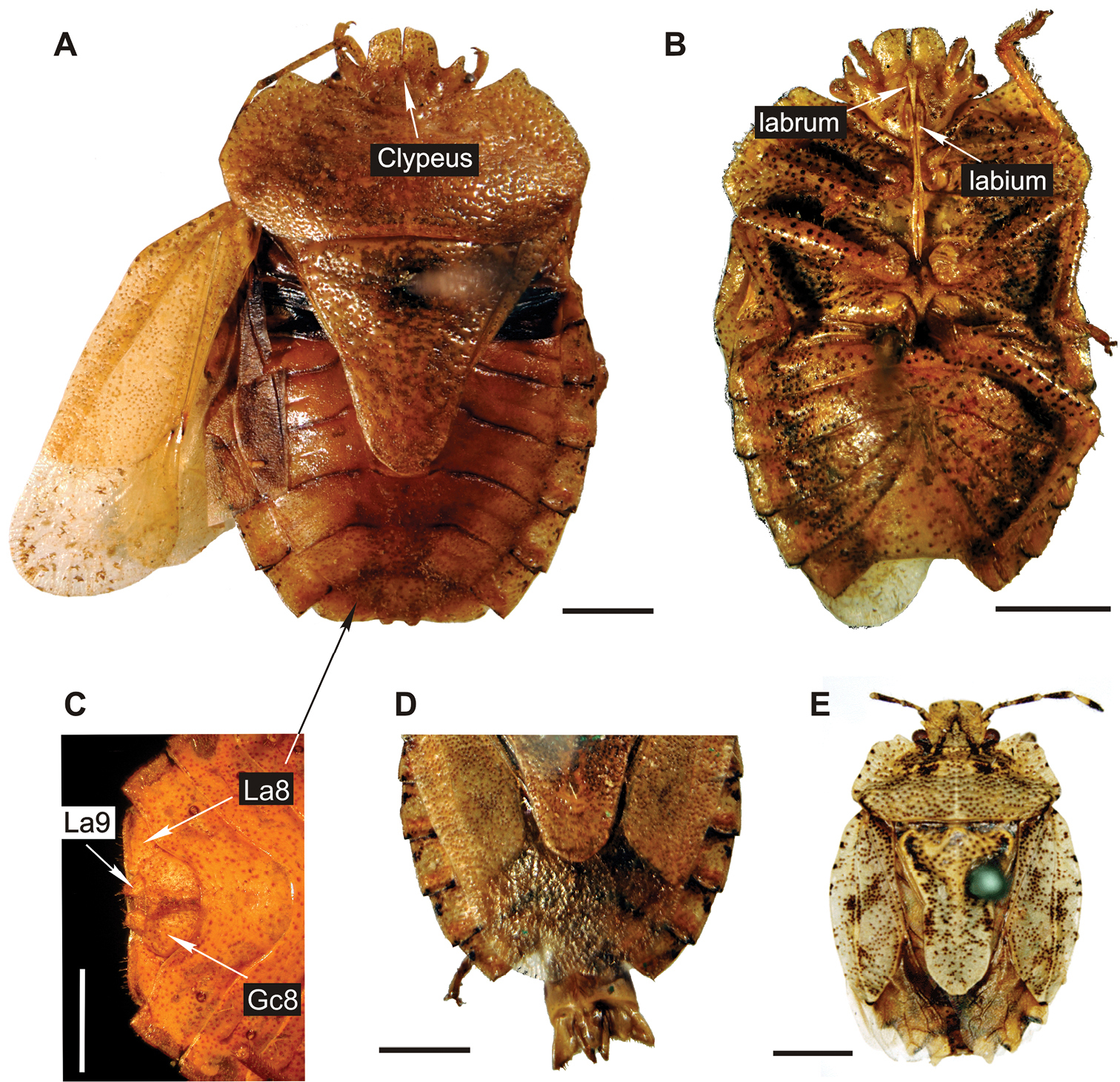
Photographs of habitus of extant genera of Discocephalini A-D specimens of Dryptochephala lurida Erichson, 1848 A female specimen, dorsal view, Tucumán, Argentina B male specimen without terminalia, ventral view, Loreto, Misiones, Argentina C detail of female abdomen in ventral view, Tucumán, Argentina, Gc8: gonocoxite 8, La8: laterotergite 8, La9: laterotergite 9 D detail male abdomen with pygophore in dorsal view, Iguazú, Misiones, Argentina E holotype of Glyphuchus sculpturatus Stål, 1858, female specimen, dorsal view “Rio Janeiro” (Stål, 1872), Brazil. Scale bars represent 2 mm.
The specimen can be attributed to a species of Pentatomoidea by the presence of several characters (
Other genera that are not broad-headed discocephalines but share other similarities with the new species are Sympiezorhincus Spinola, 1837, Psorus Bergroth, 1914, Pelidnocoris Stål, 1867, Abascantus Stål, 1864, and Coriplatus White, 1842. They all share a developed tongue (
The Discocephalinae are considered a tropical to subtropical taxon with some species reaching a warm temperate latitudes (
Thanks are due to Rubén Cúneo and Eduardo “Dudu” Ruigómez from MEF for loaning the specimen for study, María del Carmen Coscarón, María Celina Digiani and Peter Wilf for valuable help and discussion, ; and the three reviewers, Jocélia Grazia, Pavel Štys and Dong Ren, who strongly enhanced the contribution. We also thank the Swedish Naturhistoriska riksmuseet and their curator G. Lindberg for permission to use photos of holotypes from Naturhistoriska riksmuseet and to send me some supplementary data on the holotype of Glyphuchus sculpturatus. Funding support came from grants: PIP 6393 and PIP 0377 from the National Research Council of Argentina (CONICET); PICT-2012-1555 from the National Agency of Scientific and Technological Promotion of Argentina (ANPCyT); and DEB-0345750 and DEB-0919071 from the National Science Foundation of USA (NSF).