


Citation: Yin Z-W, Li L-Z (2014) A new species of Diartiger Sharp (Staphylinidae, Pselaphinae, Clavigeritae) from the Fengyangshan – Baishanzu Nature Reserve, East China. ZooKeys 419: 129–135. doi: 10.3897/zookeys.419.7867
A new clavigerine pselaphine, Diartiger zhejiangensis Yin & Li, sp. n., from the Fengyangshan – Baishanzu Nature Reserve, southern Zhejiang, is described, illustrated, and compared with congeners. The species is hosted by ants in the genus Lasius. The key to Diartiger species from China is modified to accommodate the new species.
Taxonomy, Clavigeritae, Diartiger, new species, myrmecophily, Lasius, Asia
In a recent publication (
During a survey (26.iv–03.v.2014) of the staphylinid beetles at the Fengyangshan – Baishanzu Nature Reserve, Zhejiang, three adults of an undetermined Diartiger were discovered, two in Lasius colonies, and one from a sifted litter sample. This species can be separated from all known congeners by a unique combination of external characters, and is formally described herein. The key to Diartiger species occurring in China is modified to accommodate the new species.
All material treated in the present study is housed in the Insect Collection of Shanghai Normal University, Shanghai, China (SNUC).
This study is based on three adults collected during a recent survey of the staphylinid beetles at the Fengyangshan – Baishanzu Nature Reserve by our lab students Zhong Peng and Xiao-Bin Song. The two Diartiger adults and host ants from Baishanzu Nature Reserve were transported to our lab alive for behavioral observations and photos.
Observations and dissections were carried out using an Olympus SZ61 Stereo microscope. Dissected parts were preserved in Euparal mounting medium (BioQuip Products, Inc., CA, U.S.A.) on plastic slides that were placed on the same pin with the specimen. Digital habitus images of dead and live adults were created using a Canon 7D digital camera in conjunction with a Canon MP-E 65mm f/2.8 1-5X Macro Lens, a Canon MT-24EX Macro Twin Lite Flash, and a flash light diffuser made by parchment paper. Images of the dissected parts were made using a Canon G9 camera mounted by hand on a Olympus CX31 microscope. Zerene Stacker version 1.04 was used for image stacking, and all images were edited and grouped in Adobe Photoshop CS5 Extended (version 12.0).
The following abbreviations are applied: AL–length of the abdomen along the midline; AW–maximum width of the abdomen; EL–length of the elytra along the sutural line; EW–maximum width of the elytra; HL–length of the head from the anterior clypeal margin to the occipital constriction; HW–width of the head across eyes; PL–length of the pronotum along the midline; PW–maximum width of the pronotum. Length of the body is a combination of HL + PL + EL + AL.
http://zoobank.org/20113B64-96ED-4879-AB1D-D23243DBEF12
Figs 1–3(2 ♂♂, 1 ♀). Holotype: CHINA: ♂, labeled ‘China: S. Zhejiang, Qingyuan, Baishanzu N.R., 27°45'14"N, 119°11'55"E, Fagaceae forest, ant nest in rotten wood, 1650 m, 01.v.2014, X.-B. Song leg. / HOLOTYPE [red], Diartiger zhejiangensis sp. n., det. Yin & Li, 2014, SNUC’. Paratypes: CHINA: 1 ♀, same area and date, except ‘27°45'27"N, 119°12'05"E, mixed leaf litter, sifted, 1700 m, Z. Peng leg.’; 1 ♂ [disarticulated specimen preserved in Euparal on plastic slides], labeled ‘China: S. Zhejiang, Longquan, Fengyang Shan, creek valley nr. hotel, 27°54'42"N, 119°11'52"E, Rhododendron and fir forest, ant nest in erect rotten fir trunk, 1250 m, 28.iv.2014, Peng leg.’. Each paratype bears a type label similar to that of the holotype except ‘PARATYPE [yellow]’.
Male. Body (Fig. 1A) length 2.14–2.15 mm; reddish brown. Head longer than wide, HL 0.39–0.40 mm, HW 0.27–0.28 mm; clypeus with slightly angularly rounded anterior margin; eyes each composed of about 20 facets; antennomeres IV (Fig. 2A) more than twice length of III, with truncate apex. Pronotum about as long as wide, PL 0.37–0.38 mm, PW 0.39–0.41 mm. Elytra (Fig. 2E) wider than long, EL 0.53–0.56 mm, EW 0.80–0.82 mm; lacking microsculpture; with small tuft of setae at posterolateral margins, and bigger triangular, posteriorly-narrowed trichomes at posterior margins. Metathoracic wings fully-developed. Mesoventrite (Fig. 2F) lacking median carina, metaventrite (Fig. 2F) slightly convex, both meso- and metaventrites with row of setae along midline. Profemora (Fig. 2B) with long ventral setae at base, protibiae narrowed at base and thickened from basal third toward apex; mesotrochanters (Fig. 2C) with large, bluntly triangular ventral spine, mesofemora lacking spine; mesotibiae with small apical spur; metatibiae (Fig. 2D) lacking spine or modification. Abdomen slightly wider than long, AL 0.82–0.84 mm, AW 0.88–0.92 mm; composite tergite with transverse basolateral trichomes curved mesally, first pair of paratergites with linear trichomes (Fig. 2G). Aedeagal length 0.50–0.52 mm; median lobe (Fig. 2I–K) with sinuate apical margin in dorso-ventral view.
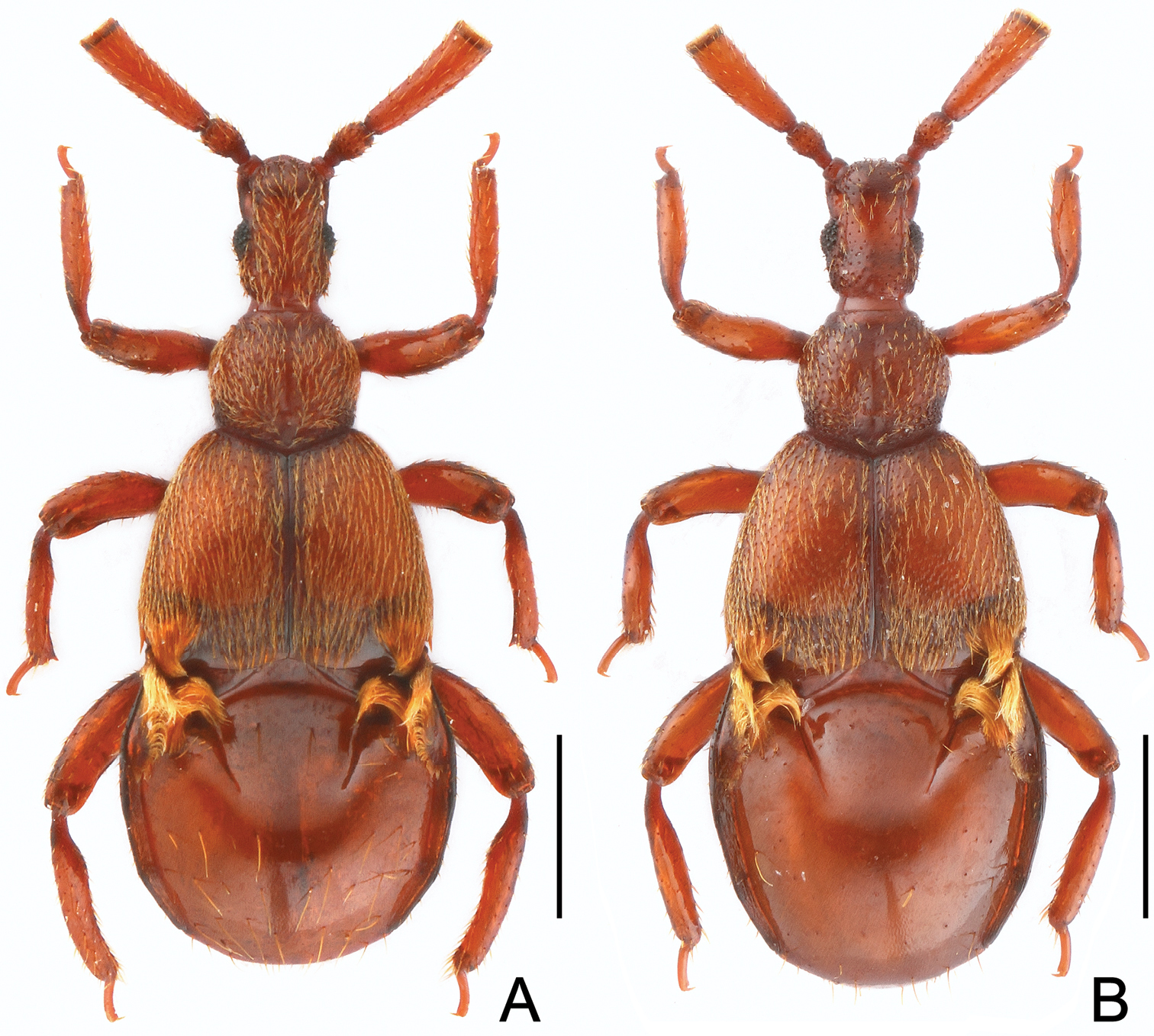
Habitus of Diartiger zhejiangensis. A Male B Female. Scales: 0.5 mm.
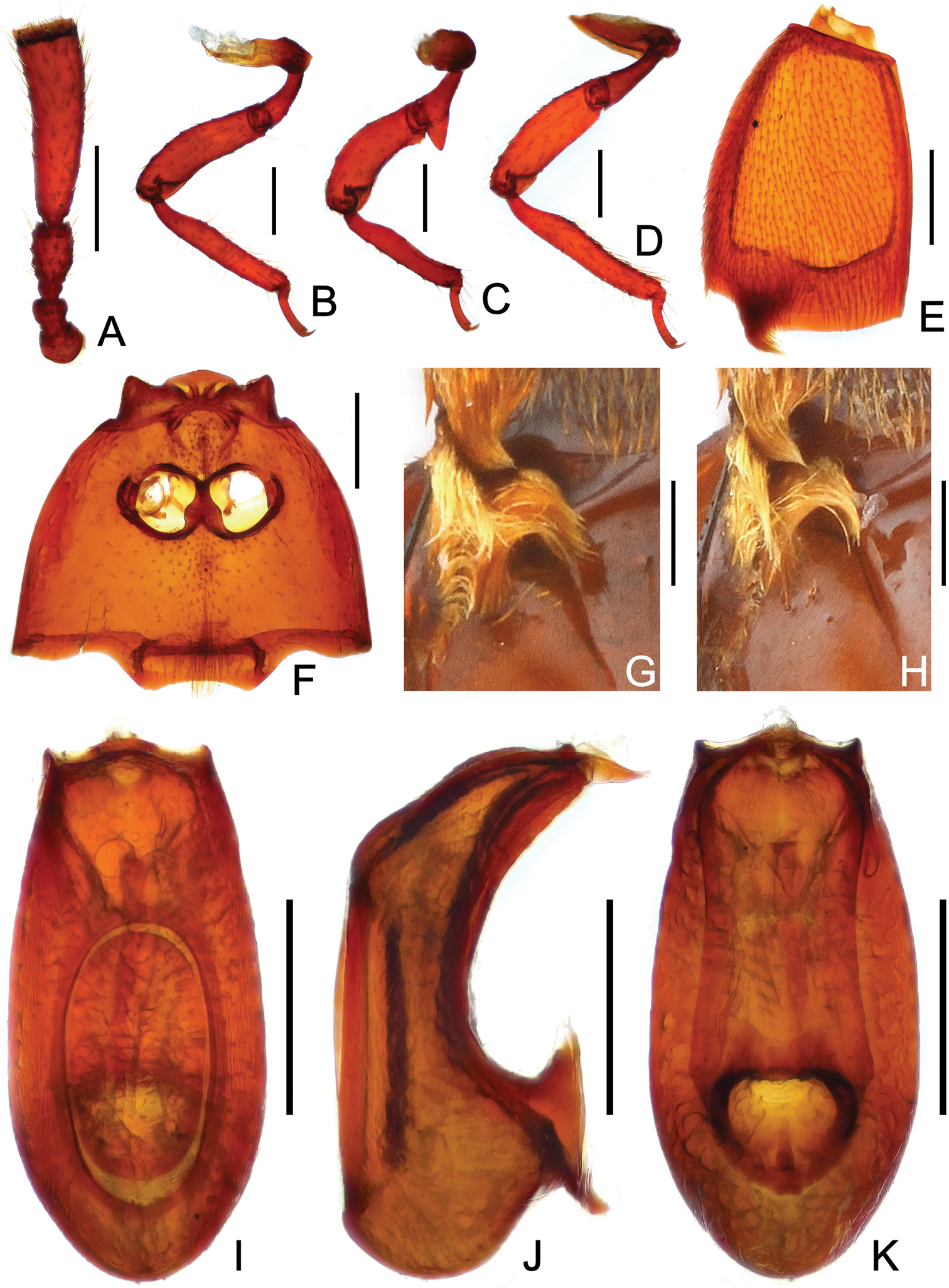
Diagnostic features of Diartiger zhejiangensis (A–G, I–K male H female). A Antenna B Fore leg C Mid leg D Hind leg E Left elytron F Meso- and metaventrites G–H Trichomes of elytra and composite tergite I Aedeagus, in dorsal view J Same, in lateral view K Same, in ventral view. Scales: 0.2 mm.
Female. General form (Fig. 1B) and trichomes on elytra and composite tergite (Fig. 2H) similar to those of male; fourth antennomeres slightly shorter, mesotrochanters and mesotibiae lacking spine; abdomen relatively larger. Each eye composed of about 20 facets. Measurements: BL 2.23 mm, HL 0.41 mm, HW 0.28 mm, PL 0.39 mm, PW 0.43 mm, EL 0.55 mm, EW 0.83 mm, AL 0.88 mm, AW 0.92 mm.
This species is placed as a member of the Diartiger fossulatus group. Males are most similar to those of Diartiger dentatus in possessing relatively short antennomeres III, and in similarities of trichomes on the posterior margins of the elytra and the base of the composite tergite, and in the aedeagal form. The two species can be readily separated by the lack of spines on the mesofemora and middle of the mesotibiae, and lack of a small tubercle on the ventral margin of the metacoxa in Diartiger zhejiangensis, while Diartiger dentatus possesses a sharp basal spine on the ventral margin of the mesofemur, the mesotibia possesses a triangular spine at the middle, and the metacoxa possesses a small tubercle on the ventral margin.
East China: Zhejiang.
The male holotype was collected from a colony of Lasius cf. koreanus (det. Maruyama, pers. comm. 2014) nesting in rotten wood in a predominantly Fagaceae forest (Fig. 3C, D). The female paratype from Baishanzu was collected from a sifted litter sample. The other female paratype collected from Fengyang Shan was found in a small colony of the same Lasius species nesting under bark of a standing rotten fir trunk in a Rhododendron and fir forest (Fig. 3A, B). During a two-day observation period, the adults of Diartiger zhejiangensis (Fig. 3E, F) were totally ignored by the ant workers, and vice versa. This may have been a consequence of the disturbance of being transported and housed in the artificial environment.
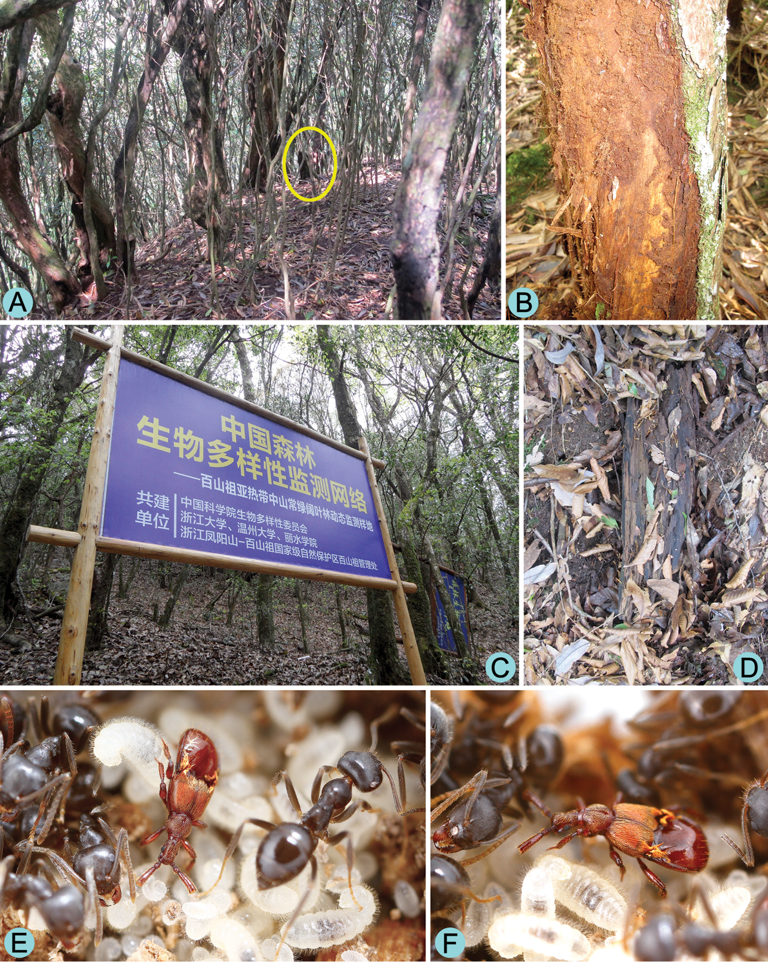
Habitat and host of Diartiger zhejiangensis. A General habitat of the Rhododendron and fir forest at Fengyang Shan Nature Reserve B Rotten trunk where the female paratype was collected C General habitat of the Fagaceae forest at Baishanzu Nature Reserve D Rotten wood where the holotype was collected E–F Living male adult of Diartiger zhejiangensis with host ants.
The specific epithet refers to the province where the type locality of the new species lies.
| 2 | Fourth antennomeres (Fig. 2A; |
2a |
| – | Fourth antennomeres ( |
3 |
| 2a | Mesofemora with a sharp ventral spine at base; mesotibiae with a triangular spine at middle ( |
Diartiger dentatus Yin & Li, 2013 |
| – | Mesofemora and middle area of mesotibiae (Fig. 2C) lacking spine, metacoxae (Fig. 2D) lacking tubercle. (East China: Zhejiang) | Diartiger zhejiangensis sp. n. |
Our colleagues Zhong Peng and Xiao-Bin Song collected the material used in this study. Munetoshi Maruyama (Kyushu University Museum, Japan) identified the host ants. Christopher E. Carlton (Louisiana State Arthropod Museum, U.S.A.) reviewed a previous draft and corrected the English narrative before submission. Two anonymous reviewers critically read the manuscript and provided helpful suggestions. The present study is supported by the National Science Foundation of China (No. 31172134) awarded to LZL.