ZooKeys 429: 47–61, doi: 10.3897/zookeys.429.8116
Status of the enigmatic Oriental genus Rhithrogeniella Ulmer, 1939 (Ephemeroptera, Heptageniidae)
Michel Sartori 1,2
1 Zoologisches Museum und Biozentrum Grindel, Martin-Luther-King-Platz 3, D-20146 Hamburg, Germany
2 Museum of Zoology, Palais de Rumine, Place Riponne 6, CH-1005 Lausanne, Switzerland

Corresponding author: Michel Sartori (michel.sartori@vd.ch)

Academic editor: E. Dominguez

received 16 June 2014 | accepted 23 July 2014 | Published 29 July 2014
(C) 2014 Michel Sartori. This is an open access article distributed under the terms of the Creative Commons Attribution License (CC BY 4.0), which permits unrestricted use, distribution, and reproduction in any medium, provided the original author and source are credited.
For reference, use of the paginated PDF or printed version of this article is recommended.

Citation: Sartori M (2014) Status of the enigmatic Oriental genus Rhithrogeniella Ulmer, 1939 (Ephemeroptera, Heptageniidae). ZooKeys 429: 47–61. doi: 10.3897/zookeys.429.8116

Abstract

Based on historic collections and new material from Sumatra and Java, the species Rhithrogeniella ornata Ulmer, 1939, type species of the genus Rhithrogeniella, is reinvestigated. The nymph is described for the first time and is closely related to the continental Southeast Asian species Rhithrogeniella tonkinensis Soldán and Braasch, 1986. Rhithrogeniella belongs to the subfamily Ecdyonurinae, and is related to the genera Nixe Flowers, 1980 and/or Paracinygmula Bajkova, 1975 based on characters of the nymphal stage. Species described from Taiwan in the genus Nixe are transferred to the genus Rhithrogeniella: Rh. littoralis (Kang and Yang, 1994) comb. n., Rh. mitifica (Kang and Yang, 1994) comb. n. and Rh. obscura (Kang and Yang, 1994) comb. n.

Keywords

Rhithrogeniella ornata, Rhithrogeniella tonkinensis, Nixe, Paracinygmula, new combinations, Sumatra, Java

Introduction

Ulmer (1939) established the genus Rhithrogeniella to accommodate the species Rhithrogeniella ornata Ulmer, 1939 known from imagos and subimagos from Java and Sumatra. According to Ulmer (1939), the genus was characterized by genitalia resembling those of Rhithrogena, with two simple lobes lacking spines or titillators. On the other hand, the new genus differed from Rhithrogena in the tarsal proportions on fore- and hind legs. Interestingly, in the key to the genera (Ulmer 1939), Rhithrogeniella is keyed with the genus Afronurus Lestage, 1924 from which it differs by the tarsal composition of the foreleg. During the following years, nothing substantial was added to the knowledge of the genus, and keys which included Rhithrogeniella were based on Ulmer’s description and drawings (Tomka and Zurwerra 1985; Tshernova 1974).

Major advancement was made by Soldán and Braasch (1986) who described a new species from Vietnam (Rh. tonkinensis) based on subimagos, together with the first description of the nymphal stage. This nymph exhibits typical Ecdyonurinae morphology, with scattered setae on the maxillae. The genus can be distinguished from all relatives by the peculiar structure of the cerci and terminal filament which possess a row of stout setae in the proximal part and bunches of long and thin setae in the medial and distal parts. Later, Rhithrogeniella tonkinensis was reported from Thailand and the male imago described (Braasch 1990). According to figures of Braasch (1990), the genitalia bear median titillators, the penis lobes are much more rounded than in Rhithrogeniella ornata, and the styliger fig is of an unusual shape with two triangular sublateral processes. In their revision of worldwide Heptageniidae, Wang and McCafferty (2004) proposed several nomenclatorial changes; in particular, they combined Rhithrogeniella ornata with the genus Rhithrogena, hence placing Rhithrogeniella in synonymy with Rhithrogena, and they assigned Rhithrogeniella tonkinensis to the genus Ecdyonurus (E. tonkinensis) based on subimaginal, larval and egg morphology. This account was never discussed later on and Braasch and Boonsoong (2010) mentioned the presence of Rhithrogeniella ornata in West Malaysia based on male subimagos, and Boonsoong and Braasch (2013) listed Rhithrogeniella tonkinensis in the Heptageniidae fauna of Thailand.

Two questions need to be resolved. Are Wang and McCafferty (2004) correct in synonymizing Rhithrogeniella with Rhithrogena (subfamily Rhithrogeninae), and assigning Rhithrogeniella tonkinensis to the genus Ecdyonurus (subfamily Ecdyonurinae)? Is the association between unreared nymphs and subimagos of Rhithrogeniella tonkinensis accurate or not?

The type material of Rhithrogeniella ornata, deposited in the collection of the Zoological Museum of Hamburg University, Germany (ZMH) has been reinvestigated together with new material from Sumatra. It is now possible to provide the first description of the nymph of Rhithrogeniella ornata.

Material and methods

Material studied here is deposited in the following institutions:

Zoologisches Museum und Biozentrum Grindel, Hamburg, Germany [ZMH]

Musée cantonal de zoologie, Lausanne, Switzerland [MZL]

Lembaga Ilmu Pengetahuan Indonesia (Indonesian Institute of Sciences), Museum of Zoology, Bogor, Indonesia [LIPI] (Bogor was formerly known as Buitenzorg)

Drawings were made with the help of a camera lucida taken from stereomicroscope Leica DM 750 and pictures from microscope Zeiss Axioscop 2 or Visionary Digital Passport II. Final digital drawings were performed on Adobe Illustrator CS6. For scanning electronic microscope (SEM) pictures, the eggs were dehydrated, carbon coated, and observed under a LEO 1525 at 5.00kV; maxillae were dehydrated, critical point dried, and then platinum coated, and observed under a FEI Quanta 250 at 5.00kV. Final figs were assembled in Adobe Photoshop CS6.

Nymphs and adults were associated with the help of the egg structure (Fig. 5).

Results
Rhithrogeniella ornata Ulmer, 1939
Rhithrogeniella ornata Ulmer, 1939, male, female imagos and subimagos
Rhithrogena ornata Wang & McCafferty, 2004
Material.

One male holotype, one female allotype: Indonesia, Java, Buitenzorg, VII 1932, Dr. Lieftinck leg. [ZMH]

Paratypes: 4 female subimagos, 1 male subimago: Indonesia, Java, Buitenzorg, Bellevue, caught at light, VII.1929, Prof. Thienemann leg. [ZMH]; 4 female imagos, 2 male subimagos: Indonesia, Sumatra, Padang, VII 1925, Prof. Fulmek leg. [ZMH]; 1 male subimago: Indonesia, Sumatra, Pangkalang, Kota baru, X 1925, Prof. Fulmek leg. [ZMH]

All specimens in ethanol, except fore- and hind legs, fore- and hind wings of the male subimago from Buitenzorg mounted on slide in Canada balsam.

Other material: 5 nymphs: Indonesia, Sumatra Barat, Sawahlunto, stream, 275m, 00°41.33'S, 100°46.72'E, (UN5), 10.XI.2011, M. Balke leg. [ZML]; 26 nymphs, of which two entirely mounted on microscopic slides: Indonesia, Sumatra Barat, Talawi, Ombilin River, 277m, 00°34.15'S, 100°43.54'E, (UN4), 8.XI.2011, M. Balke leg. [ZMH, MZL, LIPI]

Complementary description of the male imago (holotype).

Specimen completely faded; for color patterns see Ulmer (1939).

Mesonotum with transverse suture; medial depression of furcasternum sub parallel anteriorly.

Foreleg with tarsi sub equal in length to the tibia, which is 1.25x longer than the femur. Tarsal composition: 2>3>4>5>1.

Genitalia (Fig. 1): margin of the styliger fig straight to slightly convex, with two small sub-lateral rounded processes; last gonopod segment ca 0.7× the length of the previous, both together ca 0.75× the length of the antepenultimate. Penis constituted of two kidney-shape lobes, separated by a “U” incision, i.e. the inner margin of each lobe is concave and slightly hooked near the apex. No lateral or median titillators, no apical spines visible.

Complementary description of the male subimago.

Fore leg (Fig. 2) with femur ca 1.15x the length of tibia, which is subequal in length to tarsi. Tarsal composition 4≥2>3≥1>5.

Hind leg (Fig. 3) with femur ca 1.35x the length of tibia, which is ca 1.45× the length of tarsi. Tarsal composition 1=2=5>3≥4.

Genitalia (Fig. 4) with penis lobes rounded, ellipsoid, without any spine or titillators; in median position, a pair of membranous processes ending with a spine like sclerotization present in ventral view.

Complementary description of the female imago (allotype).

Thoracic structures similar to the male.

Eggs (Fig. 5): ovoid, ca 130 µm × 90 µm; chorion regularly covered with hexagonal mesh ridges, with KCT in-between, not larger at poles; micropyle rounded to slightly oval in equatorial area.

First description of the nymph.

Size: Body length: up to 5.2 mm and 5.6 mm for male and female respectively; cerci and terminal filament subequal and ca ¾ the length of the body.

Coloration similar to Figs 6 and 7.

Labrum (Fig. 8) moderately expended laterally, ca 2.6× wider than long; lateral margins regularly rounded; no anteromedian emargination; dorsal face covered with long and thin setae anteriorly; ventral face with shorter and stout setae along the anterior margin. Mandibles covered with numerous long and thin setae on the outer margin; right mandible with outer incisor saw-like, inner one with a trifid apex with 2–3 pectinate setae below it, and 2–3 long and simple setae below the mola; left mandible with outer incisor saw-like, inner one with a bifid apex with 3–4 pectinate setae below it, and 3–4 long and simple setae below the mola. Maxillary palp three-segmented; first segment covered with thin setae on inner and outer margin; second segment with thin setae on the outer margin; third segment slightly pointed, only with long and thin setae. Maxillae with fimbriate scattered setae on the ventral surface (Fig. 13): 13–14 comb-shape setae on the crown of the galea, median ones with 10–11 teeth (Fig. 14); proximal dentiseta bifid, outer margin feathered; distal dentiseta simple, entire and unbranched (Fig. 12). Labium (Fig. 9) with glossae rhomboid, inner margin covered with long and thin setae, apex characteristic with scale-like margin (Fig. 10); paraglossae moderately expended laterally. Hypopharynx (Fig. 11) with rhomboid lingua bearing a tuft of short and thin setae at apex; superlinguae well developed and expended laterally with rounded apex and setae on the outer margin extended beyond the apex.

Pronotum moderately expended laterally. Foreleg with femur ca 2.6× longer than wide; outer margin covered with long and stout setae, becoming thinner near the apex; inner margin with only few spine-like setae on the distal third. Outer margin of tibia with very few thin and short setae (Fig. 15), inner margin with few spine-like setae in the middle; tarsi with only a few spine-like setae in the middle of the inner margin. Hind leg similar, except the spine-like setae on inner margin of the femur present on the whole margin; outer margin of tibia with a row of long and thin setae (Fig. 16) and inner margin with more numerous spine-like setae. Middle leg similar to hind leg, except spine-like setae on the inner margin of the femur only present on the distal half. Bristles on the upper face of femora variable in length, always with divergent margins and rounded apically (Fig. 17). Tarsal claw moderately hooked, bearing 4–6 teeth (Fig. 18). No supracoxal spurs present.

Abdomen with posterolateral extensions weakly developed, visible only on segments V–VIII. Gills present on abdominal segments I–VII. Gill I banana-shape (Fig. 20), with fibrillar part well developed, gill IV ca 1.5× longer than wide, strongly asymmetrical (Fig. 21), gill VI with well-developed fibrillar part, more elongated and slightly asymmetrical (Fig. 22), gill VII ca 2.5× longer than wide, without fibrillar part and slightly asymmetrical (Fig. 23). Posterior margin of abdominal terga with weakly developed spines of different size and shape (Fig. 19). Cerci and terminal filament with long and stout setae in whorls on the proximal part (Fig. 24), together with long and thin setae in the median and distal part (Fig. 25).

Sequence data.

One specimen has been used for the study by Vuataz et al. (2013) under the name “Heptageniidae 1” in figures and “Heptageniidae sp. 1” in table S1, with one mitochondrial (CO1) and two nuclear genes (H3, wg) sequenced. Access numbers in GenBank are for CO1: HF536605, for wg: HF536598, for H3: HF536591.

Figures 1–4.

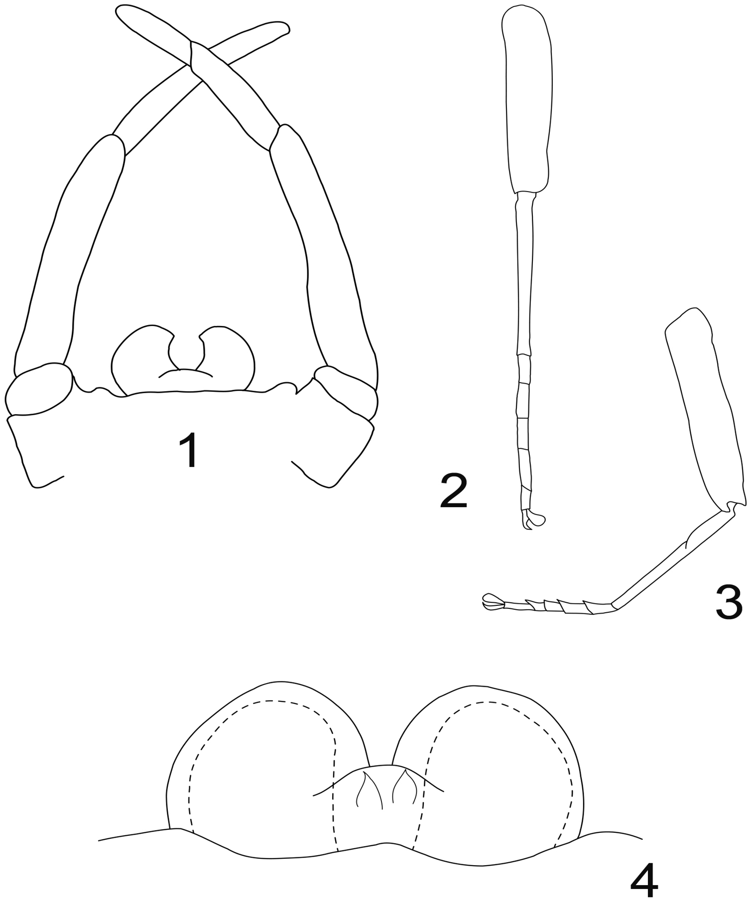
Rhithrogeniella ornata Ulmer, 1939. 1 Genitalia of the male imago (holotype) in ventral view 2 Foreleg of a male subimago (paratype) 3 Hindleg of a male subimago (paratype) 4 Penis lobes of a male subimago (paratype): plain line, cuticular structures of the subimago; dotted line, outline of the imago penis lobes.

Figure 5.

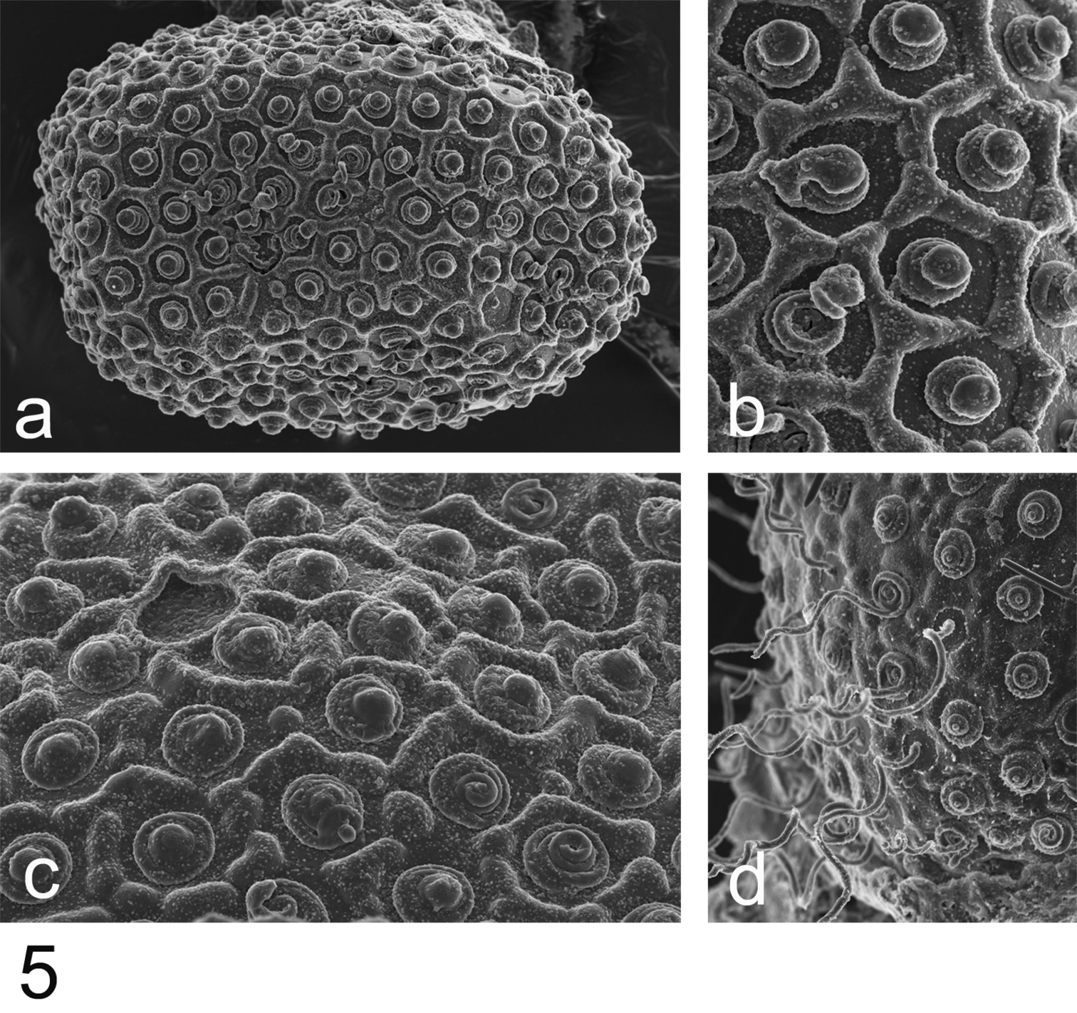
Rhithrogeniella ornata Ulmer, 1939, SEM pictures of egg structures. 5a Egg extracted from a female subimago paratype from Padang, Sumatra 5b Details of the chorionic structure of a female nymph from Ombilin River, Sumatra 5c Details of the chorionic structure and micropyle of a female subimago paratype from Buitenzorg [Bogor], Java 5d chorionic surface of the female allotype from Buitenzorg [Bogor], Java.

Figures 6–7.

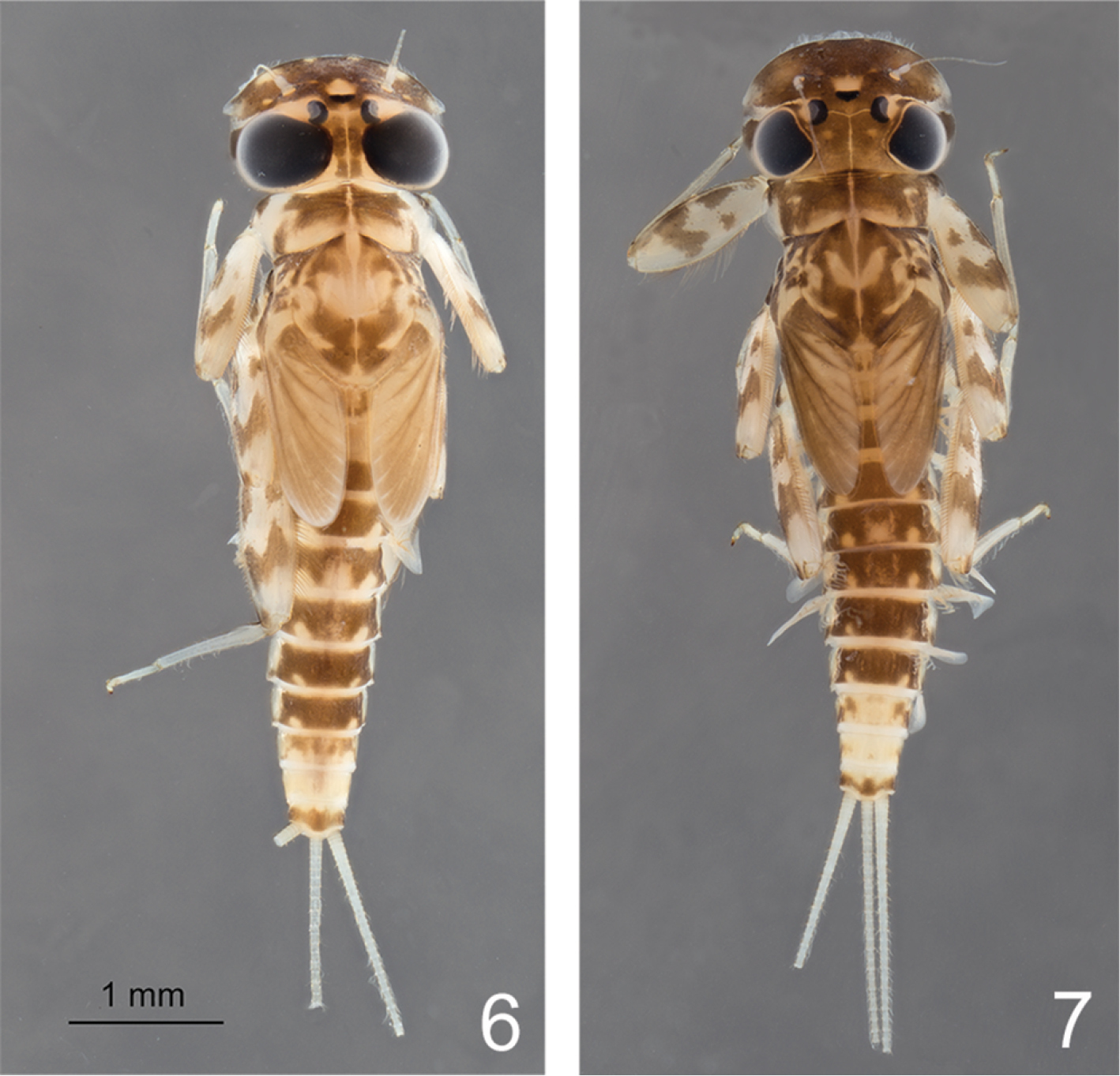
Rhithrogeniella ornata Ulmer, 1939. 6 Male nymph 7 Female nymph with slight color variations.

Figures 8–11.

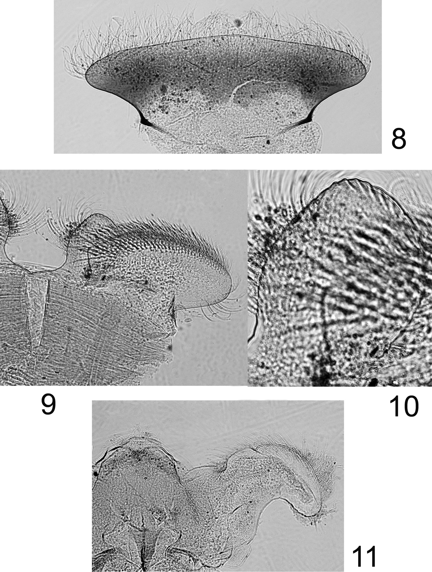
Rhithrogeniella ornata Ulmer, 1939, nymphal mouthparts. 8 Labrum in dorsal view 9 Left glossae and paraglossae of the labium 10 Detail of the glossae from 9 11 Hypopharynx, ventral view lingua and left superlingua.

Figures 12–14.

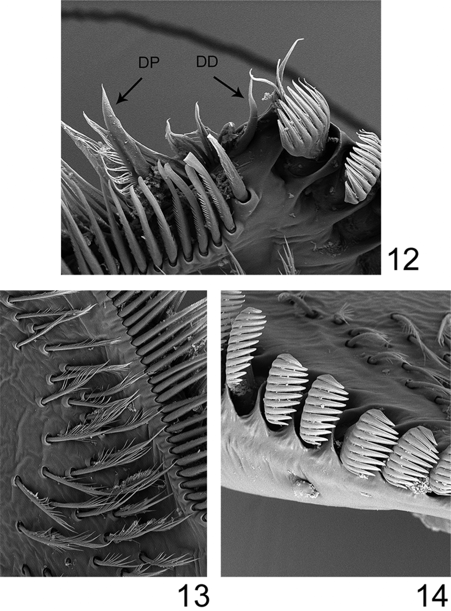
Rhithrogeniella ornata Ulmer, 1939, SEM pictures of the maxilla. 12 Dentisetae (DP: proximal dentiseta, DD: distal dentiseta) 13 Fimbriate setae on the ventral surface 14 Comb-shape setae on the crown of the galea-lacinia.

Figures 15–19.

Rhithrogeniella ornata Ulmer, 1939. 15 Outer margin of the fore tibia 16 Outer margin of the hind tibia 17 Bristles on the dorsal surface of hind femur 18 Tarsal claw 19 Posterior margin of tergite V.

Figures 20–23.

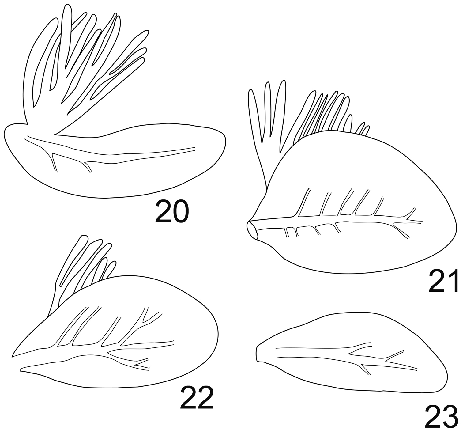
Rhithrogeniella ornata Ulmer, 1939. 20 Gill I 21 Gill IV 22 Gill VI 23 Gill VII.

Figures 24–25.

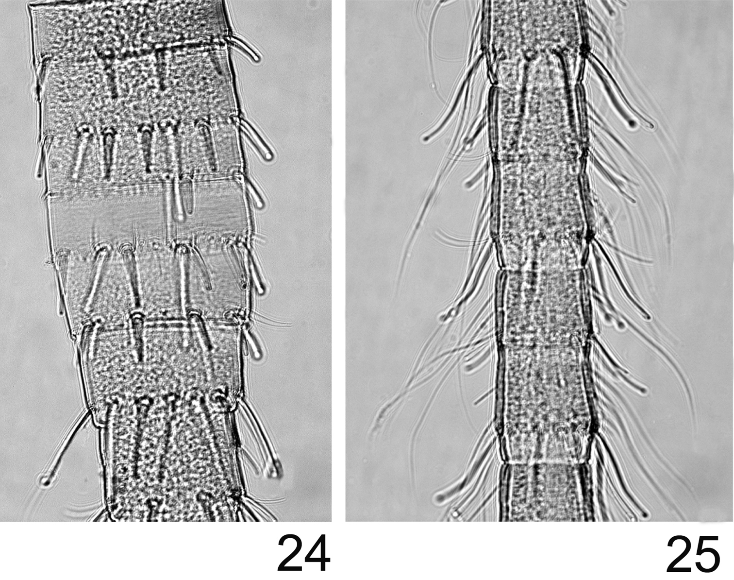
Rhithrogeniella ornata Ulmer, 1939. 24 Proximal part of the terminal filament 25 Median part of the terminal filament.

Discussion

The genitalia of the male imago differ slightly from those described by Ulmer (1939, page 577, Fig. 169), being wider and less cylindrical than illustrated. The presence of a transverse suture on the mesonotum together with the shape of the depression of the furcasternum (not narrowed anteriorly) indicates that Rhithrogeniella ornata cannot be a member of Rhithrogeninae; thus, it is not a synonym of Rhithrogena as suggested by Wang and McCafferty (2004). Moreover, the presence of scattered setae on the ventral side of the maxilla is a character only found among members of Ecdyonurinae, as suggested already by Soldán and Braasch (1986). When using the key of Webb and McCafferty (2008), Rhithrogeniella will key to the genus Afronurus Lestage, 1924 for the adults and to the genus Nixe Flowers, 1980 for the nymphs. Nearctic workers consider the genus Nixe as valid (Flowers 1986; McCafferty 2004; Wang and McCafferty 2004; Webb and McCafferty 2008), but European authors think that its concept is similar to Paracinygmula Bajkova, 1975 (Jacob et al. 1996). The Holarctic species joernensis (Bengtsson, 1909) is therefore treated either as Paracinygmula joernensis (Bauernfeind and Soldán 2012) or as Nixe joernensis (Kjaerstad et al. 2012). Nymphs of Rhithrogeniella share some characters with this concept, including the presence of swimming setae on cerci and terminal filament, and the chorionic structures of the eggs (Flowers 1980). Nymphs of Nixe/Paracinygmula however present gills with a weakly developed fibrillose part, either absent or reduced to a single filament in gill VI, which is not the case in Rhithrogeniella (Fig. 22 and Soldán and Braasch, 1986, fig. 4). Contrary to Nixe/Paracinygmula, the male genitalia have a very different shape and lack well developed median titillators as well as basal sclerite spines.

Three species of Nixe known only from the nymphal stage are reported from Taiwan (Kang and Yang 1994). Bauernfeind and Soldán (2012) transferred them to the genus Paracinygmula without new data, because they considered Nixe as a subjective junior synonym of Paracinygmula. Examination of paratypes of these species, deposited in the collections of MZL, revealed that they perfectly match the concept of Rhithrogeniella developed here, and therefore the following new combinations are proposed:

Rhithrogeniella littoralis (Kang and Yang 1994) comb. n. (= Nixe (Nixe) littoralis Kang and Yang 1994 =Paracinygmula littoralis Bauernfeind and Soldán 2012;

Rhithrogeniella mitifica (Kang and Yang 1994) comb. n. (= Nixe (Nixe) mitificus Kang and Yang 1994 =Paracinygmula mitifica Bauernfeind and Soldán 2012;

Rhithrogeniella obscura (Kang and Yang 1994) comb. n. (= Nixe (Nixe) obscurus Kang and Yang 1994 =Paracinygmula obscura Bauernfeind and Soldán 2012.

Nixe/Paracinygmula is therefore restricted to the Holarctic Realm, whereas Rhithrogeniella is Oriental, reported from Taiwan, continental Southeast Asia and from Java and Sumatra in the Sunda Islands. The genus is presently recorded neither from Borneo (Braasch 2011; Sartori et al. 2003) nor from the Philippines (Braasch 2011).

Based on the Bayesian majority-rule consensus tree reconstructed from the combined data set in Vuataz et al. (2013), Rhithrogeniella appears more related to the tribe Compsoneuriini sensu Sartori (2014) (Compsoneuria, Compsoneuriella and Notonurus), than to other Ecdyonurinae (Thalerosphyrus, Asionurus, Atopopus, Afronurus), although low posterior probability and bootstrap support does not allow to determine its exact relationships. It is possible that further studies may show that a new tribe should be established to accommodate this genus.

One remaining question concerns the presence or absence of titillators on Rhithrogeniella male genitalia. These structures are mentioned by Soldán and Braasch (1986) in the male subimago of Rhithrogeniella tonkinensis as well as in the subimago of Rhithrogeniella ornata (Soldán and Braasch 1986, page 204). Although we have not dissected the holotype (the only male imago of Rhithrogeniella known at the moment), we feel confident that this specimen lacks median titillators. The structures of the subimago male genitalia, illustrated in Fig. 4, are not “well-developed, cylindrical medial titillators with sclerotized apices” (Soldán and Braasch 1986), because they are only cuticular processes, weakly sclerotized except at the apex which is spine-like. In all Ecdyonurinae subimagos which do possess true titillators, these structures are deeply sclerotized, profoundly rooted inside the penis lobes, and are present in the imaginal stage after the subimaginal molt. The cuticular processes mentioned in Rhithrogeniella are thus likely to disappear with the subimaginal molt. We conclude therefore that, to our present knowledge, Rhithrogeniella lacks true titillators. The supposed male imago of Rhithrogeniella tonkinensis briefly described by Braasch (1990) possesses median titillators as well as a very curious styliger fig, with two large triangular processes. These processes should already be present in the subimago and easily visible; but because they are present neither in the male subimago of Rhithrogeniella ornata nor Rhithrogeniella tonkinensis, we can conclude that the male of Braasch (1990) is misassociated and possibly belongs to a species of Afronurus.

Differential diagnosis

Rhithrogeniella ornata appears to be closely related to Rhithrogeniella tonkinensis, known from Vietnam and Thailand. It differs from the latter mainly by the ornamentation of the crown of the galea-lacinia, with 13–14 comb-shape setae, median ones with 10–11 teeth, whereas Rhithrogeniella tonkinensis bears only 10–11 comb-shape setae, median ones with 6–8 teeth. Additional nymphal characters, and egg chorionic structure are also very similar. Differences between subimagos of both species proposed by Soldán and Braasch (1986) are tenuous, and rely mainly on the tarsal composition of the hind leg (1=2=5>3≥4 in Rhithrogeniella ornata compared to 1=5>2=3>4 in Rhithrogeniella tonkinensis). Tibia of foreleg is distinctly shorter than the femur in Rhithrogeniella ornata, whereas it is reported as subequal to the femur in Rhithrogeniella tonkinensis. Subimaginal male genitalia are rather similar, although penis lobes appear more rounded in Rhithrogeniella ornata than in Rhithrogeniella tonkinensis.

Compared to the Taiwan species, Rhithrogeniella ornata can be easily separated from Rhithrogeniella littoralis and Rhithrogeniella obscura by the shape of the mandibles with inner and outer incisors subequal in length (inner incisor much shorter in Rhithrogeniella littoralis and Rhithrogeniella obscura), from Rhithrogeniella mitifica and Rhithrogeniella obscura, by the higher number of teeth on the comb-shape setae of the galea-lacinia (4–5 teeth only in Rhithrogeniella mitifica and Rhithrogeniella obscura vs 10–11 in Rhithrogeniella ornata), from Rhithrogeniella mitifica by the shape of the spines on the posterior margin of the tergites (pointed in Rhithrogeniella ornata vs tabular in Rhithrogeniella mitifica), and from Rhithrogeniella littoralis by the much more elongated gill VII.

Acknowledgements

Michael Balke (Munich) is warmly thanked for donation of important material from Sumatra. I address my sincere thanks to Kai Schütte, Hossein Rajaei (ZMH) and their colleagues for facilities provided during my stay in their museum. Technical help by Renate Walter (ZMH), Geneviève L’Eplattenier and Raphael Grand (MZL) with SEM preparation and pictures was much valued. My appreciation goes to Laurent Vuataz (MZL) for useful discussions about molecular reconstructions. Thanks also to Hossein Rajaei (ZMH) for his help with the color photographs of the nymphs. Comments by Janice Peters (Tallahassee) and an anonymous reviewer were very helpful.

References
Bajkova OY (1975) [New genus of Ephemeroptera from the Primor'ye]. Izvestiya Sibirskogo Otdelrniya Akademii Nauk SSSR 1: 54–57. [in Russian]
Bauernfeind E, Soldán T (2012) The Mayflies of Europe. Apollo Books, Ollerup, 781 pp.
Bengtsson S (1909) Beiträge zur Kenntnis der paläarktischen Ephemeriden. Lunds Universitets Arsbok, NF, Afd 2 5: 1–14.
Boonsoong B, Braasch D (2013) Heptageniidae (Insecta, Ephemeroptera) of Thailand. Zookeys 272: 61–93. doi: 10.3897/zookeys.272.3638
Braasch D (1990) Neue Eintagsfliegen aus Thailand, nebst einigen Bemerkungen zu deren generischem Status (Insecta, Ephemeroptera, Heptageniidae). Reichenbachia Staatliches Museum für Tierkunde in Dresden 28: 7–14.
Braasch D (2011) New species of the family Heptageniidae (Ephemeroptera) from Borneo and the Philippines. Deutsche Entomologische Zeitschrift 58: 201–219. doi: 10.1002/mmnd.201100024
Braasch D, Boonsoong B (2010) A contribution to the Heptageniidae (Insecta, Ephemeroptera) of Thailand and Malaysia. Zootaxa 2610: 1–26.
Flowers RW (1980) Two new genera of Nearctic Heptageniidae (Ephemeroptera). The Florida Entomologist 63: 296–307. doi: 10.2307/3494626
Flowers RW (1986) Holarctic distributions of three taxa of Heptageniidae (Ephemeroptera). Entomological News 97: 193–197.
Jacob U, Dorn A, Haybach A (1996) Systematik und Verbreitung der Gattung Heptagenia und nahestehender Taxa in Europa. Verhandlungen der Westdeutschen Entomologischen Tagung 1995: 93–105.
Kang SC, Yang CT (1994) Heptageniidae of Taiwan (Ephemeroptera). Journal of Taiwan Museum 47: 5–36.
Kjaerstad G, Webb JM, Ekrem T (2012) A review of the Ephemeroptera of Finnmark - DNA barcodes identify Holarctic relations. Norwegian Journal of Entomology 59: 182–195.
Lestage J-A (1924) Les Ephémères de l'Afrique du Sud. Catalogue critique et systématique des espèces connues et description de trois genres nouveaux et de sept espèces nouvelles. Revue de Zoologie africaine 12: 316–352.
McCafferty WP (2004) Contribution to the systematics of Leucrocuta, Nixe, and related genera (Ephemeroptera : Heptageniidae). Transactions of the American Entomological Society 130: 1–9.
Sartori M (2014) The concept of Compsoneuria Eaton, 1881 revisited in light of historical and new material from the Sunda Islands (Ephemeroptera, Heptageniidae, Ecdyonurinae). Zootaxa 3835 (1): 1–32. doi: 10.11646/zootaxa.3835.1.1
Sartori M, Derleth P, Gattolliat JL (2003) New data about the mayflies (Ephemeroptera) from Borneo. In: Gaino E (Ed). Research Update on Ephemeroptera and Plecoptera. University of Perugia, Italy, Perugia, 403–406.
Soldán T, Braasch D (1986) Rhithrogeniella tonkinensis sp.n. (Ephemeroptera, Heptageniidae) from Vietnam, with descriptions of the nymphal stages and biology of the genus. Acta Entomologica Bohemoslovaca 83: 202–212.
Tomka I, Zurwerra A (1985) Key to the genera of the Heptageniidae (Ephemeroptera) of the Holarctic, Oriental and Ethiopian region. Entomologische Berichte, Luzern 14: 113–126.
Tshernova OA (1974) The generic composition of the mayflies of the family Heptageniidae (Ephemeroptera) in the Holarctic and Oriental Region. Revue d'Entomologie de l'URSS 53: 801–814.
Ulmer G (1939) Eintagsfliegen (Ephemeropteren) von den Sunda-Inseln. Archiv für Hydrobiologie 16: 443–692.
Vuataz L, Sartori M, Gattolliat JL, Monaghan MT (2013) Endemism and diversification in freshwater insects of Madagascar revealed by coalescent and phylogenetic analysis of museum and field collections. Molecular Phylogenetics and Evolution 66: 979–991. doi: 10.1016/j.ympev.2012.12.003
Wang T-Q, McCafferty WP (2004) Heptageniidae (Ephemeroptera) of the world. Part I: Phylogenetic higher classification. Transactions of the American Entomological Society 130: 11–45.
Webb JM, McCafferty WP (2008) Heptageniidae of the world. Part II: Key to the Genera. Canadian Journal of Arthropod Identification 7: 1–55. doi: available online at http://www.biology.ualberta.ca/bsc/ejournal/wm_07/wm_07.html