


Citation: Wang X, Shi Y, Wang Z, Che Y (2014) Revision of the genus Salganea Stål (Blattodea, Blaberidae, Panesthiinae) from China, with descriptions of three new species. ZooKeys 412: 59–87. doi: 10.3897/zookeys.412.7134
Three new species of Salganea Stål, 1877 are described and illustrated: S. quinquedentata sp. n., S. anisodonta sp. n. and S. flexibilis sp. n. S. taiwanensis Roth, 1979, S. guangxiensis (Feng & Woo, 1990), S. incerta (Brunner von Wattenwyl, 1893) and S. raggei Roth, 1979 are redescribed. Panesthia concinna Feng & Woo, 1990 is synonymized with S. taiwanensis Roth, 1979 and Panesthia guangxiensis Feng & Woo, 1990 is transferred to the genus Salganea for the first time. As well, a key to species from China is presented.
New species, new synonym, new combination, cockroaches, Panesthia
The blaberid genus Salganea belongs to the subfamily Panesthiinae (tribe Salganeini), which is subsocial and xylophagous. Some members live in biparental families (
This genus is recognized by its T6 with lateral margin even, T7 with lateral margin serrated, and holes associated with setae in the anterolateral corners of abdominal terga.
Herein, we redescribe Salganea, describe 3 new species from China, redescribe 4 species and give a key to the Chinese species. After the examination of type specimens, Panesthia concinna Feng & Woo, 1990 is synonymized with Salganea taiwanensis and Panesthia guangxiensis Feng & Woo, 1990 is transferred to the genus Salganea.
The terminology of the head, body and male genitalia used in this paper mainly follows
http://species-id.net/wiki/Salganea
(modified after
Because the irregularities on lateral margin of T7 maybe subobsolete, this genus is similar to Panesthia. With a notch existing anterior to the laterocaudal angle of T7 sometimes, this genus resembles Ancaudellia Shaw, 1925. But it can be distinguished by the following characteristics: 1) anterolateral corners of 6th and 7th abdominal terga usually with holes and holes associated with well spaced setae (Panesthia rarely with holes and without setae, Ancaudellia with grooves and associated with dense patches of contiguous setae); 2) lateral margin of S7 with a ridge extending from anterior margin to hind margin (with a feeble and short ridge, or without a ridge in Panesthia and Ancaudellia).
Although Salganea amboinica, Salganea morio and Salganea passaloides passaloides (Walker, 1868) were recorded from China, none of them were found after our wide survey of collecting and examining loans from other museums. Moreover,
Few places of Palaearctic Region (China, Japan), Oriental Region, Australian Region.
| 1 | L2d bifurcated (nigrita species-group) | 2 |
| – | L2d not bifurcated (raggei species-group) | 8 |
| 2 | Tegmina and wings reduced, not reaching apex of metanotum | 3 |
| – | Tegmina and wings fully developed, surpassing apex of abdomen | 4 |
| 3 | Anterior margin of pronotum with a V-shaped mesal excision and a small reflexed tubercle on each side of the indentation, anterolateral corners of T5–T6 with holes and associated setae | Salganea biglumis |
| – | Anterior margin of pronotum hardly excised mesally or smooth, with a small unobvious tubercle on each side of the indentation, anterolateral corners of T6–T7 with holes but lacking associated setae | Salganea gressitti |
| 4 | R2 reduced, usually not hook-like or absent | 5 |
| – | R2 developed, hook-like | 6 |
| 5 | Hind margin of supra-anal plate with 8-13 triangular teeth (Fig. 45) | Salganea taiwanensis |
| – | Hind margin of supra-anal plate with 10-11 obtuse rounded teeth (Fig. 22) | Salganea guangxiensis comb. n. |
| 6 | Hind margin of supra-anal plate with 9-13 contiguous triangular teeth (Fig. 55) | Salganea incerta |
| – | Hind margin of supra-anal plate with relatively spaced teeth | 7 |
| 7 | Pronotum with a pair of tubercles mesially (Fig. 33), hind margin of supra-anal plate with 7-8 stubby subacute teeth (Fig. 36) | Salganea anisodonta sp. n. |
| – | Pronotum without tubercles (Fig. 25), hind margin of supra-anal plate with 5 relatively slender teeth, sometimes fused together or with small acute spines between them (Fig. 28) | Salganea quinquedentata sp. n. |
| 8 | Lateral margin of T7 with 5-6 obtuse teeth (Fig. 62), hind margin of supra-anal plate with 8-16 subequal teeth (Fig. 64) | Salganea raggei |
| – | Lateral margin of T7 with 3 subacute teeth (Fig. 70), hind margin of supra-anal plate with 8 unequal teeth (Fig. 72) | Salganea flexibilis sp. n. |
http://zoobank.org/863217EA-CB48-41C7-9AC0-980E1A0B775C
http://species-id.net/wiki/Salganea_quinquedentata
Figs 1–4, 21–28, 80–81, 89–90Male. Body dark reddish brown, darker or black on caudal segments (Fig. 1). Eyes brown and ocelli yellowish. Antennae, upper lip, mandible, labial palpi and maxillary palpomeres brown. Legs reddish brown with coxae and trochanter brown. Abdominal sternites reddish brown with the middle of anterior three sternites brown (Fig. 2).
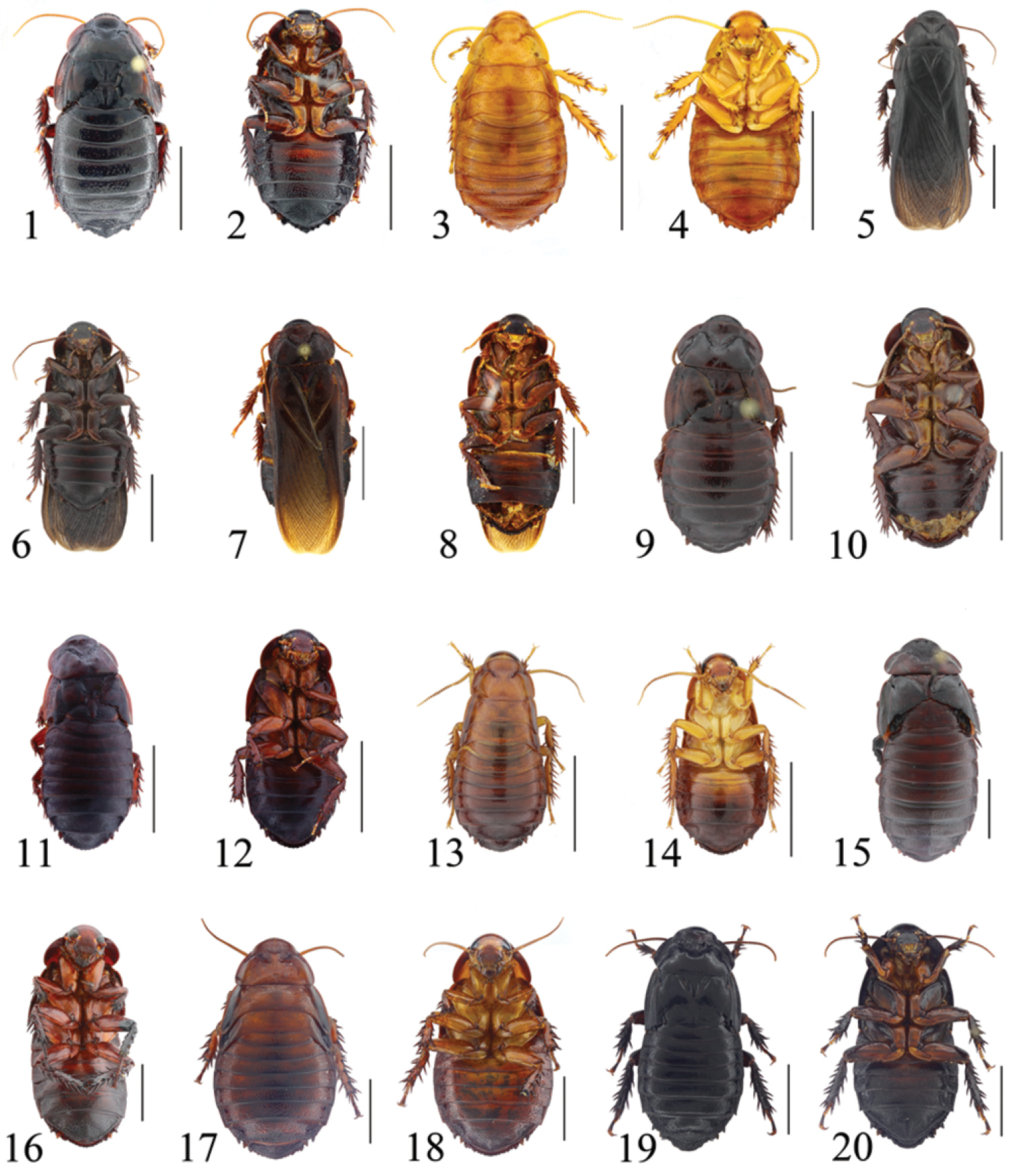
1–2 Salganea quinquedentata sp. n., male: 1 holotype, dorsal view 2 same, ventral view 3–4 Salganea quinquedentata sp. n., nymph: 3 paratype, dorsal view 4 same, ventral view 5–6 Salganea anisodonta sp. n., male: 5 holotype, dorsal view 6 same, ventral view 7–8 Salganea taiwanensis Roth, 1979, male: 7 holotype of Panesthia concinna Feng & Woo, 1990, dorsal view 8 same, ventral view 9–10 Salganea guangxiensis (Feng & Woo, 1990), male: 9 holotype of Panesthia guangxiensis Feng & Woo, 1990, dorsal view 10 same, ventral view 11–12 Salganea incerta (Brunner von Wattenwyl, 1893), male: 11 dorsal view 12 ventral view 13–14 Salganea incerta (Brunner von Wattenwyl, 1893), nymph: 13 dorsal view 14 ventral view 15–16 Salganea raggei Roth, 1979, male: 15 dorsal view 16 ventral view 17–18 Salganea raggei Roth, 1979, nymph: 17 dorsal view 18 ventral view 19–20 Salganea flexibilis sp. n., male: 19 holotype, dorsal view 20 same, ventral view. Scale bars = 1.0 cm.
Vertex and face punctate, the former exposed. Anterior margin of pronotum smooth, or weakly concave; anterior half of pronotum slightly depressed, the floor punctured, denser laterally; posterior half punctured sparsely and almost evenly, without tubercles (Fig. 21). Tegmina and wings well developed, extending beyond end of abdomen, sometimes mutilated (Fig. 1). Radius of tegmen with a long apical posterior branch, which has accessory branches, or apical posterior branch absent; median vein is simple or branched (Figs 80–81). Radial vein of hind wing with posterior branch medially; median vein branched terminally or not; cubitus with 4–5 complete and 5–6 incomplete branches (Figs 89–90). Anterior ventral margin of front femur with 1–3 spines and a small distal spine, hind margin with a large distal spine. Abdominal tergites punctured, the punctures denser laterally and caudally; T5–T7 with gradually increased holes on the anterolateral corners, minute sparse hairs sometimes visible on the surfaces; caudal angles of T6 weakly explored; lateral margins of T7 slightly uneven, caudal angles oblique, large and tapering (Fig. 22). Abdominal sternites densely punctured, the punctations larger and denser caudally; hind margin of the last sternite entire (Fig. 23). Supra-anal plate densely punctured, coarser than abdominal tergites; hind margin with 5 subacute and symmetrically slender teeth, which are deflexed and widely spaced, the largest one situated in the middle; teeth with margin smooth or small acute spines between the teeth, sometimes teeth fused together; lateral angles larger than the medial tooth. Cercus without setae dorsally, ventral surface convex with dense hairs (Fig. 24). Anterior margin of subgenital plate concave, anterolateral corners rounded; lateral margins concave (Fig. 25).
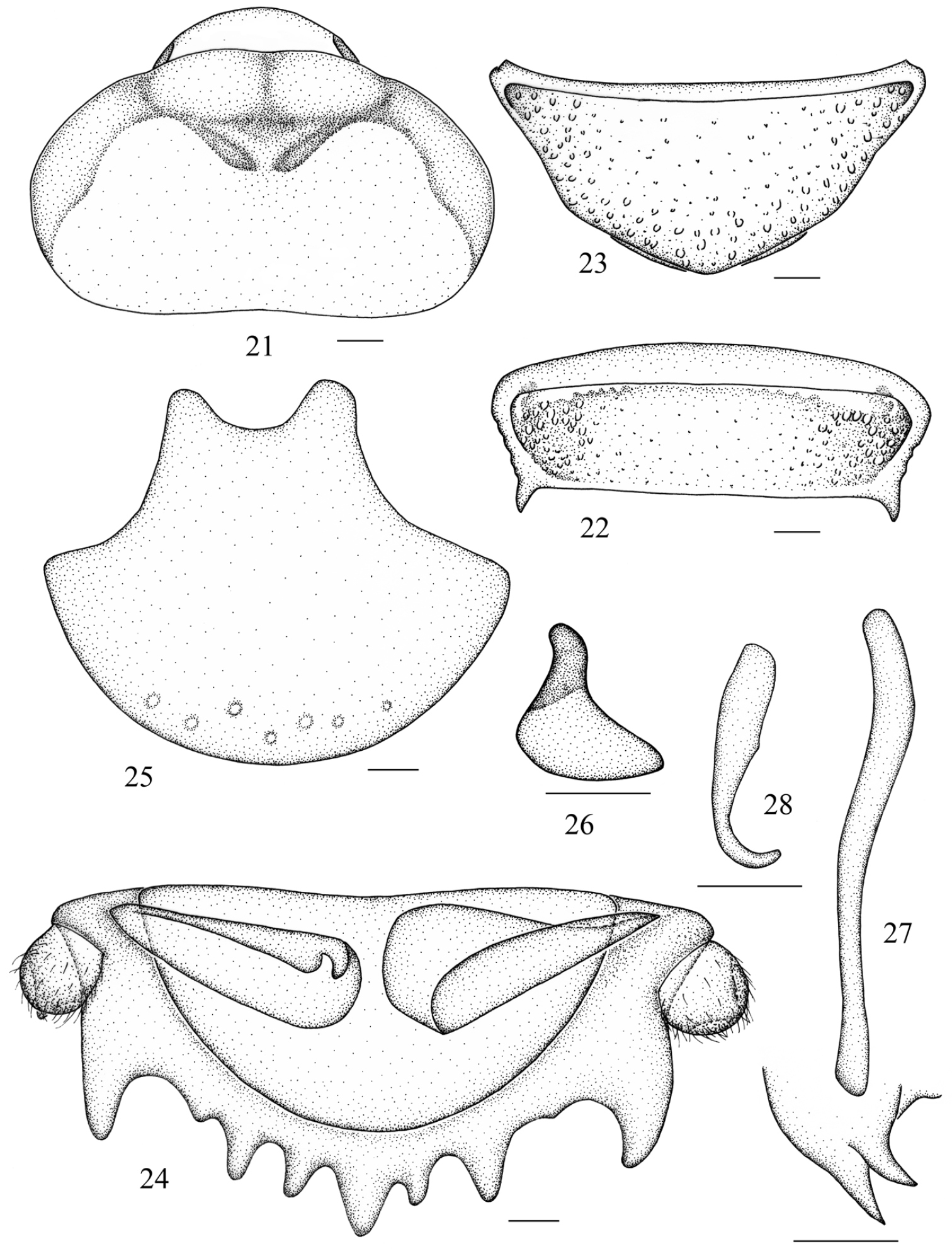
Salganea quinquedentata sp. n. 21 vertex and pronotum 22 abdominal tergum 7, dorsal view 23 abdominal sternite 7, ventral view 24 supra-anal plate and paraprocts, ventral view 25 subgenital plate, dorsal view 26 left phallomere (L1) 27 median phallomere (L2vm and L2d) 28 right phallomere (R2). Scale bars = 1.0 mm (Figs 21–23), 0.5 mm (Figs 24–28).
Male genitalia. Genital phallomere L1 reduced, only a short lobe remaining, or absent (Fig. 26); L2d tapering at apex, with a relatively large lateral lobe (Fig. 27); R2 weakly curved, hook-shaped (Fig. 28).
Female. Essentially similar to male, difficult to distinguish externally.
Nymph. Body yellowish brown and eyes dark. Hind margin of the supra-anal plate with 5 contiguous and triangular teeth, sometimes separated by tiny tines. Remaining external morphological features are characteristic of the adult (Figs 3–4).
Male, 3th–5th maxillary segments: 0.57–0.67/0.48–0.87/0.61–1.00mm; pronotum: length × width: 5.2–5.5 × 8.8–9.3mm; tegmen: 24.1–25.0mm; body length: 26.9–29.5mm; fore leg: coxae: 2.29–2.51mm, trochanter: 1.46–1.79mm, femur: 3.24–3.85mm, tibia: 1.19–2.61mm, 1st–5th tarsus: 0.57–0.69/0.20–0.24/0.19–0.24/0.28–0.32/1.00–1.28mm; mid leg: coxae: 2.48–2.90mm, trochanter: 2.33–2.58mm, femur: 5.01–5.17mm, tibia: 3.84–4.06mm, 1st–5th tarsus: 1.03–1.12/0.25–0.28/0.24–0.27/0.33–0.35/1.00–1.24mm; hind leg: coxae: 2.14–2.53mm, trochanter: 2.70–2.75mm, femur: 5.23–5.81mm, tibia: 5.47–6.14mm, 1st–5th tarsus: 1.18–1.22/0.27–0.29/0.27–0.31/0.27–0.30/1.07–1.15mm; cerci: 0.64–0.97mm.
Female, 3th–5th maxillary segments: 0.73–0.79/0.84–0.85/0.93–1.08mm; pronotum: length × width: 5.0 × 8.8–9.5mm; body length: 26.5–27.5mm; fore leg: coxae: 2.17–2.29mm, trochanter: 0.61–2.30mm, femur: 1.94–2.07mm, tibia: 2.16–2.64mm, 1st–5th tarsus: 0.63–0.80/0.24–0.28/0.23–0.28/0.32–0.27/0.91–1.20mm; mid leg: coxae: 2.81–2.89mm, trochanter: 2.55–2.87mm, femur: 5.31–5.76mm, tibia: 4.52–4.60mm, 1st–5th tarsus: 1.22–1.23/0.28–0.30/0.25–0.27/0.32–0.34/1.06–1.45mm; hind leg: coxae: 2.06–2.28mm, trochanter: 2.53–2.92mm, femur: 5.00–6.23mm, tibia: 5.47–7.32mm, 1st–5th tarsus: 1.36/0.35/0.29/0.36/1.39mm; cerci: 0.87–0.91mm.
Holotype, male, China: Hainan Prov., Lingshui County, Mt. Diaoluoshan, 18°43.462'N, 104°52.105'E, 4 May 2013, coll. Yan Shi and Shunhua Gui (SWU). Paratypes, two males, three females and six nymphs, same data as holotype (SWU); one female, Hainan Prov., Mt. Wuzhishan, 2 May 1964, coll. Yuliang Luo (SWU).
This species is assigned into the Salganea nigrita species group by the forked L2d. It resembles Salganea incerta, but can be distinguished by the following characteristics: 1) anterior margin of pronotum entire and without tubercles, indented and with tubercles in Salganea incerta; 2) the floor of pronotum without tubercles, with tubercles in Salganea incerta; 3) hind margin of seventh abdominal sternite entire, the latter with a medial excision; 4) hind margin of supra-anal plate with 5 distinct, subacute and slender teeth, with 9-13 triangular teeth in Salganea incerta.
The specific epithet is derived from the Latin word “quinquedentatus”, referring to the posterior margin of supra-anal plate with 5 distinct and slender teeth.
http://zoobank.org/45FEEBFC-E670-492B-8FB2-6E9CB92C01B4
http://species-id.net/wiki/Salganea_anisodonta
Figs 5–6, 29–36, 82, 91Male. Body dark reddish brown (Fig. 5). Face black, eyes dark brown, ocelli yellowish, upper lip and mandible brown; antennae, labial palpi and maxillary palpomeres dark brown. Legs reddish brown with coxae and trochanter brown. Abdominal sternites reddish brown, darker caudally (Fig. 6).
Vertex exposed and without punctures. Face punctulated, ocelli round and with border distinct. Anterior margin of pronotum with a V-shaped excision mesially, a small recurved tubercle on the each side of the indentation; anterior 1/3 half of pronotum depressed, the floor densely granular; lateral and posterior half punctured, with a pair of small tubercles in the middle (Fig. 29). Tegmina and wings mutilated, probably well developed (Fig. 5). Radial vein of tegmen with a long apical posterior branch mesially, which is branched at apex and with an accessory branch; the median vein branched before the midline (Fig. 82). Hind wing with subcostal vein branched at apical part; radial vein bifurcated at apex and forked medially; median vein simple; cubitus with 6 complete and 5 incomplete branches (Fig. 91). Anterior ventral margin of front femur without spines, hind margin with a large distal spine. Abdominal tergites punctured, the punctures denser and larger laterally; T7 with coarse surface scattered with sparse setae, and with circular depressions laterally, lateral margin crenulate, caudal angles oblique and tapering (Fig. 30). T6–T7 with small holes on the anterolateral corners, which is associated with minute sparse hairs in the openings. Abdominal sternites densely punctured; hind margin of S7 slightly convex and subgenital plate weakly exposed (Fig. 31). Supra-anal plate extremely coarse, hirsute and covered with depressions similar to T7 in density; hind margin with 7-8 relatively separated teeth, which have an uneven border with lateral ones larger than teeth in the middle, or two fused together; lateral angles equal or larger than the biggest tooth between them. Cercus basiconic, with ventral side swollen and with setae ventrally and dorsally (Fig. 32). Anterior margin of subgenital plate slightly concave or more or less straight, lateral margin curved inwards (Fig. 33).
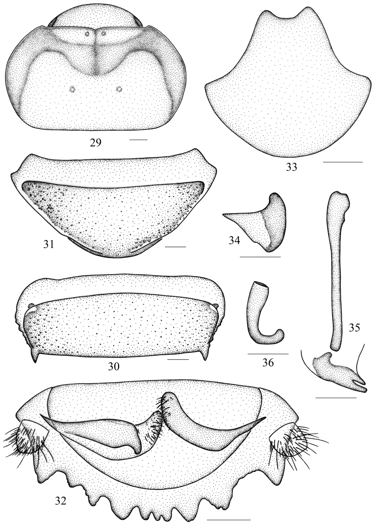
Salganea anisodonta sp. n. 29 vertex and pronotum 30 abdominal tergum 7, dorsal view 31 abdominal sternite 7, ventral view 32 supra-anal plate and paraprocts, ventral view 33 subgenital plate, dorsal view 34 left phallomere (L1) 35 median phallomere (L2vm and L2d) 36 right phallomere (R2). Scale bars = 1.0 mm (Figs 29–33), 0.5 mm (Figs 34–36).
Male genitalia. Genital phallomere L1 reduced to a small plate (Fig. 34); L2d tapering and bifurcated apically, major branch with apex rounded and a little larger than the lateral one (Fig. 35); R2 developed and curved, hook-shaped, with apex obtuse and slightly swollen (Fig. 36).
Female. Anterior margin of pronotum with smaller tubercles than male and the tubercles not recurved.
Nymph. Unknown.
Male, 3th–5th maxillary segments: 0.53–0.69/0.76–0.78/0.61–0.97mm; pronotum: length × width: 5.0–5.1 × 8.4–8.6mm; distance between disc tubercles: 1.9–2.1mm; tegmen: 28.5mm; body length: 27.5–28.0mm; fore leg: coxae: 2.63–2.83mm, trochanter: 1.50–1.97mm, femur: 3.24–3.76mm, tibia: 1.73–2.33mm, 1st–5th tarsus: 0.73/0.35/0.26/0.34/1.30mm; mid leg: coxae: 2.70–2.93mm, trochanter: 2.19–2.72mm, femur: 4.54–4.85mm, tibia: 3.72–3.98mm, 1st–5th tarsus: 1.02–1.16/0.26–0.27/0.24–0.28/0.29–0.34/1.02–1.22mm; hind leg: coxae: 2.69–2.74mm, trochanter: 2.55–2.65mm, femur: 5.13–5.38mm, tibia: 5.37–5.76mm, 1st–5th tarsus: 1.24/0.28/0.27/0.32/1.10mm; cerci: 0.67–0.74mm.
Female, 3th–5th maxillary segments: 0.60/0.82/0.92mm; pronotum: length × width: 5.6 × 9.1mm; distance between disc tubercles: 2.2mm; body length: 29.6mm; fore leg: coxae: 2.45mm, trochanter: 2.05mm, femur: 3.44mm, tibia: 2.42mm, 1st–5th tarsus: 0.65/0.32/0.29/0.32/0.95mm; mid leg: coxae: 2.99mm, trochanter: 2.84mm, femur: 5.20mm, tibia: 3.90mm, 1st–5th tarsus: 1.03/0.27/0.26/0.29/1.19mm; hind leg: coxae: 2.72mm, trochanter: 3.12mm, femur: 5.75mm, tibia: 6.30mm, 1st–5th tarsus: 1.41/0.37/0.30/0.40/1.37mm; cerci: 0.66mm.
Holotype, male, China: Yunnan Prov., Longling County, Longxin Township, Heishan Village, 2300m, 23–25 December 2008, coll. Jishan Xu and Zhenhua Gao (HBU). Paratypes, one male and one female, same data as holotype (HBU).
This species is placed into the Salganea nigrita species group and is similar to Salganea incerta, but can be distinguished by: 1) anterior margin of pronotum with tubercles in female, without or weakly indicated in female of Salganea incerta; 2) anterior margin with two reflexed tubercles mesially, tubercles in Salganea incerta not reflexed; 3) hind margin of supra-anal plate with 7-8 relatively separated teeth, lateral ones larger than teeth in the middle, teeth in Salganea incerta contiguous and subequal; 4) L2d with rounded apex and lateral sclerite relatively larger, apex acute and lateral sclerite smaller in Salganea incerta.
The scientific epithet of this species is derived from the Latin word “anisodontus” which refers to the different teeth in the hind margin of the supra-anal plate.
http://species-id.net/wiki/Salganea_taiwanensis
Figs 7–8, 37–45, 83–84, 92–93, 98–113Male. Body dark reddish brown, darker on caudal segments, or totally black (Fig. 7). Eyes black-brown and ocelli yellowish. Antennae, upper lip, mandible, labial palpi and maxillary palpomeres brown. Legs dark reddish brown, paler on coxae and trochanter. Abdominal sternites reddish brown with the middle of the first and second sternites brown (Fig. 8).
Vertex with few punctations, exposed; face densely punctulated; ocelli round and distinct. Anterior margin of pronotum thickened, with a small mesial V-shaped indentation, a small reflexed protuberance on the each side of the excision; anterior half of pronotum depressed, with sparsely granular surface; posterior half punctured, with 2 oblique mounds armed with 2 tubercles at apex (Fig. 37). Tegmina and wings well developed, extending beyond the abdominal terminal (Fig. 7). In tegmen, the radius with 3 short or 1 relatively long posterior branches; median vein simple or branched (Figs 83–84). Radius in hind wing with apical posterior branch near the middle, which is associated with 2 branches and one of them sometimes branched, some veins fused partially; median vein branched or not; cubitus with 5–6 complete and 6-8 incomplete branches (Figs 92–93). Anterior ventral margin of front femur with 0-4 spines and a small distal spine, hind margin with a large distal spine. Abdominal tergites punctured, and the punctures denser laterally; T4–T7 with holes on the anterolateral corners which are surrounded with fine hairs, the holes on T4 small or absent; lateral margins of T7 feebly crenulate, sometimes the irregularities subobsolete and indistinct, caudal angle oblique (Fig. 38). Abdominal sternites densely punctured, hind margin with a mesial concavity (Fig. 39). Supra-anal plate densely punctate, hind margin with 8–13 subequal teeth (10 in most cases), which are broad basally, triangular, or fused together, lateral angles obtuse and about the same size as the largest tooth between them. Cercus conical, very few or no setae dorsally, ventral surface swollen, densely setose (Fig. 41). Anterior margin of subgenital plate slightly concave, lateral margins sunken (Fig. 42).

Salganea taiwanensis Roth, 1979 37 vertex and pronotum 38 abdominal tergum 7, dorsal view 39 abdominal sternite 7 of male, ventral view 40 abdominal sternite 7 of female, ventral view 41 supra-anal plate and paraprocts, ventral view 42 subgenital plate, dorsal view 43 left phallomere (L1) 44 median phallomere (L2vm and L2d) 45 right phallomere (R2). Scale bars = 1.0 mm (Figs 37–40), 0.5 mm (Figs 41–45).
Male genitalia. Genital phallomere L1 reduced or absent (Fig. 43); L2d elongate, apically acute and forked, with a small lateral lobe (Fig. 44); R2 variably reduced and usually not hook-shaped, or absent (Fig. 45).
Female. Essentially similar to male, but larger than male and with hind margin of the seventh sternite rounded (Fig. 40).
Male, 3th–5th maxillary segments: 0.65–0.75/0.63–0.89/0.86–1.01mm; pronotum: length × width: 4.7–6.2 × 7.7–10.5mm; distance between disc tubercles: 1.8–2.8mm; tegmen: 24.0–27.0mm; body length: 24.0–30.5mm; fore leg: coxae: 2.60–3.19mm, trochanter: 1.61–1.71mm, femur: 2.71–4.07mm, tibia: 1.30–2.33mm, 1st–5th tarsus: 0.62–0.67/0.21–0.22/0.19–0.22/0.24–0.32/1.37–1.44mm; mid leg: coxae: 2.76–3.00mm, trochanter: 2.33–2.39mm, femur: 4.35–5.01mm, tibia: 3.81–4.84mm, 1st–5th tarsus: 0.80–0.82/0.26–0.28/0.26–0.27/0.28/1.07–1.11mm; hind leg: coxae: 2.44–2.94mm, trochanter: 2.52–2.60mm, femur: 4.71–5.58mm, tibia: 5.67–7.21mm, 1st–5th tarsus: 1.06–1.22/0.26–0.35/0.26–0.34/0.28–0.41/1.07–1.33mm; cerci: 0.68–0.79mm.
Female, 3th–5th maxillary segments: 0.68–0.85/0.66–0.75/0.59–0.94mm; pronotum: length × width: 4.7–5.8 × 8.0–13.0mm; distance between disc tubercles: 1.8–2.7mm; tegmen: 24.1–27.5mm; body length: 25.0–29.5mm; fore leg: coxae: 3.30–3.78mm, trochanter: 1.99–2.01mm, femur: 3.19–3.41mm, tibia: 2.05–2.06mm, 1st–5th tarsus: 0.68–0.78/0.23–0.29/0.22–0.27/0.25–0.29/0.77–1.00mm; mid leg: coxae: 2.85–3.39mm, trochanter: 2.29–2.37mm, femur: 4.37–4.39mm, tibia: 3.80–4.03mm, 1st–5th tarsus: 0.79–0.96/0.24–0.30/0.23–0.27/0.28–0.33/1.05–1.14mm; hind leg: coxae: 2.67–3.03mm, trochanter: 2.44–2.45mm, femur: 4.90–5.04mm, tibia: 6.29–6.47mm, 1st–5th tarsus: 1.13–1.28/0.28–0.31/0.30–0.32/0.25–0.37/1.11–1.16mm; cerci: 0.61–0.68mm.
One male (holotype of Panesthia concinna Feng & Woo, 1990), Fujian Prov., Mt. Wuyishan, 10–17 July 1982, coll. Feng Xia; one male, Fujian Prov., Mt. Wuyishan, 15 July 1984, coll. Sizheng Wang; two males and one female, Jiangxi Prov., Mt. Jiulianshan, Gongtang, 30 April 1986, coll. Jianzhong Zheng; four males and one female, Jiangxi Prov., Mt. Jiulianshan, 4 May 1986, coll. Liu Luo; two females, Fujian Prov., 30 June 1982, coll. Fan Jiang; one female, Guangxi Prov., Huaping Nature Preserves, Mt. Tianpingshan, coll. Kun Yang; two females, Guizhou Prov., Ceheng County, 800–950m, 23–27 July 1979, coll. Shaokun Du; one male, Guangdong Prov., Meizhou City, Mt. Wuzhishan, May 2007, coll. Lijun Cai. (SWU)
Considering the Panesthia-like lateral margin on T7 and the disappearance of sclerite R2,
China (Jiangxi, Fujian, Guangxi, Guizhou, Guangdong, Taiwan); Japan; Vietnam.
http://species-id.net/wiki/Salganea_guangxiensis
Figs 9–10, 21–24, 46–49Male. Body dark reddish, with the coloration similar to Salganea taiwanensis.
Vertex sparsely punctate, exposed; face densely punctulated; ocelli round and distinct. Anterior margin of pronotum weakly thickened, with a small mesial emargination, a protuberance on the each side of the excision; anterior half of pronotum depressed, the surface sparsely granular; posterior half punctured, with 2 tubercles (Fig. 9). Tegmina and wings mutilated, probably fully developed (Fig. 9). Anterior ventral margin of front femur with 2 spines and a small distal spine, hind margin with a large distal spine. T4–T7 with holes on the anterolateral corners which are surrounded by fine hairs, the holes on T4 are very small; lateral margins of T7 Panesthia-like (Fig. 46). Supra-anal plate densely punctate, hind margin with 10 subequal teeth, obtuse rounded (Fig. 47). The last abdominal sternite and subgenital plate were broken.
Male genitalia. Genital phallomere L1 lost; L2d elongate, apically acute and forked, with a lateral lobe (Fig. 48); R2 reduced, clavate (Fig. 49).

Salganea guangxiensis (Feng et Woo, 1990) 46 abdominal tergum 7, dorsal view 47 supra-anal plate and paraprocts, ventral view 48 median phallomere (L2vm and L2d) 49 right phallomere (R2). Scale bars = 0.5 mm.
Male, 3th–5th maxillary segments: 0.73/0.76/1.05mm; pronotum: length × width: 5.1 × 9.5mm; distance between disc tubercles: 2.2mm; body length: 27.4mm; fore leg: coxae: 2.42mm, trochanter: 1.64mm, femur: 3.47mm, tibia: 2.48mm, 1st–5th tarsus: 0.70/0.30/0.19/0.26/1.34mm; mid leg: coxae: 3.31mm, trochanter: 2.74mm, femur: 4.33mm, tibia: 3.96mm; hind leg: coxae: 2.57mm, trochanter: 2.72mm, femur: 4.43mm, tibia: 5.92mm; cerci: 0.71mm.
One male (holotype of Panesthia guangxiensis Feng & Woo, 1990), China: Guangxi Prov., Mt. Jinxiulaoshan, 6 September 1981, collector unknown. (SWU)
Although the lateral margin of T7 is Panesthia-like, the characters of the holes in anterolateral corners of T4–T7 associated with setae, and the ridge along with the lateral margin of S7 are more typical of the genus Salganea. Thus, we place this species in genus Salganea. It is very similar to Salganea taiwanensis, only differing in the rounded teeth in the hind margin of supra-anal plate. To be rigorous, the relationship between this two species requires more specimens to provide an absolute determination.
China (Guangxi).
http://species-id.net/wiki/Salganea_incerta
Figs 11–14, 50–60, 85–86, 94–95Male. Body reddish brown, darker caudally (Fig. 11). Eyes blackish brown; ocelli yellowish. Antennae, upper lip, mandible, labial palpi and maxillary palpomeres reddish brown and only a little paler than body. Legs reddish brown, paler on coxae and trochanter. Abdominal sternites reddish brown with the middle of the former two sternites brown (Fig. 12).
Face sparsely punctate and vertex exposed. Anterior margin of pronotum slightly concave mesially, with a small tubercle on each side of the excision; anterior half slightly depressed, the floor with sparse pustules; few punctations on posterior half, with a pair of small tubercles (Fig. 50). Tegmina and wings extending beyond the end of abdomen, sometimes mutilated (Fig. 11). Radial vein of tegmen with 1 forked posterior branch at base, or with 2 simple posterior branches; median vein branched before the midline (Figs 85–86). Radial vein of hind wing simple and branched apically, or with 3 small branches which terminate in the anterior apical angle; median vein branched; cubitus with 5 complete branches, which may be forked or not, and 7–8 incomplete branches (Figs 94–95). Anteroventral margin of front femur armed with 1 spine or unarmed, with or without a minute distal spine, hind margin with a distal spine. Abdominal tergites 1–6 sparsely punctate; T7 denser punctulated caudally, lateral margin weakly crenulate, laterocaudal angle slightly produced (Fig. 52); anterolateral corners with small holes in T6 and T7, some minute and indistinct in T5. Abdominal sternites punctulate, the last sternite densely punctuated and hind margin indented (Fig. 53). Supra-anal plate densely punctate, hind margin varied with 9–13 triangular teeth, border of teeth smooth or crenulate (Figs 55–56). Cercus conical, with few setae near the border dorsally and ventral surface setose (Fig. 55). Anterior margin of subgenital plate slightly concave, lateral margins slightly curved (Fig. 57).
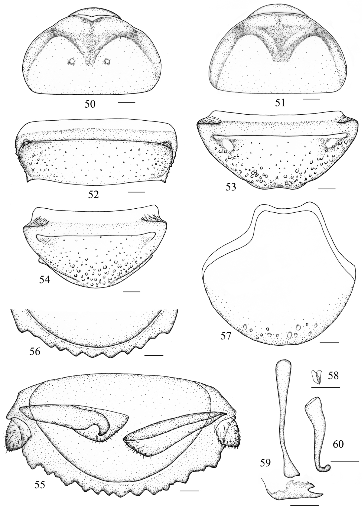
Salganea incerta (Brunner von Wattenwyl, 1893) 50 vertex and pronotum, male 51 vertex and pronotum, female 52 abdominal tergum 7, dorsal view 53 abdominal sternite 7 of male, ventral view 54 abdominal sternite 7 of female, ventral view 55 supra-anal plate and paraprocts, ventral view 56 hind margin of supra-anal plate, ventral view 57 subgenital plate, dorsal view 58 left phallomere (L1) 59 median phallomere (L2vm and L2d) 60 right phallomere (R2). Scale bars = 1.0 mm (Figs 50–54), 0.5 mm (Figs 55–60).
Male genitalia. L1 reduced, only two small lobes remained (Fig. 58); L2d forked apically and apex acute (Fig. 59); R2 hook-shaped, with weak hook portion and the apex not curved upwards (Fig. 60).
Female. Differs from male as follows: anterior margin of pronotum smooth or indented mesially and the tubercles absent or weakly indicated (Fig. 51); hind margin of S7 entire (Fig. 54).
Nymph. Body brown, darker caudally (Figs 13–14), the depression of pronotum punctate.
Male, 3th–5th maxillary segments: 0.36–0.59/0.64–0.78/0.90–0.94mm; pronotum: length × width: 3.6–5.2 × 6.5–8.7mm; distance between disc tubercles: 1.4–2.2mm; tegmen: 24.0–26.5mm; body length: 17.7–26.2mm; fore leg: coxae: 1.53–2.29mm, trochanter: 1.65–1.86mm, femur: 2.81–3.19mm, tibia: 1.37–1.55mm, 1st–5th tarsus: 0.34–0.63/0.17–0.20/0.19–0.23/0.24–0.29/1.00–1.18mm; mid leg: coxae: 2.22–2.51mm, trochanter: 2.12–2.58mm, femur: 4.48–4.90mm, tibia: 3.73–3.79mm, 1st–5th tarsus: 0.76–0.99/0.22/0.23/0.26–0.30/1.00–1.03mm; hind leg: coxae: 2.02–2.84mm, trochanter: 2.34–2.56mm, femur: 4.43–5.16mm, tibia: 5.43–5.91mm, 1st–5th tarsus: 1.02–1.20/0.25–0.32/0.26–0.28/0.30–0.32/1.12–1.17mm; cerci: 0.73–0.89mm.
Female, 3th–5th maxillary segments: 0.70–0.74/0.59–0.66/0.96–0.99mm; pronotum: length × width: 4.4–5.8 × 7.6–9.1mm; body length: 20.0–27.0mm; fore leg: coxae: 2.27–2.54mm, trochanter: 1.68–1.93mm, femur: 3.52–3.54mm, tibia: 1.64–2.00mm, 1st–5th tarsus: 0.54–0.56/0.16–0.21/0.20–0.26/0.27–0.30/1.10–1.17mm; mid leg: coxae: 2.34–3.02mm, trochanter: 1.63–1.99mm, femur: 4.67–4.75mm, tibia: 4.25–4.46mm, 1st–5th tarsus: 1.08–1.11/0.29–0.30/0.25/0.30–0.31/1.01–1.13mm; hind leg: coxae: 2.11–2.51mm, trochanter: 2.01–2.30mm, femur: 5.02–5.60mm, tibia: 6.04–6.19mm, 1st–5th tarsus: 1.02–1.17/0.30–0.34/0.27–0.28/0.32–0.33/1.00–1.13mm; cerci: 0.54–0.94mm.
Two males, Guangxi Prov., Jinxiu County, Mt. Yangjiaoshan, 25 September 1981, collector unknown; one male and two females, Chongqing, Mt. Simianshan, Dawopu, 11 July 2008, coll. Zongqing Wang; one male and one female, Sichuan Prov., Hongya County, Mt. Wawushan, 30 June 2013, coll. Yang Li and Jinjin Wang; one male, Yunnan Prov., Yingjiang County, 1418m, 24°61'N, 97°62'E, 4–5 June 2008, coll. Weiwei Zhang; two males, Yunnan Prov., Yingjiang County, Xima Township, Menglaihe River 2nd Hydroelectric Power Station, 1470m, 24°78.404'N, 97°67.493'E, 27–29 May 2009, coll. Weiwei Zhang; two males, Yunnan Prov., Yingjiang County, Xima Township, Menglaihe 2nd Hydroelectric Power Station, 1470m, 6–9 June 2008, coll. Weiwei Zhang; two males, Yunnan Prov., Yingjiang Country, Taiping Town, Longpen Village, 30 May-9 June 2009, coll. Weiwei Zhang; one male and one female, Hainan Prov., Mt. Diaoluoshan, 18°43.462'N, 108°52.105'E, 4 May 2013, coll. Yan Shi and Shunhua Gui; one male and one female, China, 4 May 1980, coll. Qiaosheng Yuan; two males, two females and 3 nymphs, Chongqing, Mt. Simianshan, 2 October 2013, coll. Hao Xu and Jianyue Qiu. (SWU)
China (Guangxi, Chongqing, Sichuan, Yunnan, Hainan); India; Myanmar; Thailand.
http://species-id.net/wiki/Salganea_raggei
Figs 15–18, 61–68, 87–88, 96–97Male. Body reddish brown, darker caudally, or black (Fig. 15). Eyes black and ocelli yellowish. Antennae, upper lip, mandible, labial palpi and maxillary palpomeres reddish brown. Legs brown, reddish on coxae, trochanter and anterior half of femora. Abdominal sternites reddish brown with the middle of the former several sternites brown (Fig. 16).
Face punctulated and vertex exposed. Ocelli small and round. Anterior margin of pronotum with a V-shaped emargination in the midline, a small recurved tubercle behind the margin on each side of the excision; anterior half depressed, the floor densely granular, lateral area punctate; region behind grooves convex and densely punctate, with one or two pairs of relatively large tubercles on the mounds symmetrically (Fig. 61). Tegmina and wings well developed, sometimes mutilated and remaining an uneven base (Fig. 15). Radius of tegmen with or without 1 long posterior branch which is forked; median vein branched (Figs 87–88). Radial vein of hind wing with one or more branches near the apex, some branches reforked, the posterior branch branched or not; median vein with two branches or simple; cubitus with 7–8 complete and 8–10 incomplete branches (Figs 96–97). Anterior ventral margin of fore femur equipped with 1–3 spines, and with or without a minute distal spine, posterior margin with a large distal spine. Abdominal tergites punctated, the punctures denser laterally and dorsoposteriorly, anterolateral corners usually without holes, rarely with small holes on T6 and T7 only; lateral angle of T6 produced into a rounded and oblique spine; lateral margin of T7 with 5–6 obtuse teeth, laterocaudal angle stout and subacute (Fig. 62). Abdominal sternites densely punctured, S7 with a depression on the caudal margin (Fig. 63), subgenital plate more or less exposed. Supra-anal plate densely covered with coarse punctations, sometimes with minute hairs; hind margin with 8-16 subequal teeth, whose apex obtuse; some teeth contiguous. Cerci bulbous, dorsal surface hairless and ventral surface setose (Fig. 64). Subgenital plate with anterior margin concave, lateral margin with a mesal indentation (Fig. 65).

Salganea raggei Roth, 1979 61 vertex and pronotum 62 abdominal tergum 7, dorsal view 63 abdominal sternite 7, ventral view 64 supra-anal plate and paraprocts, ventral view 65 subgenital plate, dorsal view 66 left phallomere (L1) 67 median phallomere (L2vm and L2d) 68 right phallomere (R2). Scale bars = 1.0 mm (Figs 61–64), 0.5 mm (Figs 65–68).
Male genitalia. L1 developed but slightly sclerotized (Fig. 66); L2d not bifurcated, elongate and tapering towards the apex, mostly with a weak concavity in hind margin (Fig. 67); R2 well developed, hook-shaped, apex subacute and curved upwards (Fig. 68).
Female. Differs only slightly from male in partial specimens as follows: anterior margin of pronotum slightly excised, and the tubercles indistinct behind the indentation.
Nymph. Similar to adult, but body yellowish-brown, anterior margin of the pronotum without excision and protuberance, the tubercles on the floor absent; depression of anterior half densely punctuate; meso- and metanotum with produced laterocaudal angle, the protrusion dark or black (Figs 17–18).
Male, 3th–5th maxillary segments: 0.86–0.90/0.88–1.29/1.15–1.67mm; pronotum: length × width: 6.3–10.1 × 10.0–16.0 mm; distance between anterior disc tubercles: 5.4–7.7 mm; distance between posterior disc tubercles: 2.4–4.7mm; tegmen: 38.0–51.0mm; body length: 29.0–49.0 mm; fore leg: coxae: 4.26–4.93mm, trochanter: 2.67–3.17mm, femur: 4.98–5.65mm, tibia: 2.65–3.23mm, 1st–5th tarsus: 0.88–0.99/0.27–0.36/0.30–0.35/0.36–0.55/1.51–1.65mm; mid leg: coxae: 4.35–5.04mm, trochanter: 2.87–4.25mm, femur: 6.39–6.84mm, tibia: 5.47–5.87mm, 1st–5th tarsus: 1.18–1.47/0.31–0.40/0.31–0.43/0.40–0.58/1.34–1.98mm; hind leg: coxae: 3.81–5.02mm, trochanter: 3.13–4.39mm, femur: 7.26–7.44mm, tibia: 7.54–9.98mm, 1st–5th tarsus: 1.42–1.47/0.44–0.51/0.37–0.45/0.43–0.66/1.60–2.10mm; cerci: 1.00–1.22mm.
Female, 3th–5th maxillary segments: 0.95–1.14/0.73–0.98/1.12–1.37mm; pronotum: length × width: 7.0–12.0 × 11.0–16.5 mm; distance between posterior disc tubercles: 2.5–4.7 mm; tegmen: 33.0–50.0mm; body length: 30.5–49.0 mm; fore leg: coxae: 2.29–5.01mm, trochanter: 2.97–3.33mm, femur: 4.62–4.91mm, tibia: 2.39–2.67mm, 1st–5th tarsus: 0.89–1.11/0.31–0.32/0.30–0.32/0.31–0.42/1.57–1.85mm; mid leg: coxae: 3.99–4.43mm, trochanter: 4.08–4.29mm, femur: 6.58–6.69mm, tibia: 5.54–5.85mm, 1st–5th tarsus: 1.10–1.24/0.29–0.38/0.27–0.39/0.42–0.45/1.30–1.50mm; hind leg: coxae: 3.94–4.56mm, trochanter: 3.66–4.27mm, femur: 6.54–7.44mm, tibia: 7.91–9.35mm, 1st–5th tarsus: 1.22–1.64/0.32–0.40/0.32–0.38/0.38–0.52/1.15–2.19mm; cerci: 0.97–1.20mm.
One male and two females, Yunnan Prov., Damenglong Town, 650m, 13 April 1958, coll. Chunpei Hong; one male and one female, Yunnan Prov., Damenglong Town, 650m, 16 March 1958, coll. Zhizi Chen; one male, one female and one nymph, Yunnan Prov., Damenglong Town, 650m, 18 April 1958, coll. Fuji Pu; three males, Yunnan Prov., Yingjiang County, Tongbiguan Township, 1418m, 24°61'N, 97°62'E, 4–5 June 2008, coll. Weiwei Zhang; one male, Xizang Prov., Motuo County, 1300m, 10 September 1979, coll. Gentao Jin and Jianyi Wu; one female, Xizang Prov., Motuo County, Gedang Township, 2080m, 15-18 April 1980, coll. Gentao Jin and Jianyi Wu; two males, Hainan Prov., Mt. Jianfengling, 22 February 1982, collector unknown; one female, Hainan Prov., Mt. Jianfengling, 10 May 1964, coll. Sikong Liu; two males and one nymph, Hainan Prov., Mt. Jianfengling, 4 May 2013, coll. Yan Shi and Shunhua Gui; one male, Hainan Prov., Wuzhishan city, shuiman Township, 740m, 18°51'N, 109°40', 28–30 June 2008, coll. Weiwei Zhang; one male, Hainan Prov., Mt. Jianfengling, 15 June 1983, collector unknown; one male, Hainan Prov. Mt. Jianfengling, Tianchi, 25 April 1981, coll. Shaoying Liang. (SWU).
China (Yunnan, Xizang, Hainan, Taiwan); Bhutan; India; Laos; Nepal; Vietnam; Sikkim; Thailand.
http://zoobank.org/F372D347-53DE-4495-8E4A-252371FA93B1
http://species-id.net/wiki/Salganea_flexibilis
Figs 19–20, 69–76Male. Body black (Fig. 19). Head black, dark reddish brown between eyes, ocelli and eyes with yellowish border. Antennae, upper lip, mandible, labial palpi and maxillary palpomeres dark brown. Legs black, coxae, trochanter and the former half of femora yellowish brown. Abdominal sternites black with the middle of the former five segments reddish brown, cercus reddish brown (Fig. 20).
Vertex exposed, punctured; face densely punctured; ocelli circular and distinct. Pronotum convex (anterior margin damaged, it is probably a developmental error), anterior and lateral area depressed, densely granular and equally distributed; posterior half with a distinct tubercle on each side of the midline, the floor densely punctured (Fig. 69). Tegmina and wings mutilated, but probably fully developed (Fig. 19). Anterior ventral margin of front femur with 2 spines, and a minute distal spine, posterior margin with a spine. Centre region of abdominal tergites sparsely punctured, more and larger laterally, and with dense setae; lateral angle of T6 produced; T7 hirsute, with large disc pits densely; round holes only present in the anterolateral corners of T6 and T7; lateral margin of T7 with 3 subacute and distinct teeth, sometimes also with subobsolete papulas, caudal angle produced into a strong and oblique spine (Fig. 70). Abdominal sternites densely punctured; S7 densely covered with hairs, hind margin emarginated (Fig. 71), subgenital plate slightly exposed. Supra-anal plate convex, hirsute, with large disc pits densely; middle of hind margin with 8 unequal teeth, deflexed, which are triangular or apically truncate; caudal angles tapering, and same length as the largest tooth between them. Cerci conical, dorsal and ventral surfaces densely setose (Fig. 72). Anterior margin of subgenital plate depressed, anterolateral corners subacute, lateral margins straight and not concave (Fig. 73).

Salganea flexibilis sp. n. 69 vertex and pronotum 70 abdominal tergum 7, dorsal view 71 abdominal sternite 7, ventral view 72 supra-anal plate and paraprocts, ventral view 73 subgenital plate, dorsal view 74 left phallomere (L1) 75 median phallomere (L2vm and L2d) 76 right phallomere (R2). Scale bars = 1.0 mm (Figs 69–72), 0.5 mm (Figs 73–76).
Male genitalia. L1 well developed (Fig. 74); L2d not bifurcated with acute apex (Fig. 75); R2 hook-shaped (Fig. 76).
Female. Unknown.
Nymph. Unknown.
Male, 3th–5th maxillary segments: 0.90/0.89/0.99mm; pronotum: length × width: 6.9 × 11.5mm; distance between disc tubercles: 2.8mm; body length: 32.2mm; fore leg: coxae: 2.99mm, trochanter: 2.54mm, femur: 4.04mm, tibia: 2.00mm, 1st–5th tarsus: 0.90/0.32/0.32/0.38/1.36mm; mid leg: coxae: 3.50mm, trochanter: 2.88mm, femur: 5.76mm, tibia: 4.34mm, 1st–5th tarsus: 1.09/0.33/0.34/0.42/1.30mm; hind leg: coxae: 3.32mm, trochanter: 3.36mm, femur: 6.77mm, tibia: 6.59mm, 1st–5th tarsus: 1.27/0.42/0.38/0.45/1.46mm; cerci: 0.93mm.
Holotype, male, China: Yunnan Prov., Nujiang State, Gongshan County, Dulongjiang Township, Kongmu Village, 1391m, 27°44.79'N, 98°20.19'E, 25 May 2013, coll. Hao Xu and Jianyue Qiu (SWU).
Owing to L2d not being bifurcated, this species should be placed under the Salganea raggei species group. It is superficially similar to Salganea aperturifera, but can be differentiated by the following characteristics: 1) posterior half of pronotum with 1 pair of tubercles, 2 pairs in Salganea aperturifera; 2) abdominal tergites 6 and 7 with holes in the anterolateral corners, T3–T7 with holes in Salganea aperturifera; 3) supra-anal plate convex, hind margin with 8 deflexed and unequal teeth, the teeth but in Salganea aperturifera the number of teeth is 8–10, which are subequal and undeflexed.
The specific epithet “flexibilis” is derived from Latin, which means that the teeth on the hind margin of the supra-anal plate are deflexed.
The members of Salganea are known to be burrowers in rotten logs (Figs 78–79), with a hard, rigid, pitted exoskeletion and a thick, scoop-shaped pronotum (
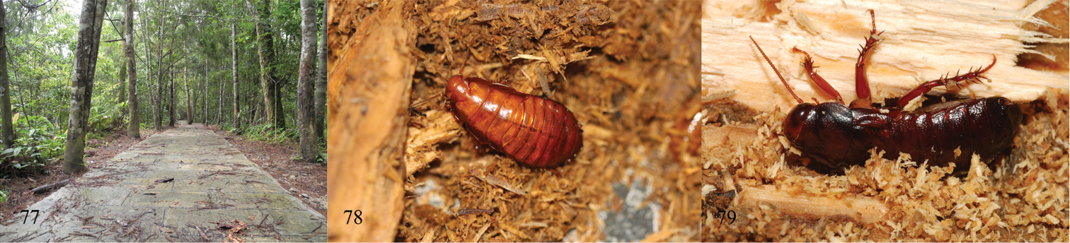
77 ecotope of Mountain Diaoluoshan, Hainan Province (Photographs by Keliang Wu) 78 nymph of Salganea quinquedentata sp. n. 79 Salganea incerta (Brunner von Wattenwyl, 1893), in Mountain Simianshan, Chongqing, 2 October 2013 (Photographs by Jianyue Qiu).
Owing to this unique behavior of shedding their tegmina and wings, venation as an important taxonomic character has not been used widely in the classification of Panesthiinae. But a large number of species of Panesthiinae have fully developed tegmina and wings (
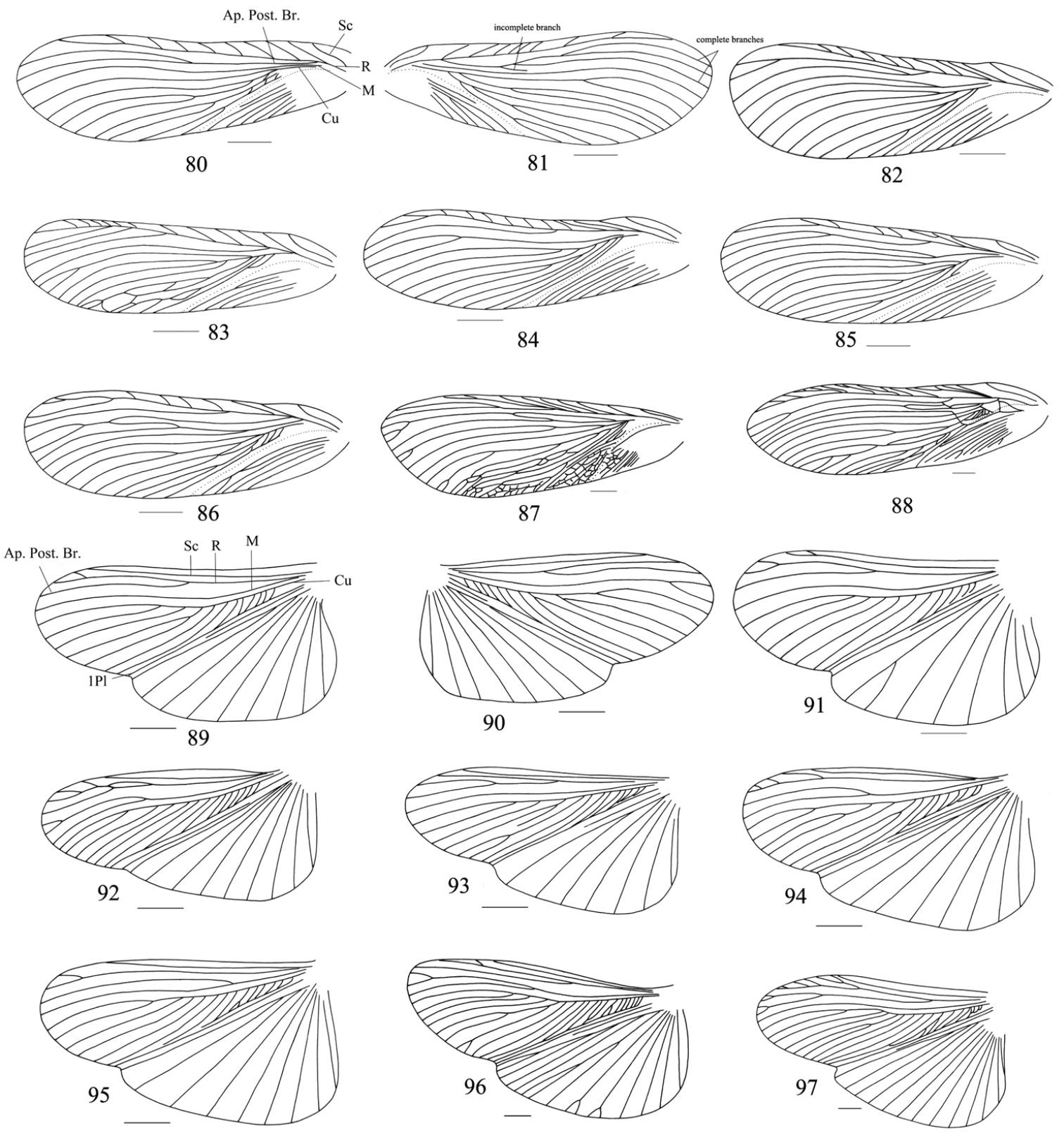
80–88 tegmina: 80–81 left and right tegmina of one specimen (Salganea quinquedentata sp. n.), dorsal view 82 Salganea anisodonta sp. n. 83–84 Salganea taiwanensis Roth, 1979 85–86 Salganea incerta (Brunner von Wattenwyl, 1893) 87–88 Salganea raggei Roth, 1979 89–97 wings: 89–90 left and right wings of one specimen (Salganea quinquedentata sp. n.), dorsal view 91 Salganea anisodonta sp. n. 92–93 Salganea taiwanensis Roth, 1979 94–95 Salganea incerta (Brunner von Wattenwyl, 1893) 96–97 Salganea raggei Roth, 1979. Scale bars = 4.0 mm.
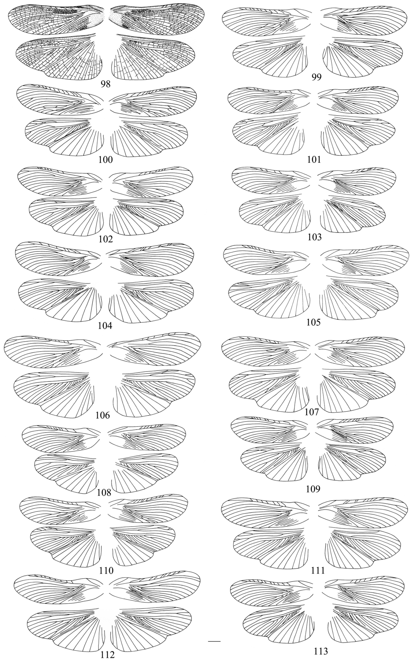
four wings of Salganea taiwanensis Roth, 1979 of one specimen, dorsal view 98 wings with cross-veins 99–113 cross-veins of wings omitted. Scale bars = 4.0 mm.
Tegmen venation variability of Salganea taiwanensis for 16 specimens The second line numbers are data of left and right tegmina separately. Post. + M – posterior branch of radius + media; Total – total number of veins without radius and anal veins; CV – coefficient of variation. (
| Sc | R | Post. + M | Cu | Total | |
|---|---|---|---|---|---|
| Min | 1 1;1 |
7 6;7 |
2 2;2 |
7 7;8 |
11 11;11 |
| Max | 1 1;1 |
14 12;14 |
6 5;6 |
12 12;11 |
17 17;17 |
| Median | 1 1;1 |
9 9;9 |
3 3.5;3 |
9 9;9 |
13 13;13 |
| Mode | 1 1;1 |
9 8;9 |
3 4;3 |
9 9;8 |
13 13;14 |
| Average | 1 1;1 |
9.563 9.313;9.563 |
3.344 3.313;3.344 |
9.063 8.938;9.188 |
13.406 13.438;13.375 |
| Deviation | 0 0;0 |
1.590 1.815;1.590 |
1.066 1.250;1.066 |
1.243 1.167;1.243 |
1.500 1.548;1.500 |
| CV in % | 0 0;0 |
16.631 19.495;16.631 |
31.877 26.224;37.736 |
13.713 14.994;12.705 |
11.186 11.519;11.215 |
Hindwing venation variability of Salganea taiwanensis for 16 specimens. The second line numbers are data of left and right tegmina separately. Total – total number of veins without anal veins; CV – coefficient of variation. (
| Sc | R | M | Cu | Total | |
|---|---|---|---|---|---|
| Min | 1 1;1 |
1 2;2 |
1 1;1 |
4 5;4 |
10 10;10 |
| Max | 1 1;1 |
7 5;7 |
3 3;2 |
8 8;8 |
15 15;15 |
| Median | 1 1;1 |
3 3.5;3 |
1 1;1 |
6 6.5;6 |
12 12;12 |
| Mode | 1 1;1 |
3 3;3 |
1 1;1 |
6 7;6 |
11 12;11 |
| Average | 1 1;1 |
3.625 3.625;3.625 |
1.188 1.188;1.188 |
6.406 6.438;6.375 |
12.188 12.188;12.188 |
| Deviation | 0 0;0 |
1.157 0.885;1.408 |
0.471 0.544;0.403 |
0.837 0.814;0.885 |
1.355 1.274;1.471 |
| CV in % | 0 0;0 |
31.918 24.415;38.850 |
39.657 45.803;33.946 |
13.066 12.644;13.883 |
11.114 10.473;12.066 |
Overall, we find venation to be of little value as a specific character; but the venation variation is more stable and distinct at higher taxonomic levels. Additional investigation will be required to search for more stable wing characteristics to support our view.
We are sincerely grateful to Prof. J. R. Schrock (Department of Biological Sciences, Em- poria State University, USA) for revising the manuscript and also thanks to Prof. Guodong Ren (Hebei University, China) and Dr. Weiwei Zhang for their kindness in loaning specimens to us. This study is supported by the National Natural Sciences Foundation of China (Nos. 30900146, 31093430), and also partly by the Project-sponsored by SRF for ROCS, SEM and the Fundamental Research Funds for the Central Universities (XDJK2012B025, XDJK2013B013).