


Citation: Logunov DV, Marusik YM (2014) Taxonomic notes on the genus Eupoa Żabka, 1985 (Arachnida, Araneae, Salticidae). ZooKeys 410: 63–93. doi: 10.3897/zookeys.410.7548
The south-east Asian genus Eupoa is redescribed and diagnosed. Seven new species are diagnosed, described and illustrated: E. daklak sp. n. (♀) from Viet-Nam; E. lehtineni sp. n. (♂♀) from India, Thailand and Viet-Nam; E. lobli sp. n. (♂) from Malaysia; E. pappi sp. n. (♂) from Thailand; E. pulchella sp. n.(♂) from Thailand; E. schwendingeri sp. n. (♂♀) from Thailand; and E. thailandica sp. n. (♂♀) from Thailand. Eupoa prima Żabka, 1985 and E. yunnanensis Peng & Kim, 1997 are redescribed and illustrated on the basis of type and/or newly collected materials. The female of E. yunnanensis Peng & Kim, 1997 is found and described for the first time.
SE Asia, jumping spiders, Aranei, Eupoa, new species, (re)descriptions
The genus Eupoa Żabka, 1985 was described to accommodate a single species Eupoa prima Żabka, 1985 from northern Viet-Nam (
By their general appearance, members of the genus Eupoa are superficially similar to those of Neon Simon, 1876. Both groups are small to very small spiders, with males having the swollen and relatively large palps for the size of the spiders (Figs 42–43, 86, 90). Both Eupoa and Neon are typical dwellers of forest leaf litter. However, the conformation of male copulatory organs in Eupoa is much more complex leaving no doubts that both genera are not related. The phylogenetic relationships of Eupoa remain poorly resolved, although the genus is indeed a basal salticid; see below under ‘Diagnosis and Affinities’.
The aim of the present paper is twofold: (1) to provide a comprehensive description of the genus Eupoa on the basis on newly collected materials; and (2) to describe seven new species from various regions of SE Asia.
A total of 42 adult museum specimens belonging to nine species has been examined. The material studied in this paper was borrowed from the following museums: HNHM = Hungarian Natural History Museum, Budapest, Hungary (curator: Dr T. Szuts); MHNG = Museum d’Historie Naturelle, Geneve, Switzerland (curator: Dr P. Schwendinger); SMFM = Naturmuseum und Forschungsinstitutt Senckenberg, Frankfurt am Main, Germany (curator: Dr P. Jäger and Ms J. Altmann); ZMTU = Zoological Museum, Turku University, Turku, Finland (curator: Mr S. Koponen); ZMUM = Zoological Museum of the Moscow State University, Moscow, Russia (curator: Dr K. G. Mikhailov).
Digital photographs were taken using an Olympus E-520 camera attached to an Olympus SZX16 stereomicroscope, and prepared using CombineZP image stacking software. Photographs were taken with the specimens secured in dishes with paraffin on bottom. SEM microphotographs were made by means of a JEOL JSM-5200 in the Zoological Museum, University of Turku. Epigynes were cleared in a 10% KOH solution.
Abbreviations used in the text and figures are as follows: Eyes: AME – anterior median eye, ALE – anterior lateral eye, PME – posterior median eye, PLE – posterior lateral eye. Copulatory organs: CTA – compound terminal apophysis; E – embolus; ID – insemination ducts; MA – median apophysis; PA – patellar apophysis; R – receptacle; TA – tegular apophysis; TbA – tibial apophysis. Leg segments: Fm – femur, Pt – patella, Tb – tibia, Mt – metatarsus, Tr – tarsus. Position of leg spines: d – dorsal, pr – prolateral, rt – retrolateral, v – ventral. For the leg spination the system adopted is that used by
http://species-id.net/wiki/Eupoa
Small to very small spiders ranging from about 1.65 to 2.45 mm in length. Sexes similar in general body form; sexual dimorphism is poorly marked and can be seen in the following characters: dorsal scutum presents in males (absent in females), body coloration in males is slightly darker or more contrastingly coloured, anterior and posterior pairs of spinnerets are of contrasting colours in males (brown/dark grey vs. yellow; e.g., Eupoa prima, Eupoa yunnanensis), and legs I and II in males are often with no spines or just with 2/3 pairs of ventral spines on metatarsi (always with spines on tibiae and metatarsi in females; e.g., Eupoa schwendingeri sp. n., Eupoa yunnanensis). Carapace: rather high, with abruptly declining, practically vertical thoracic part (Figs 6, 59, 90); sparsely covered with white elongated pinnate scales (Figs 1–3); fovea absent (Figs 73, 75); lateral sides of carapace, near ALEs, with vertical rows of skin structures (50-60 in a group) looking like either as rounded or elongated smooth bare patches, slightly risen above the surrounding skin (Figs 20, 22), or as flat elongated patches with what looks like several micro-pustules situated on them (Fig. 18); similar bare skin structures occur on leg patellae (see below; Figs 20–21). Eyes: in three rows, with large black areas around eyes (Figs 42–46, 58–59); anterior eye row wider in both sexes, so the quadrangle is as an inverted trapezium; second row midway between ALE and PLE; quadrangle length 52–66% of carapace length. Clypeus: narrow, about 17–47% of AME diameter (from frontal view; Figs 4–5), visibly backward sloping (Figs 6, 59). Chelicerae: small and vertical (Figs 4–5, 7; promargin with two small teeth; retromargin with three small teeth (Fig. 8). Maxillae: slightly convergent; usual shape. Labium: transverse-ovoid. Sternum: as inverted cone with swollen lateral sides (Figs 30, 72, 76). Pedicel: short, in live specimens not visible in dorsal view. Abdomen: elongate, covered with elongated pinnate scales (Fig. 32); dorsal scutum present in males; colour markings on dorsum simple, either consisting of a median yellow stripe (Figs 41, 43, 45) or two rows of spots with a pair of largest sports at the rear of dorsum (Figs 85–86). Book-lung covers: usual shape, not sclerotized. Spinnerets: posterior pair almost two times longer than anterior pair (Figs 31–32). Legs: subequally developed (Fig. 9); legs I in males usually with dark brown longitudinal stripes; trichobothrial bases relatively flat and striated (Fig. 14); tarsal organ as a rounded or ovoid pit (Figs 12–13); tarsal claws narrow, with poorly developed teeth (Figs 10–11); skin structures of two kinds present anteriorly on the dorsal surface of leg patellae, situated in a longitudinal row of about 20–30 pores (arrowed in Figs 19, 21, 23–24): structures of the first kind represents flat elongated and smooth bare patches with what looks like several micro-pustules situated on them (Figs 17, 24); structures of the second kind look like a rounded or elongated or circular smooth bare patch, slightly risen above the surrounding skin (Figs 21; similar bare structures occur on the carapace, see above, Figs 20, 22), these structures resemble the second kind of skin pores described in Neon (
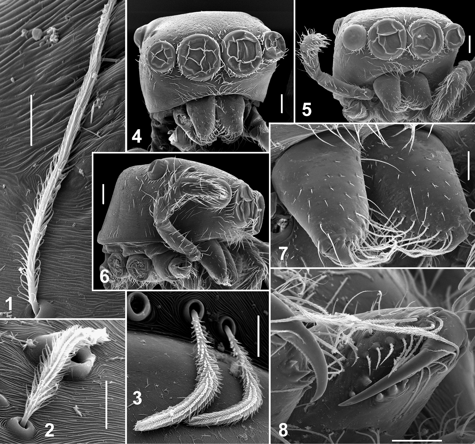
Somatic characters of Eupoa lehtineni sp. n. 1–3 plumose scales on female carapace. 4 male carapace, frontal view 5 female carapace, frontal view 6 ditto, lateral view 7 female chelicerae, frontal view 8 female fang and cheliceral teeth. Scale bars: 10 μm (1–3), 50 μm (7–8), 0.1 mm (4–6).
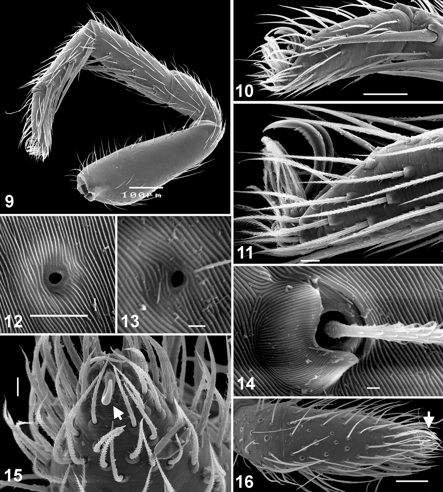
Somatic characters of Eupoa lehtineni sp. n. (9–11, 13–16) and Eupoa thailandica sp. n. (12). 9 female leg I, median view 10 female tarsus I, lateral view 11 female tarsus III, lateral view 12–13 tarsal organ on female tarsus I, dorsal view 14 trichobotrial base, female tarsus I, dorsal view 15–16 female palp and the claw at its tip (arrowed). Scale bars: 1 μm (13–14), 5 μm (12), 10 μm (11, 15), 50 μm (10, 16), 0.1 mm (9).
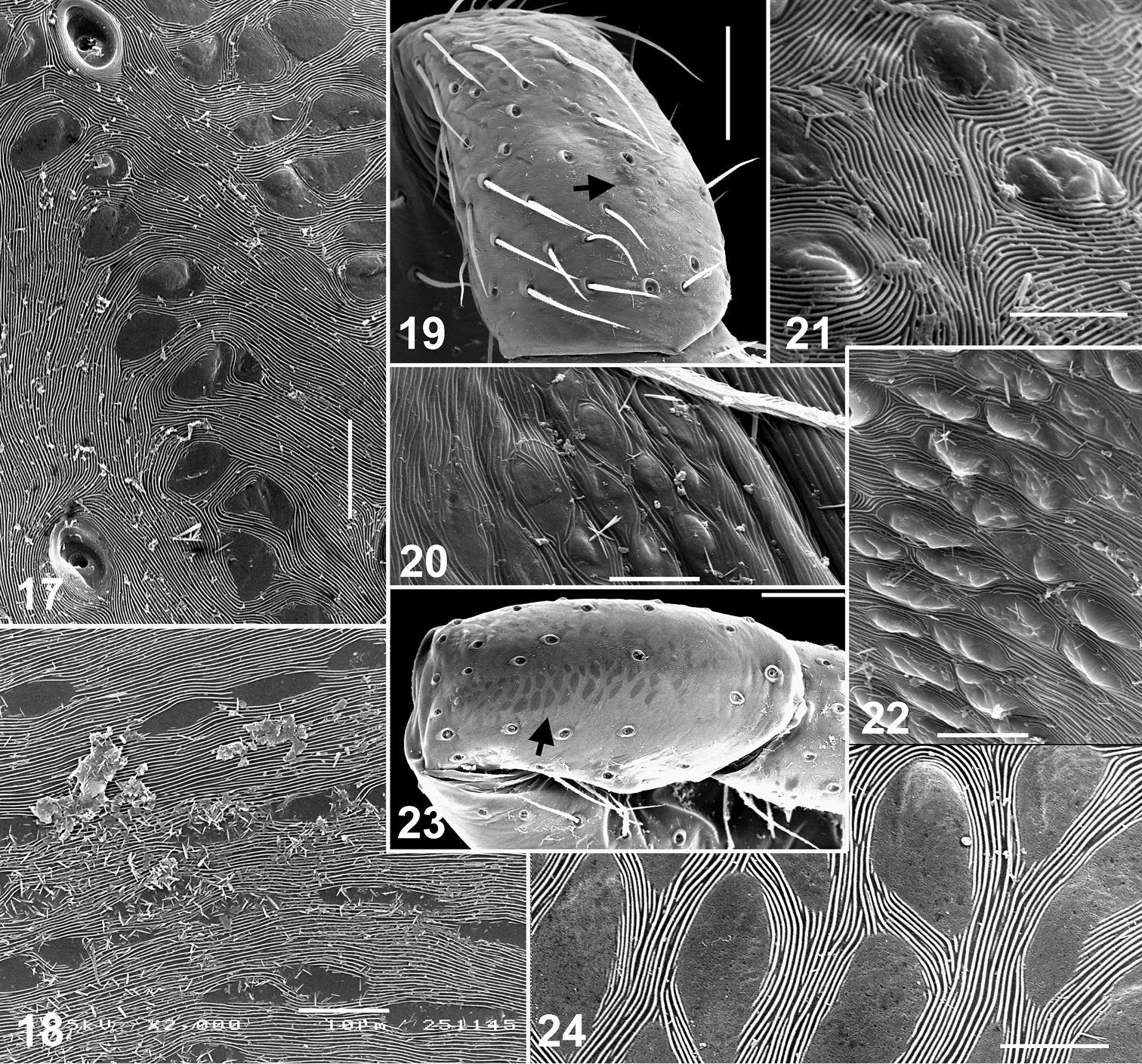
Skin structures of Eupoa lehtineni sp. n. (17, 19–22) and Eupoa thailandica sp. n. (18, 23, 24). 17 male patella I, dorsal view 18, 20, 22 female carapace, lateral view 19, 21, 23–24 female patella I, dorsal view. Scale bars: 5 μm (21, 24), 10 μm (17–18, 20, 22), 50 μm (19, 23).
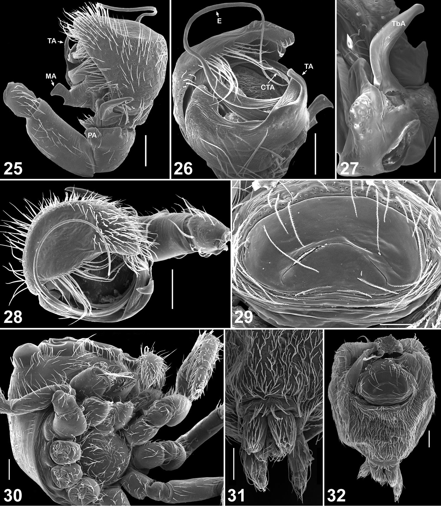
Copulatory organs and somatic characters of Eupoa lehtineni sp. n. 25 male palp, retrolateral view 26 ditto, median view 27 male tibial apophysis, retrolateral view 28 male palp, apical view 29 epigyne, ventral view 30 female carapace, ventral view 31 female spinnerets, ventral view 32 female abdomen, ventral view. Abbreviations as explained in ‘Material and methods’. Scale bars: 50 μm (27, 29), 0.1 mm (25–26, 28, 30–32).
Of the described salticid genera, Eupoa is closest to Corusca Zhou & Li, 2013 (10 species) and Sinoinsula Zhou & Li, 2013 (12 species) known from Hainan Island of China (
The phylogenetic relationships of Eupoa remain poorly resolved. On the basis of two morphological characters (presence of the median apophysis and a tarsal claw in the female palp; Figs 15–16) and molecular data,
Currently, 12 species are included in Eupoa (
Eupoa seems to be restricted to the Oriental Region, for all the described species thereof as well as those of two closely related genera, Corusca and Sinoinsula, are restricted to SE Asia (
http://zoobank.org/EF8169C1-C2D9-45C3-BAC0-38DD65DBBD40
http://species-id.net/wiki/Eupoa_daklak
Figs 33–35Holotype ♀ (ZMUM) from Viet-Nam, Dak Lak Prov., c. 25 km SSW of Buon Ma Thuot (c. 12°26'48"N, 107°58'E), Dak Linh, 500 m a.s.l., 28-29.04.1986, L.N. Medvedev & S.I. Golovatch.
The species epithet is a noun taken in apposition of the type locality: Daklak province of Viet-Nam.
The central shallow depression of the epigyne of Eupoa daklak sp. n. (Fig. 33) is unique among all the Eupoa species known to us. The male of Eupoa daklak sp. n. remains unknown.
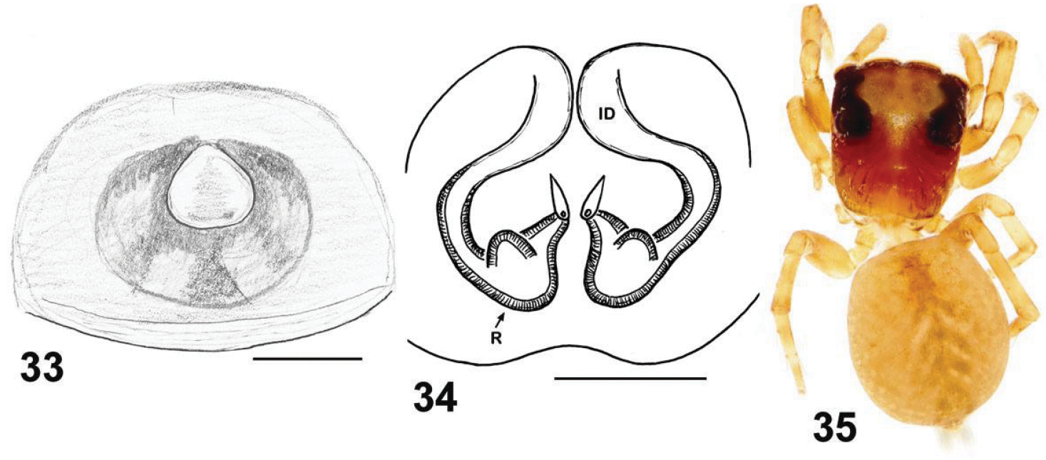
Copulatory organs and somatic characters of Eupoa daklak sp. n. 33 epigyne, ventral view 34 vulva, dorsal view 31 female body, dorsal view. Abbreviations as explained in ‘Material and methods’. Scale bars: 0.1 mm.
Although this species is described on the basis of a single female, it is highly unlikely that it could belong to one of the three other new species described below on the basis of males (Eupoa lobli sp. n. from Malaysia; Eupoa pappi sp. n. and Eupoa pulchella sp. n. from Thailand). All Eupoa species, as well as those of two closely related genera (Corusca and Sinoinsula), are litter-dwellers, with most of them (except for Eupoa lehtineni sp. n.) having small, local ranges. For instance, 22 distinct species of Corusca and Sinoinsula were recently described from Hainan Island of China alone (
The type locality only.
Male unknown.
FEMALE. Measurements. Carapace 0.89 long, 0.76 wide and 0.49 high at PLE. Ocular area 0.59 long, 0.77 wide anteriorly and 0.71 wide posteriorly. Diameter of AME 0.24. Clypeus height 0.07, chelicera length 0.29. Abdomen 1.06 long, 0.89 wide. Length of leg segments: I: 0.47 + 0.22 + 0.29 + 0.29 + 0.19; II: 0.41 + 0.19 + 0.23 + 0.23 + 0.19; III: 0.39 + 0.17 + 0.26 + 0.27 + 0.19; IV: 0.61 + 0.23 + 0.43 + 0.34 + 0.23. Leg spination. Leg I: Tb v 2-2-2ap; Mt v 2-2-2ap. Leg II: Tb v 2-2; Mt v 2-2-2ap. Leg III: Tb pr and rt 0-1-0; Mt pr 1-1ap, rt and v 1-0. Leg IV: Tb pr and rt 0-1-0; Mt pr 1-0-2ap, rt 1-0-1ap, v 1-0. Coloration (Fig. 35). Carapace yellow-brown, with brownish radial veins on thorax and yellow eye field. Blackened around eyes. Clypeus naked, yellow, with a brown marginal line. Sternum, maxillae, labium and chelicerae yellow, tinged with brown. All abdomen brownish yellowish, without a colour pattern. Book-lung covers yellow, tinged with brown. Spinnerets: anterior pair brownish, posterior pair pale yellow. All legs yellow, with poorly marked brownish patches at segment joints. Palps: femora and patellae brownish, metatarsi and tarsi yellow. Epigyne and vulva as in Figs 33–34: epigynal plate as a rounded shallow depression; insemination ducts thin and transparent; pear-shaped receptacles are close to each other, meeting up along the median line.
http://zoobank.org/ACCC403B-039D-4B0E-8EE1-4C9AA4458999
http://species-id.net/wiki/Eupoa_lehtineni
Figs 1–11, 13–17, 19–22, 25–32, 36–54Holotype ♂ (ZMTU) from India, Meghalaya, East Khasi Hills, Umran, riverside forest, 1200 m a.s.l., 4.05.1979, P.T. Lehtinen.
Paratypes: INDIA: 2♂5♀ (ZMTU), together with the holotype. – THAILAND: 3♂5♀ (MHNG, one male without abdomen and palp), Trat Prov., Ko Chang, west side (12°03'N, 102°18'E), WINKLER-extraction in secondary forest with primary spots (AS-T-5), 50-200 m a.s.l., 3-23.12.1999, A. Schultz; 1♀ (MHNG), Kanchanaburi Prov., Sai Yok Distr., Sai Yok National Park, near HQ, 100 m a.s.l., 21.07.1987, P.J. Schwendinger. – VIET-NAM: 1♀ (ZMUM), Dong Nai Prov., Vinh Cuu Dist., Vinh Cuu Nature Reserve (= Ma Da Forest), c. 6 km N of Ba Hao Vil. (c. 11°19'N, 107°05'E), 80 m a.s.l. (sample-5), 10.05.1995, T. Sergeeva; 1♀ (ZMUM), same locality (sample-11), 6.06.1995, T. Sergeeva.
The species is named in honour of our colleague and friend, Dr Pekka Lehtinen (Turku, Finland), who collected the holotype.
By the conformation of the large, round tegulum, the tibial apophysis and the thin compound terminal apophysis that is hidden in the apical cavity of tegulum (Figs 26, 52), the male of Eupoa lehtineni sp. n. is most similar to that of Eupoa lobli sp. n. (Figs 60–62), but can easily be distinguished by the shape of tegular and median apophyses. The female of Eupoa lehtineni sp. n. has the easily recognizable triangle epigynal plate (Figs 29, 39–40) and the mutual arrangement and shape of receptacles (Figs 53–54).
From eastern India, south-eastward to Thailand and southern Viet-Nam (present data).
MALE (from Thailand: Ko Chang). Measurements. Carapace 0.94 long, 0.79 wide and 0.51 high at PLE. Ocular area 0.61 long, 0.79 wide anteriorly and 0.77 wide posteriorly. Diameter of AME 0.24. Clypeus height 0.06, chelicera length 0.29. Abdomen 0.76 long, 0.58 wide. Length of leg segments: I: 0.44 + 0.22 + 0.30 + 0.26 + 0.16; II: 0.40 + 0.21 + 0.21 + 0.24 + 0.19; III: 0.38 + 0.18 + 0.22 + 0.27 + 0.20; IV: 0.59 + 0.23 + 0.41 + 0.30 + 0.23. Leg spination. Leg I: Mt v 2-2-2ap. Leg II with no spines. Leg III: Tb pr and rt 0-1-0. Leg IV: Tb pr and rt 0-1-0; Mt pr and rt 0-1-1-ap. Coloration (Figs 42–43). Carapace light grey-brown, with grey-yellow eye field and with poorly visible dark brown radial veins on thorax. Blackened around eyes. Clypeus naked, yellow-brown. Sternum light grey-brown. Maxillae, labium and chelicerae grey-yellow. Abdomen light grey-brown, dorsum completely covered with large scutum and with a thin longitudinal, medial yellow line. Book-lung covers light grey-brown. Spinnerets: anterior pair grey brownish, posterior pair yellow. All legs grey-yellow, with dark brown patches at segment joints, but patellae, tibiae and metatarsi I pro-ventrally with dark brown longitudinal stripe. Palp grey-yellow, its structure as in Figs 25–28, 36–38, 49–52: patellar apophysis short and obtuse; tibial apophysis blade-shaped, bent dorsad; tegulum well-developed, apically with prominent, hook-shaped tegular apophysis; median apophysis massive, directed ventrad; compound terminal apophysis thin and long, situated inside the apical cavity of tegulum; embolus coiled, making two revolutions, with its terminal end resting on top of the cymbium.
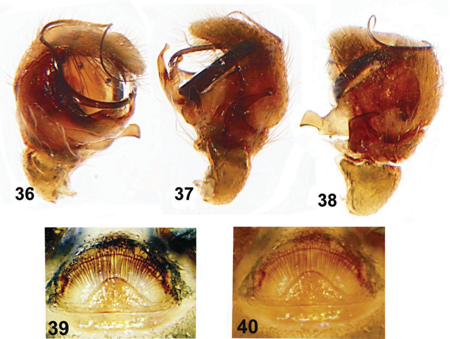
Copulatory organs of Eupoa lehtineni sp. n. from India. 36 male palp, median view 37–38 ditto, retrolateral view 39–40 epigyne, ventral view.
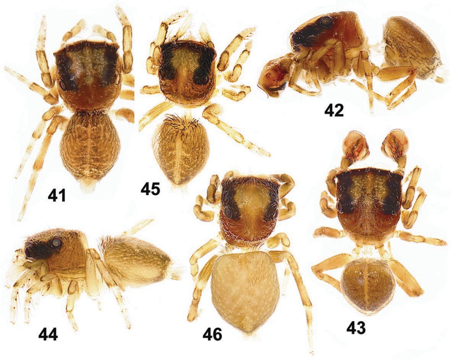
General appearance of Eupoa lehtineni sp. n. 41, 45–46 females, dorsal view 42 male, lateral view 43 ditto, dorsal view 44 female, lateral view. Specimens: 41–43 – India; 44–46 – Viet-Nam.
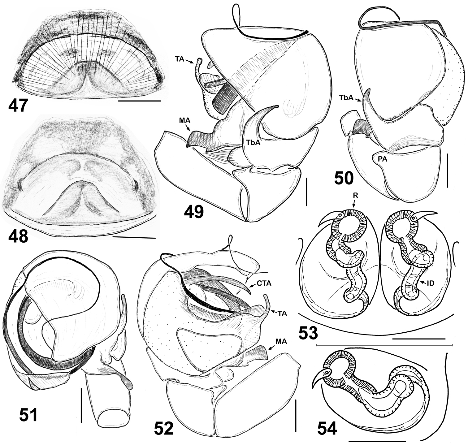
Copulatory organs of Eupoa lehtineni sp. n. 47–48 epigyne, ventral view 49 male palp, retrolateral view 50 ditto, dorsal view 51 ditto, apical view 52 ditto, median view 53–54 vulva, dorsal view. Abbreviations as explained in ‘Material and methods’. Specimens: 49–53 – India; 47–48, 54– Viet-Nam. Scale bars: 50 μm (27, 29), 0.1 mm (25–26, 28, 30–32).
FEMALE (from Viet-Nam: Dong-Nai). Measurements. Carapace 0.93 long, 0.73 wide and 0.51 high at PLE. Ocular area 0.59 long, 0.75 wide anteriorly and 0.71 wide posteriorly. Diameter of AME 0.24. Clypeus height 0.06, chelicera length 0.33. Abdomen 0.89 long, 0.71 wide. Length of leg segments: I: 0.49 + 0.26 + 0.31 + 0.31 + 0.21; II: 0.39 + 0.21 + 0.23 + 0.26 + 0.18; III: 0.39 + 0.17 + 0.24 + 0.31 + 0.21; IV: 0.63 + 0.23 + 0.49 + 0.37 + 0.23. Leg spination. Leg I: Tb v 2-2-2ap; Mt v 2-2-2ap. Leg II: Tb pr 0-1-0, v 1-1; Mt v 2-2-2ap. Leg III: Tb pr 0-1-0, Mt pr 1-1ap, rt 1ap. Leg IV: Tb pr and v 0-1-0; Mt pr 2-0-2ap, rt 1-0-1ap. Coloration (Figs 41, 44–46). Carapace pale brown, with yellow eye field and a thin longitudinal median line on thorax. Blackened around eyes. The entire carapace covered (not very densely) with white and transparent elongated scales. Clypeus naked, brown-yellow. Sternum yellow, tinged with brown. Maxillae, labium and chelicerae pale yellow. Abdomen: dorsum light brown, with a longitudinal medial yellow line; sides light brown, each with a wide yellow stripe; venter yellow. Anterior edge of carapace with a bunch of long brownish hairs. Book-lung covers yellow. Spinnerets: anterior pair brownish, posterior pair pale yellow. All legs yellow, with brownish (semi)rings at segment joints, but femora, patellae and tibiae I pro-ventrally with dark brown stripe. Palps: femora and patellae light brown, metatarsi and tarsi yellow. Epigyne and vulva as in Figs 29, 39–40, 47–48, 53–54: epigynal plate triangular, with obtuse tip directed anteriorly, covered with long whitish hairs; insemination ducts relatively short, subparallel and directed anteriorly; receptacles relatively small and rounded; receptacles and fertilization ducts situated in the anterior part of vulva.
http://zoobank.org/C5D9756D-9195-4014-A068-939D89C19F09
http://species-id.net/wiki/Eupoa_lobli
Figs 55–65MALAYSIA: 1♂ (MHNG, both palps detached), West Malaysia, Pahang, Cameron Highlands, 1520 m a.s.l., trail 14, Bukit Mentiga (tamisage), 23.03.1993, I. Löbl & F. Calame.
The species is named after the famous coleopterist, Dr. Ivan Löbl (Geneva, Switzerland), who collected the holotype.
The male of Eupoa lobli sp. n. is most similar to that of Eupoa lehtineni sp. n., but can easily be distinguished by the shape of tegular and median apophyses (cf. Figs 60–61 and 52). The female of Eupoa lobli sp. n. remains unknown.
The type locality only.
MALE. Measurements. Carapace 0.93 long, 0.74 wide and 0.51 high at PLE. Ocular area 0.54 long, 0.74 wide anteriorly and 0.67 wide posteriorly. Diameter of AME 0.23. Clypeus height 0.11, chelicera length 0.23. Abdomen 0.71 long, 0.64 wide. Length of leg segments: I: 0.46 + 0.21 + 0.31 + 0.29 + 0.21; II: 0.38 + 0.23 + 0.22 + 0.26 + 0.20; III: 0.37 + 0.19 + 0.21 + 0.22 + 0.20; IV: 0.64 + 0.22 + 0.39 + 0.31 + 0.25. Leg spination. Leg I: Tb v 0-1-0; Mt v 2-2-2ap. Leg II with no spines. Leg III: Tb rt 1-0. Leg IV: Tb pr and rt 0-1-0; Mt pr 1-0-1ap, rt 1-0-2ap. Coloration (Figs 58–59). Carapace light brown, sparsely covered with white elongated scales; eye field with a median yellow stripe. Blackened around eyes. Clypeus naked, yellow. Sternum, maxillae and labium yellow brownish. Abdomen light brown, with no colour pattern; dorsum completely covered with scutum. Book-lung covers light brown. Spinnerets: anterior pair brownish, posterior pair pale yellow. All legs yellow, with brownish patches at segment joints, but tibiae I and II pro-ventrally with dark brown longitudinal stripe. Palps brownish yellow, their structure as in Figs 55–57, 60–65: patellar apophysis short and wide, as if cut on its tip; tibial apophysis claw like, blade-shaped and directed dorsad; tegulum well-developed, with prominent, wide tegular apophysis apically possessing three hook-shaped, obtuse teeth; median apophysis massive, directed latero-ventrad; compound terminal apophysis relatively short and thin, hidden inside the cavity formed by cymbium and tegulum; embolus coiled, making two revolutions, with its tip resting on top of the cymbium.
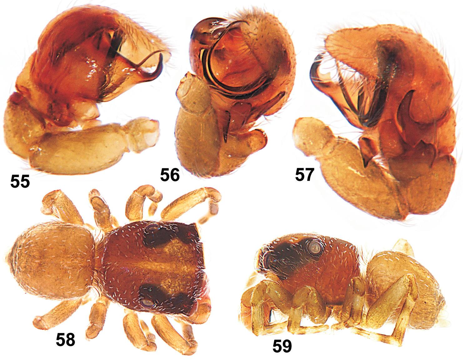
Copulatory organs and somatic characters of Eupoa lobli sp. n. (the holotype). 55 male palp, median view 56 ditto, apical view 57 ditto, retrolateral view 58 male general appearance, dorsal view 59 ditto, lateral view.
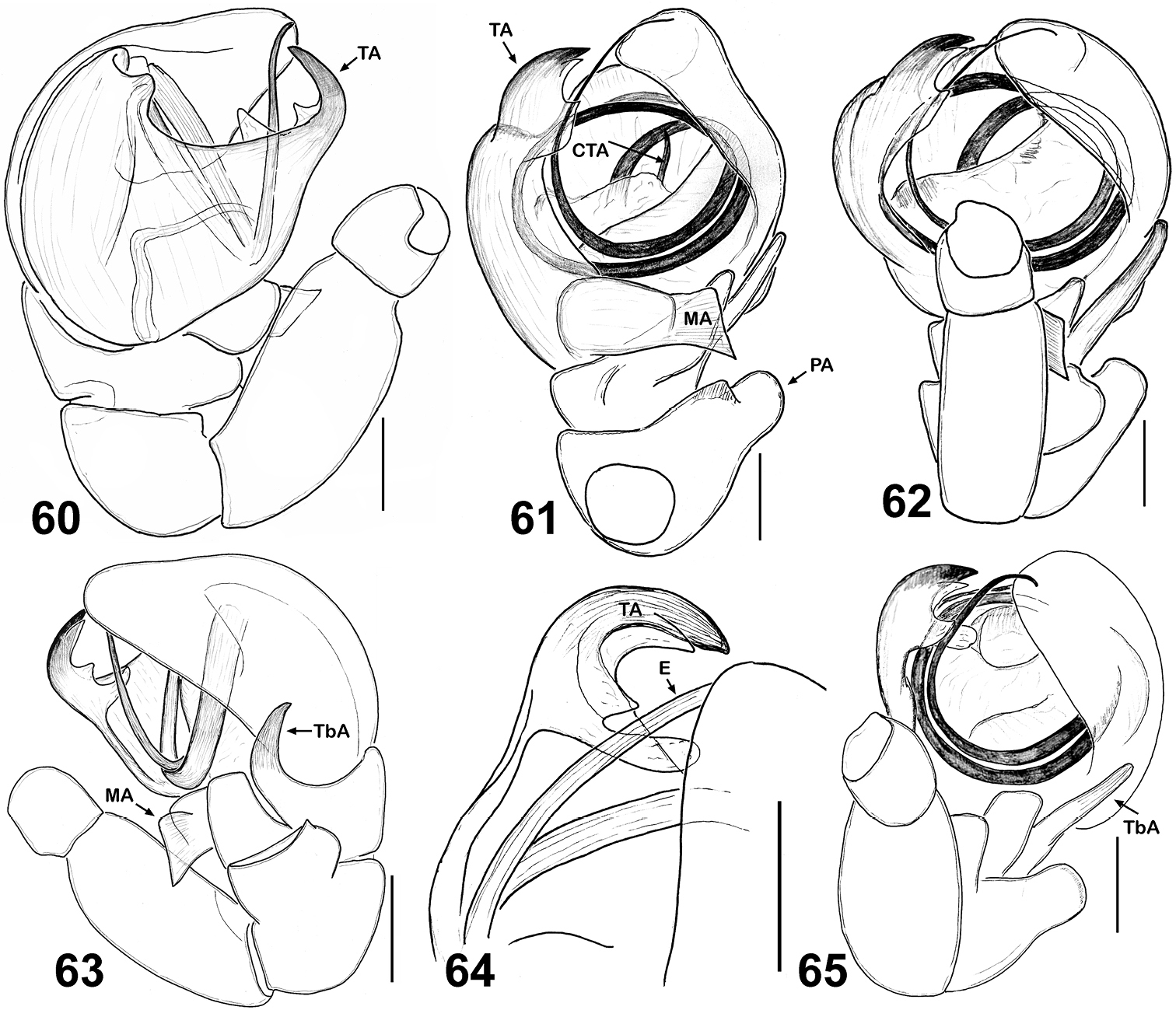
Copulatory organs of Eupoa lobli sp. n. (the holotype). 60 male palp, median view 61–62, 65 ditto, ventral view 63 ditto, retrolateral view 64 tegular apophysis, retrolateral view. Abbreviations as explained in ‘Material and methods’. Scale bars: 0.1 mm.
Female unknown.
http://zoobank.org/72FED4F4-8221-4D13-9ABE-726088FDF2A5
http://species-id.net/wiki/Eupoa_pappi
Figs 66–71Holotype ♂ (HNHM) from Thailand Trang Prov., Thung Khai Botanic Garden, primary lowland rainforest, 12.11.2004, L. Papp & M. Földvári.
The species is named after Dr. László Papp (Budapest, Hungary), who collected the holotype.
By having the massive compound terminal apophysis with a deep longitudinal groove on its apical edge (Fig. 67), the male of Eupoa pappi sp. n. is most similar to that of Eupoa thailandica sp. n. (Figs 109, 115), from which it differs in having the larger open cavity of tegulum and the shape of tegular and compound terminal apophyses. The female of Eupoa pappi sp. n. remains unknown.
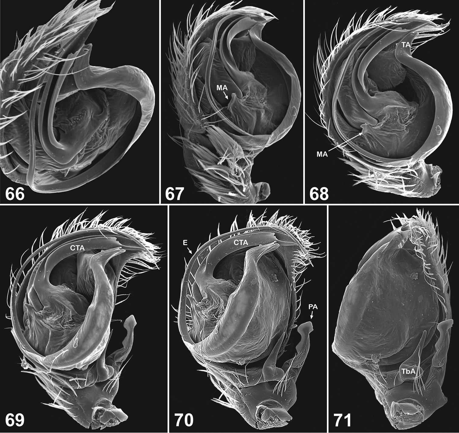
Copulatory organs of Eupoa pappi sp. n. (the holotype). 66 male palp, apical view 67–68 ditto, median view 69 ditto, ventral view 70–71 ditto, retrolateral view. Abbreviations as explained in ‘Material and methods’.
The type locality only.
MALE (one palp missing). Measurements. Carapace 0.93 long, 0.75 wide and 0.60 high at PLE. Ocular area 0.60 long, 0.80 wide anteriorly and 0.63 wide posteriorly. Diameter of AME 0.23. Clypeus height 0.10, chelicera length 0.28. Abdomen 0.88 long, 0.63 wide. Length of leg segments: I: 0.55 + 0.24 + 0.40 + 0.39 + 0.20; II: 0.49 + 0.21 + 0.29 + 0.29 + 0.17; III: 0.48 + 0.33 + 0.34 + 0.35 + 0.23; IV: 0.78 + 0.28 + 0.53 + 0.43 + 0.28. Leg spination. Leg I: Mt v 2-2ap. Leg II and III: no spines. Leg IV: Tb pr and rt 0-1-0; Mt pr and rt 1-1ap. Coloration. Carapace light yellow, with brownish margins. Eye field brownish, blackened around eyes. Clypeus naked, yellowish brownish. Sternum, maxillae, labium and chelicerae light yellow. Abdomen: dorsum yellow, with dark grey longitudinal lateral bands and two round, large dark grey spots in its rear part; venter yellow. Book-lung covers yellow. Spinnerets: anterior pair yellow, posterior pair dark grey. All legs yellow, but patellae IV with brownish sides. Palps: femora yellow, remaining segments brownish yellow. Palpal structure as in Figs 66–71: patellar apophysis long, reaching almost a half of the cymbial lenght; tibia with a median bunch of thick white hairs; tibial apophysis sharpened at its tip directed anteriorly and with a wide, rounded base; tegulum flat and developed on its pro-lateral side only; tegular apophysis situated on the apical end of tegulum and bent laterad; median apophysis poorly developed, seen as a short triangle process at the base of compound terminal apophysis (but separated from it by membrane); compound terminal apophysis massive, with a deep longitudinal groove on its apical edge; embolus whip-shaped, making one revolution.
Female unknown.
http://species-id.net/wiki/Eupoa_prima
Figs 72–84Holotype ♂ (ZMTU) from Viet-Nam, Bac Thai, Bach Tong, Duong Quang (apparently, Bach Thong Distr., Bac Kan Prov.), jungle slope, 900 m a.s.l., 17.10.1978, P.T. Lehtinen.
Paratypes: VIET-NAM: 1♂1♀ (ZMTU; epigyne missing), together with the holotype; 1♀ 5juv. (HNHM; epigyne missing), c. 5 km E of Lao Cai town, 200 m a.s.l., sieving forest litter, 1971, Topál-Matskásl.
Both sexes of Eupoa prima are most similar to those of Eupoa jingwei and Eupoa nezha known from Guangxi province of China; threes species seem to form a natural species group. Males of all three species can easily be separated by the shape of tegular and patellar apophyses; cf. Figs 80–84 with figs 1–3, 8–10 in
Unfortunately, neither the ♀ allotype deposited at the ZMTU, nor the ♀ paratype deposited at the HNHM possesses its epigyne, although in both cases there are separate micro-vials that should have contained them. Thus, our notion about the epigynal and vulval structures of Eupoa prima is based on the original illustrations by
Southern Viet-Nam (
MALE (the holotype). Measurements. Carapace 1.00 long, 0.89 wide and 0.55 high at PLE. Ocular area 0.64 long, 0.89 wide anteriorly and 0.81 wide posteriorly. Diameter of AME 0.29. Clypeus height 0.06, chelicera length 0.24. Abdomen 0.95 long, 0.61 wide. Length of leg segments: I: 0.54 + 0.26 + 0.34 + 0.36 + 0.19; II: 0.47 + 0.21 + 0.30 + 0.32 + 0.19; III: 0.46 + 0.21 + 0.27 + 0.35 + 0.23; IV: 0.71 + 0.30 + 0.51 + 0.39 + 0.29. Leg spination. Leg I: Tb v 1-1-1; Mt v 2-2-2ap. Leg II: Mt v 1-1-1ap. Leg III: Tb pr and rt 0-1. Leg IV: Tb pr 0-1; Mt pr and rt 1ap, v 0-1-0. Coloration (Figs 72–73, 78–79). Carapace yellow-brown, with a large yellow spot occupying almost the entire eye field; blackened around eyes. Clypeus naked, yellow, with a dark brown marginal line. Sternum, maxillae and labium yellow, tinged with brown. Chelicerae yellow, each with an anterior longitudinal brown stripe. Abdomen: dorsum brown, with shining scutum and a poorly marked yellow spot; sides brown; venter yellow. Book-lung covers yellow. Spinnerets: anterior pair brownish, posterior pair yellow. All legs yellow, but pro- and retrolateral sides of all segments (except for tarsi) brownish. Palps yellow-brown. Palpal structure as in Figs 74, 80–84: femur modified, with a wide proximal-ventral protuberance (as a short apophysis); patella with two apophyses, short basal apophysis and long median one, reaching almost a third of the cymbial lenght; tibial apophysis bi-ramous: its ventral branch wide, massive and visibly sclerotized and its dorsal branch short and cone-shaped; cymbium with triangular lobe in retrobasal part; tegulum well-developed; tegular apophysis situated on the median side of tegulum and directly anteriroly; median apophysis thin and hook-shaped, directed laterad; compound terminal apophysis low and wide, situated at the embolic base; embolus whip-shaped, making one revolution, its tip is resting on the dorsal side of tegular apophysis (its course is shown in Fig. 74).
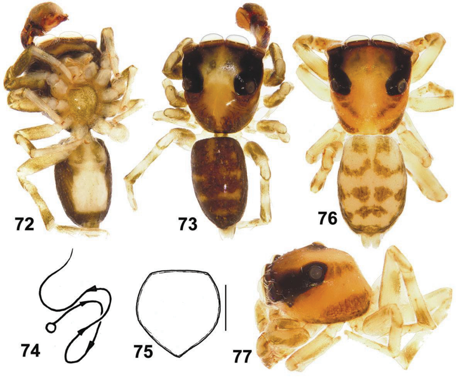
General appearance and somatic characters of Eupoa prima (♂ holotype and ♀ allotype). 60 male body, ventral view 73 ditto, dorsal view 74 diagrammatic course of the embolar path 75 female sternum, ventral view 76 female body, dorsal view 77 female carapace, lateral view. Scale bars: 0.25 mm (76).
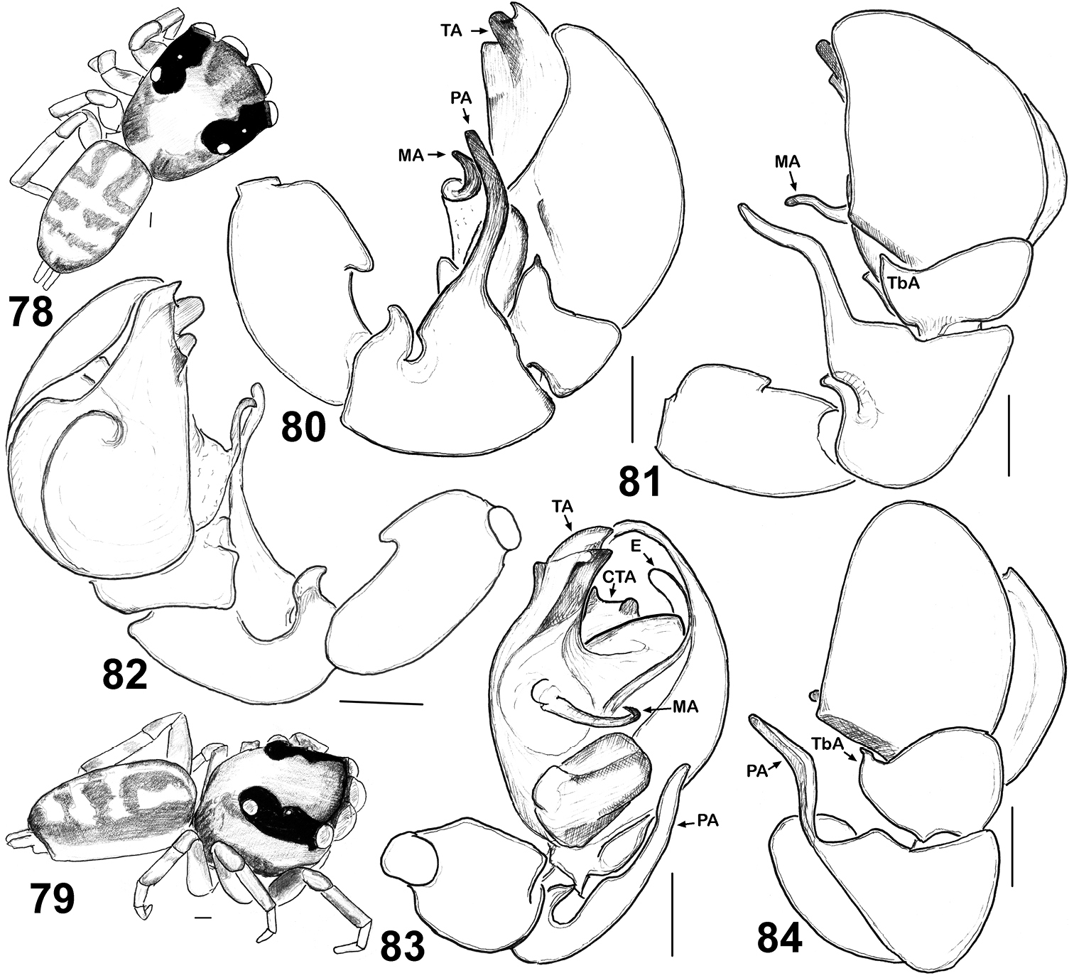
General appearance and copulatory organs of Eupoa prima (♂ paratype). 78 male body, dorsal view 79 ditto, lateral view 80 male palp, retrolateral view 81, 84 ditto, dorsal view 82 ditto, median view 83 ditto, ventral view. Abbreviations as explained in ‘Material and methods’. Scale bars: 0.1 mm.
FEMALE (the paratype). Measurements. Carapace 1.21 long, 1.01 wide and 0.60 high at PLE. Ocular area 0.76 long, 1.07 wide anteriorly and 0.96 wide posteriorly. Diameter of AME 0.36. Clypeus height 0.06, chelicera length 0.29. Abdomen 1.23 long, 0.86 wide. Length of leg segments: I: 0.70 + 0.30 + 0.43 + 0.43 + 0.21; II: 0.57 + 0.28 + 0.33 + 0.37 + 0.23; III: 0.56 + 0.21 + 0.36 + 0.43 + 0.27; IV: 0.89 + 0.29 + 0.64 + 0.56 + 0.31. Leg spination. Leg I: Tb v 2-2-2ap; Mt v 2-2-2ap. Leg II: Tb pr 0-1, v 1-1; Mt v 2-2-2ap. Leg III: Tb pr and rt 0-1-0; Mt pr 1-2ap, rt 2ap, v 1-0. Leg IV: Tb pr and rt 0-1-0, v 1-0; Mt pr 1-2ap, rt 1-1ap, v 1-0. Coloration much lighter than in the male (Figs 75, 77). Carapace yellow, with two wide brown stripes on sides. Blackened around eyes. Clypeus and ‘cheeks’ naked, yellow. Sternum, maxillae, labium and chelicerae yellow. Abdomen: dorsum yellow, with four transverse brown bands; sides yellow, each with a longitudinal brown stripe; venter yellow. Book-lung covers and spinnerets yellow. Legs I yellow, with pro- and retrolateral sides of femora, patellae and tibiae brownish. Legs II-IV yellow, with brownish patches at segment joints. Palps yellows, tinged with brown, but tarsi entirely yellow. Epigyne and vulva as in
http://zoobank.org/DB59AEEB-D36B-4440-AF08-79C284E997C1
http://species-id.net/wiki/Eupoa_pulchella
Figs 87–88, 91–96Holotype ♂ (MHNG, both palps separated) from Thailand, Prov. Chiang Mai, Chiang Dao Distr., Doi Chiang Dao Wildlife Sanctuary, 510 m a.s.l., pitfall traps, 23.12.1990–15.01.1991, P. J. Schwendinger.
Paratypes: THAILAND: 3♂ (MHNG; palps of all specimens are separated), together with the holotype; 1♂ (MHNG; with the single detached palp), same locality, 510 m a.s.l., pitfall traps, 25.10–23.11.1990, P. J. Schwendinger.
From the Latin word ‘pulchellus’ meaning ‘pretty’.
This species differs from all Eupoa species known to us in having all the bulbal sclerites in apical position, as if grouped together at the top of tegulum (Figs 91–96), and the unique shape of patellar apophysis, which is short, thick and bi-ramous (Figs 93, 96). The female of Eupoa pulchella sp. n. remains unknown.
The type locality only.
MALE (♂ paratype with the palp). Measurements. Carapace 0.94 long, 0.73 wide and 0.46 high at PLE. Ocular area 0.56 long, 0.75 wide anteriorly and 0.70 wide posteriorly. Diameter of AME 0.23. Clypeus height 0.06, chelicera length 0.20. Abdomen 0.76 long, 0.59 wide. Length of leg segments: I: 0.44 + 0.21 + 0.29 + 0.29 +0. 19; II: 0.39 + 0.19 + 0.23 + 0.24 + 0.17; III: 0.37 + 0.16 + 0.23 + 0.26 + 0.27; IV: 0.59 + 0.23 + 0.41 + 0.33 + 0.21. Leg spination. Leg I: Tb v 2-2-2ap; Mt v 2-2ap. Leg II without spines. Leg III: Tb pr and rt 0-1-0. Leg IV: Tb pr and rt 0-1-1; Mt pr 1-2ap, rt 1-1ap. Coloration. Carapace brown, with a wide median yellow stripe started from the middle part of eye field and running to thorax; blackened around eyes. Clypeus naked and brown. Sternum, maxillae, labium and chelicerae pale yellow. Abdomen: dorsum brown, with two longitudinal rows of large yellow spots; sides brown; venter yellow. Book-lung covers yellowish brownish. Spinnerets: anterior pair brownish, posterior pair pale yellow. All legs yellow, with brown patches at segment joints, but femora, patellae and tibiae pro-ventrally and retrolaterally brown. Palps dark brown. Palpal structure as in Figs 91–96: patellar apophysis short, thick and bi-ramous; tibial apophysis short and cone-shaped; tegulum well-developed; tegular apophysis not developed; median apophysis fingerlike, directed anteriorly; compound terminal apophysis wide, plate-shaped and rugose; embolus short, S-shaped.
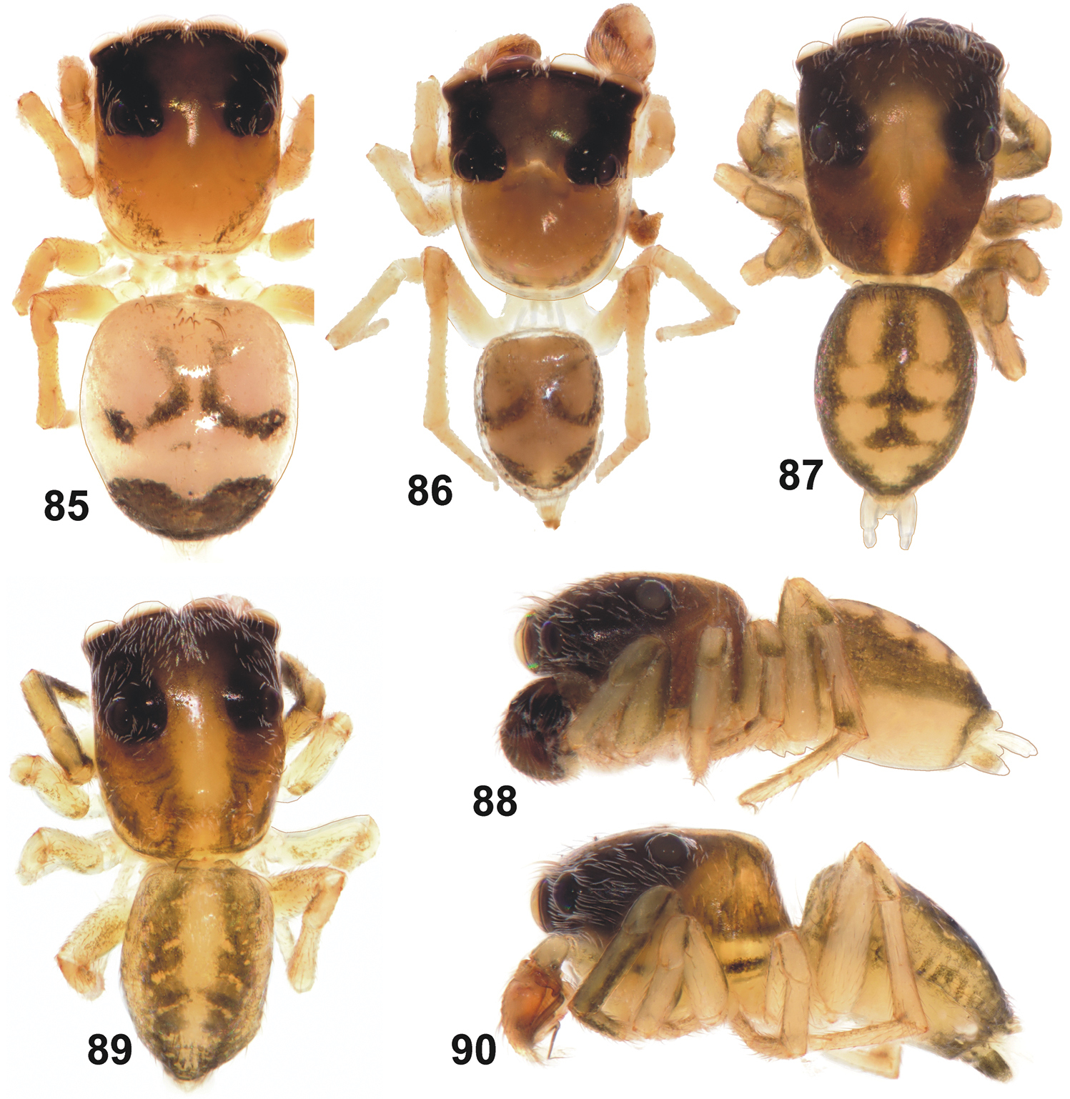
General appearance of Eupoa pulchella sp. n. (87–88, ♂ paratype), Eupoa schwendingeri sp. n. (♂ holotype, 89–90) and Eupoa thailandica sp. n. (♀ and ♂ paratypes, 85–86). 85 female body, dorsal view 86–87, 89 male body, dorsal view 88, 90 ditto, lateral view.
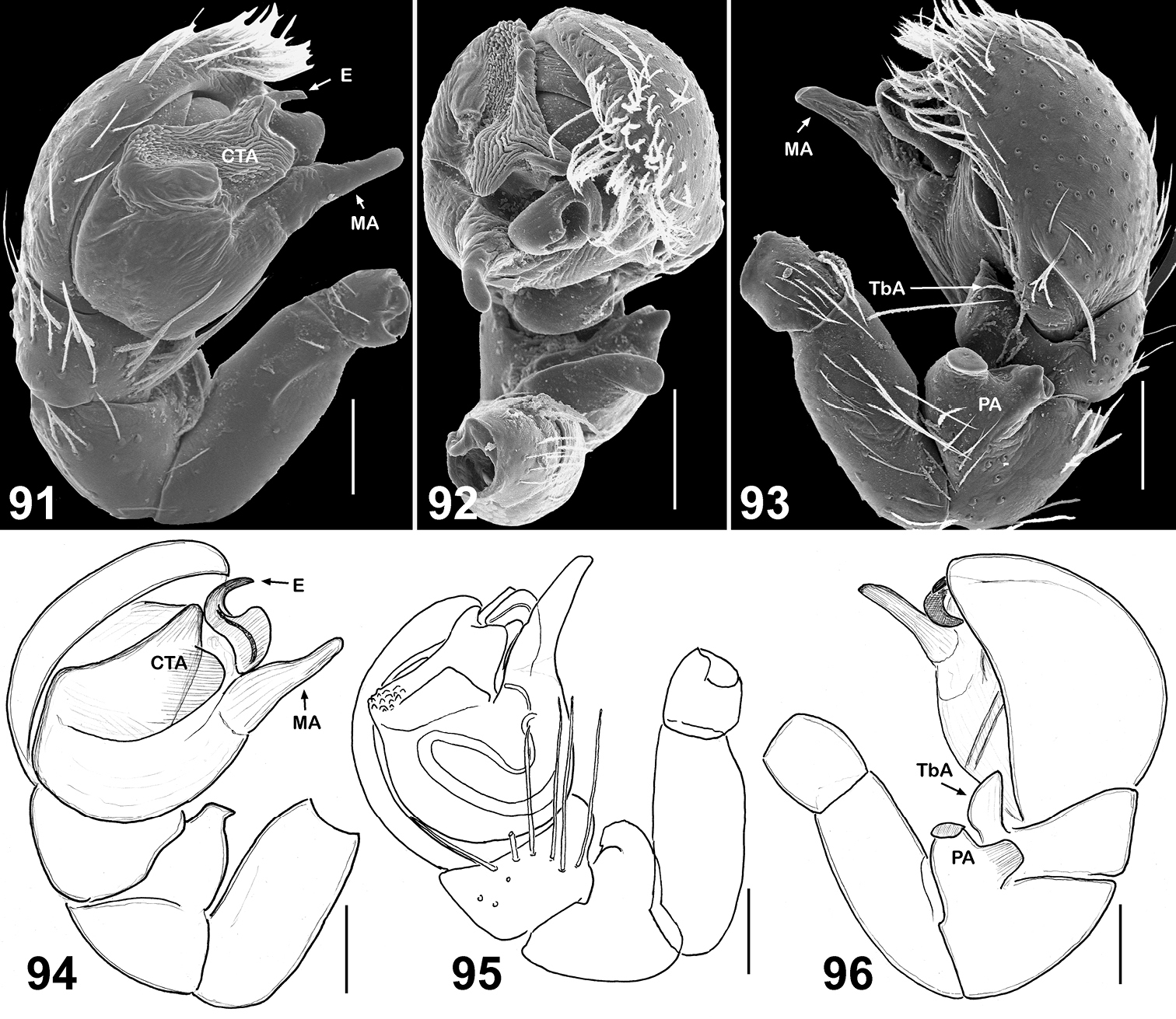
Copulatory organs of Eupoa pulchella sp. n. (91–93, the holotype; 94–96, ♂ paratype). 91, 94–95 male palp, median view 92 ditto, apical view 93, 96 ditto, retrolateral view. Abbreviations as explained in ‘Material and methods’. Scale bars: 0.1 mm.
Female unknown.
http://zoobank.org/58EF7EFD-7F8C-42FF-9719-3DA2BC5C001B
http://species-id.net/wiki/Eupoa_schwendingeri
Figs 89–90, 97–105Holotype 1♂ (MHNG) from northern Thailand, Chiang Mai Prov. and Distr., Doi Suthep-Pui National Park, Doi (=Mount) Suthep, 1180 m a.s.l., 1-30.03.1987, P. J. Schwendinger.
Paratypes: THAILAND: 2♀ (MHNG), together with the holotype; 1♂ (MHNG), northern Thailand, Chiang Mai Prov. and Distr., Doi Suthep-Pui National Park, near Pin Pak Pai Waterfall, 1155 m a.s.l., pitfall traps 10.01-11.02.1997, P. Dankittipakul.
The species is named in honour of our colleague, the well-known arachnologist, Dr Peter Schwendinger (Geneva, Switzerland), who collected the type series and who made the majority of the Eupoa specimens treated in the present work available to us.
The male palp of Eupoa schwendingeri sp. n. differs from those of all Eupoa species known to us in having the rather small and inconspicous median and compound terminal apophyses (Figs 97–99). The female of Eupoa schwendingeri sp. n. has the unique epigyne shaped as a square plate, with parallel sides (Fig. 100) and the entrances of insemination ducts directed to each other (Fig. 101). Besides, both sexes of this species have a very characteristic body colour pattern consisting of two longitudinal subparallel grey stripes on dorsum (Figs 89, 104–105).
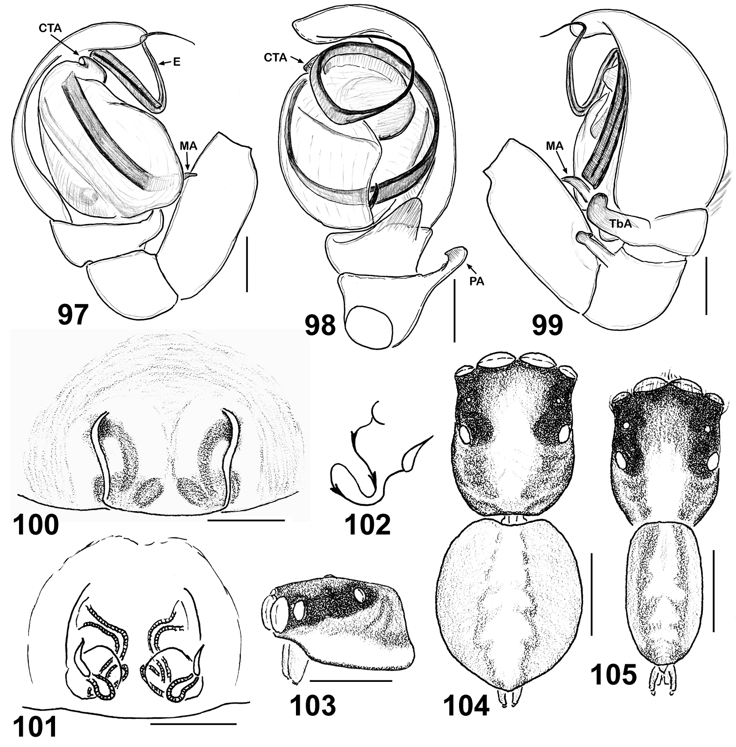
Copulatory organs and somatic characters of Eupoa schwendingeri sp. n. (♂ and ♀ paratypes). 97 male palp, median view 98 ditto, ventral view 99 ditto, retrolateral view 100 epigyne, ventral view 101 vulva, dorsal view 102 diagrammatic course of insemination ducts 103 female carapace, lateral view 104 female body, dorsal view 105 male body, dorsal view. Abbreviations as explained in ‘Material and methods’. Scale bars: 0.1 mm (97–101), 0.25 mm (103–105).
The only locality in Thailand: Doi Suthep-Pui National Park.
The ♂ paratype was lost after having been photographed, as the observation glass was incidentally dropped down to the floor. Thus, the ♂ paratype is represented by the male palp only (Figs 97–99) that is retained in the MHNG.
MALE (the holotype). Measurements. Carapace 0.90 long, 0.60 wide and 0.45 high at PLE. Ocular area 0.57 long, 0.73 wide anteriorly and 0.64 wide posteriorly. Diameter of AME 0.23. Clypeus height 0.04, chelicera length 0.30. Abdomen 0.88 long, 0.53 wide. Length of leg segments: I: 0.50 + 0.23 + 0.33 + 0.29 + 0.21; II: 0.40 + 0.16 + 0.23 + 0.26 + 0.20; III: 0.37 + 0.16 + 0.24 + 0.27 + 0.21; leg IV is missing. Leg spination. Legs I and II: no spines. Leg III: Mt pr 0-1-0. Leg IV is missing. Coloration (Figs 89–90, 104). Carapace yellow, with two wide longitudinal light brown bands. Eye field dark grey, blackened around eyes. Clypeus naked, yellowish brownish. Sternum, labium, maxillae and chelicerae light yellow. Abdomen yellow, with two longitudinal light grey stripes on dorsum. Book-lung covers light yellow. Spinnerets: anterior pair dark grey, posterior pair yellow. Leg I yellow, but femur (its distal part), patella, tibia and metatarsus with black pro-lateral longitudinal stripe, tibia and metatarsus also with light grey retro-lateral longitudinal stripe. Leg II yellow, but tibia ventrally with light grey longitudinal stripe. Legs III and IV yellow. Palps yellow, with brownish cymbia. Palpal structure as in Figs 97–99: patellar apophysis short, fingerlike; tibial apophysis thick and obtuse, directed ventrad with its tip bend dorsad; tegulum developed on its prolateral side only; tegular apophysis poorly developed, looking like an anterior ridge of tegulum; median apophysis spine-shaped, situated at the proximal end of tegulum; compound terminal apophysis poorly developed, looking like a hook-shaped process at the basal part of embolus; embolus coiled, making two revolutions.
FEMALE. Measurements. Carapace 0.98 long, 0.76 wide and 0.55 high at PLE. Ocular area 0.63 long, 0.79 wide anteriorly and 0.71 wide posteriorly. Diameter of AME 0.24. Clypeus height 0.07, chelicera length 0.29. Abdomen 0.90 long, 0.68 wide. Length of leg segments: I: 0.54 + 0.20 + 0.35 + 0.34 + 0.21; II: 0.50 + 0.20 + 0.26 + 0.29 + 0.21; III: 0.44 + 0.16 + 0.29 + 0.33 + 0.20; IV: 0.73 + 0.26 + 0.54 + 0.43 + 0.26. Leg spination. Leg I: Tb v 2-2-2ap; Mt v 2-2-2ap. Leg II: Tb pr 0-1, v 1-1; Mt v 2-2-2ap. Leg III: Tb pr and rt 0-1-0; Mt pr and rt 1-0. Leg IV: Tb pr and rt 0-1-0; Mt pr and rt 1-1. Coloration as in the male (Fig. 105), but palps and both pairs of spinnerets entirely yellow. Epigyne and vulva as in Figs 100–102: epigyne as square-shaped plate, with parallel sides; insemination ducts at their entrances directed to each other and then run posteriorly; receptacles small and pear-shaped.
http://zoobank.org/6E26B6C3-1CE6-43E4-8C17-58C4F7CCD61F
http://species-id.net/wiki/Eupoa_thailandica
Figs 85–86, 106–118Holotype 1♂ (MHNG) from Thailand, Trat Prov., Ko Chang, west side (12°03'N, 102°18'E), WINKLER-extraction in secondary forest with primary spots (AS-T-5), 50-200 m a.s.l., 3-23.12.1999, A. Schultz.
Paratypes: THAILAND: 2♂1♀ (MHNG, one male without abdomen and palp), together with the holotype.
The species epithet originates from the country of origin of the type series.
By having the massive compound terminal apophysis with a deep longitudinal groove on its apical edge (Fig. 109, 115), the male of Eupoa thailandica sp. n. is most similar to that of Eupoa pappi sp. n. (Fig. 67–68), from which it differs in having the smaller and more closed apical cavity of tegulum and the shape of tegular and compound terminal apophyses. The hook-shaped embolic tip of Eupoa thailandica sp. n. (Fig. 110) is unique among all the Eupoa species known to us. The female of Eupoa thailandica sp. n. has a characteristic, mushroom-shaped central epigynal plate (Fig. 117) and the distinct spermathecae (Fig. 118). Besides, both sexes of Eupoa thailandica sp. n. have a very characteristic colour pattern on the dorsum containing a large transverse brown spot at its rear end (Figs 85–86); of the Eupoa species known to us, only the female of Eupoa yunnanensis has got a similar colour pattern (Fig. 121).
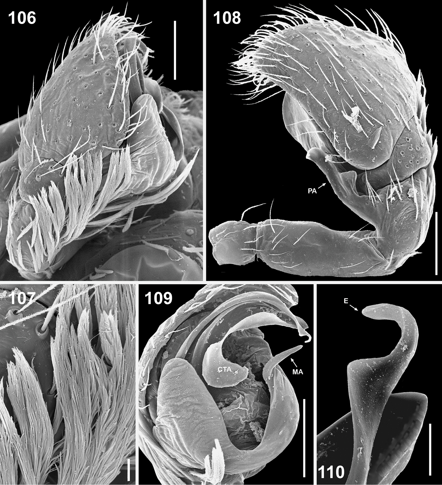
Copulatory organs of Eupoa thailandica sp. n. (♂ paratype). 106 male palp, median view 107 bunches of white hairs at the base of cymbium, median view 108 male palp, retrolateral view 109 ditto, ventral view 110 embolic tip, apical view. Abbreviations as explained in ‘Material and methods’. Scale bars: 10 μm (107, 110), 0.1 mm (106, 108–109).
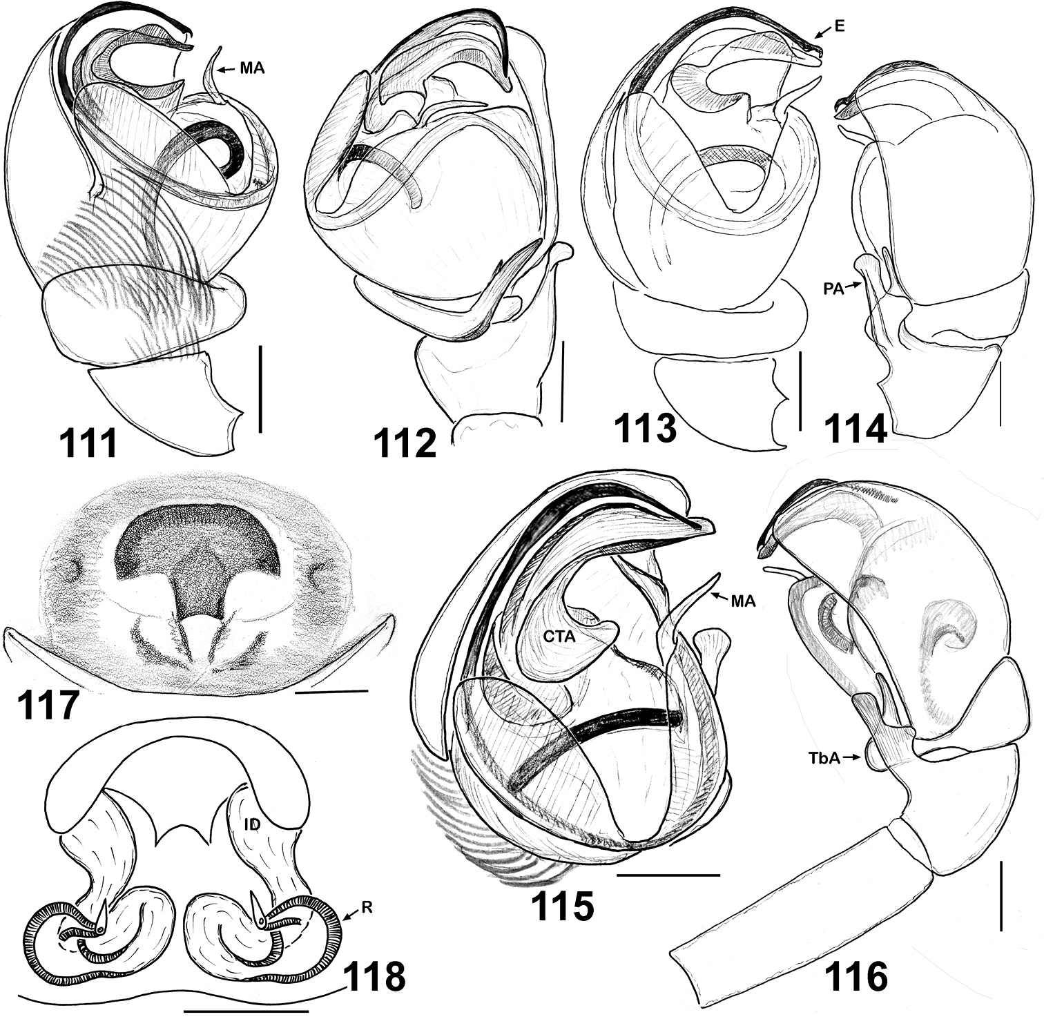
Copulatory organs of Eupoa thailandica sp. n. (♂ and ♀ paratypes). 111, 113 male palp, median view 112 ditto, ventral view 114 male palp, dorsal view 115 ditto, ventral view 116 ditto, retrolateral view 117 epigyne, ventral view 118 vulva, dorsa view. Abbreviations as explained in ‘Material and methods’. Scale bars: 0.1 mm.

General appearance of Eupoa yunnanensis from Laos. 119, 120 male body, dorsal view 121 female body, dorsal view 122 male body, lateral view.
The type locality only.
MALE (the holotype). Measurements. Carapace 0.87 long, 0.73 wide and 0.54 high at PLE. Ocular area 0.56 long, 0.77 wide anteriorly and 0.66 wide posteriorly. Diameter of AME 0.24. Clypeus height 0.13, chelicera length 0.24. Abdomen 0.67 long, 0.50 wide. Length of leg segments: I: 0.50 + 0.21 + 0.33 + 0.30 + 0.21; II: 0.39 + 0.16 + 0.31 + 0.27 + 0.20; III: 0.39 + 0.17 + 0.26 + 0.27 + 0.18; IV: 0.67 + 0.26 + 0.47 + 0.36 + 0.23. Leg spination. Leg I: Tb v 2-2-2ap; Mt v 2-2-2ap. Leg II: Tb v 1-0; Mt v 1-1. Leg III: Tb pr 0-1-0. Leg IV: Tb pr 0-1-0; Mt pr 1ap. Coloration (Fig. 86). Carapace yellow, with a brownish marginal stripe at rear end and with brownish eye filed. Blackened around eyes. Clypeus naked, yellow. Sternum, maxillae, labium and chelicerae pale yellow. Abdomen pale yellow, but dorsum with a colour pattern consisting of two brown semi-rings in its fore-half and a transverse brown spot at the rear end (just in front of the spinnerets). Book-lung covers and spinnerets pale yellow. All legs and palps yellow. Palpal structure as in Figs 106–116: femur rather long and thin, almost equal to the length of cymbium; patellar apophysis medium-sized, fingerlike, with a dorso-basal bulge, reaching almost a third of the cymbial length; tibia with set of long hairs prolaterally, tibial apophysis hook-shaped, directed ventrad with its sharp tip bend upward and directed anteriorly; cymbium with a median, basal bunch of thick white hairs; tegulum well-developed; tegular apophysis not developed; median apophysis thin, directed ventrad and situated in the central part of tegulum; compound terminal apophysis massive, as a large, thick hook having a longitudinal groove on its dorsal edge; embolus coiled, making one and a half revolutions, its tip hook-shaped.
FEMALE. Measurements. Carapace 0.96 long, 0.77 wide and 0.54 high at PLE. Ocular area 0.59 long, 0.81 wide anteriorly and 0.69 wide posteriorly. Diameter of AME 0.24. Clypeus height 0.07, chelicera length 0.31. Abdomen 1.04 long, 0.91 wide. Length of leg segments: I: 0.54 + 0.21 + 0.36 + 0.34 + 0.20; II: 0.44 + 0.19 + 0.26 + 0.27 + 0.20; III: 0.41 + 0.17 + 0.30 + 0.31 + 0.21; IV: 0.73 + 0.22 + 0.59 + 0.43 + 0.25. Leg spination. Leg I: Tb v 2-2-2ap; Mt v 2-2-2ap. Leg II: Tb pr 0-1, v 1-2; Mt pr 1-1, rt 0-1-0, v 2-0-2ap. Leg III: Tb pr and rt 0-1-0; Mt pr and rt 0-1-0. Leg IV: Tb pr 0-1-0; Mt pr and rt 1-0-1ap. Coloration as the male, but the dark brown spot on dorsum is larger (Fig. 85). Epigyne and vulva as in Figs 117–118: central epigynal plate mushroom-shaped, sclerotized and contrastingly darker than the rest of epigyne; wide transparent insemination ducts; round receptacles that are spaced up.
http://species-id.net/wiki/Eupoa_yunnanensis
Figs 119–128Laos: 2♂ (SMFM), Luang Prabang Prov., NE Luang Prabang, Nam Ou, Nong Khiao, Tham Pathok (20°33.082'N, 102°37.925'E'), 373 m a.s.l., outside cave, sieving leaf litter, 17–18.03.2007, P. Jäger & F. Steinmetz; 2♀ (SMFM), Luang Prabang Prov., SE Luang Prabang, Xieng Nguen Distr., Nam Khan, Ban Keng Koung (19°40.963'N, 102°18.442'E), 372 m a.s.l., along stream, sieving leaf litter, 22.03.2007, P. Jäger; 1♀ (SMFM), same locality, disturbed forest, dry stream bed, sieving and WINKLER-extraction, 21-23.02.2008, P. Jäger.
The male of Eupoa yunnanensis has the unique, widest and strongest patellar apophysis (Figs 124, 126) among all the Eupoa species known to us. The female has the unique conformation of its epigyne: viz., the singular atrium formed by the posterior chitinous margin and the anterior transverse pocket (Fig. 127) and the S-shaped spermathecae (Fig. 128).
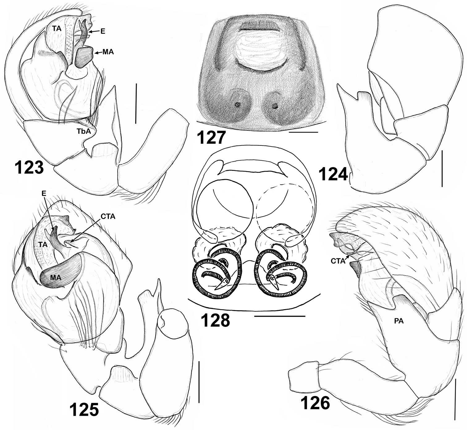
Copulatory organs of Eupoa yunnanensis from Laos. 123 male palp, median view 124 ditto, dorsal view 125 ditto, ventral view 126 ditto, retrolateral view 127 epigyne, verntral view 128 ditto, dorsal view. Abbreviations as explained in ‘Material and Methods’. Scale bars: 0.1 mm.
China (Yunnan) (
MALE. Measurements. Carapace 0.96 long, 0.79 wide and 0.50 high at PLE. Ocular area 0.60 long, 0.83 wide anteriorly and 0.75 wide posteriorly. Diameter of AME 0.29. Clypeus height 0.06, chelicera length 0.21. Abdomen 0.83 long, 0.50 wide. Length of leg segments: I: 0.49 + 0.19 + 0.30 + 0.31 + 0.21; II: 0.43 + 0.20 + 0.23 + 0.27 + 0.21; III: 0.43 + 0.19 + 0.24 + 0.31 + 0.26; IV: 0.64 + 0.24 + 0.47 + 0.39 + 0.29. Leg spination. Leg I: Mt v 2-2-2ap. Leg II: no spines. Leg III: Tb pr and rt 0-1-0. Leg IV: Tb pr and rt 0-1-0; Mt d, pr and rt 1-1ap. Coloration (Figs 119–120, 122). Carapace brownish, with a wide longitudinal median yellow stripe, the area between PLEs also yellow; blackened around eyes. Clypeus naked, brownish yellow. Sternum, labium, maxillae and chelicerae yellow. Abdomen: dorsum and sides dark grey, with two longitudinal rows of yellow spots on dorsum; dorsum with shining scutum; venter yellow. Book-lung covers yellow. Spinnerets: anterior pair dark grey, posterior pair yellow. All legs yellow, but femora I and patellae I grey on their sides, tibiae I ventrally grey and patellae II-IV grey on their sides. Palps brownish. Palpal structure as in Figs 123–126: patellar apophysis thick, massive and split at its tip, reaching almost a half of the cymbial length; tibial apophysis short and wide, poorly-developed; tegulum well-developed; tegular apophysis wide and strong, looking like a median extension; median apophysis thick and visibly sclerotized, situated in the central part of tegulum; compound terminal apophysis short, situated at the basis of embolus; embolus fingerlike, with a short hook-shaped process on its tip.
FEMALE. Measurements. Carapace 1.13 long, 0.86 wide and 0.60 high at PLE. Ocular area 0.59 long, 0.89 wide anteriorly and 0.76 wide posteriorly. Diameter of AME 0.29. Clypeus height 0.06, chelicera length 0.20. Abdomen 1.10 long, 0.85 wide. Length of leg segments: I: 0.65 + 0.28 + 0.48 + 0.40 + 0.25; II: 0.53 + 0.23 + 0.35 + 0.35 + 0.23; III: 0.51 + 0.23 + 0.38 + 0.40 + 0.25; IV: 0.90 + 0.33 + 0.73 + 0.50 + 0.30. Leg spination. Leg I: Tb v 2-2-2ap; Mt v 2-2-2ap. Leg II: Tb pr and rt 0-1-0, v 1-2; Mt v 2-2-2ap. Leg III: Tb pr and rt 0-1-0; Mt pr and rt 1-0. Leg IV: Tb pr and rt 0-1-0; Mt pr 1-0-2ap, rt 1-0-1ap. Coloration (Fig. 121). Carapace yellow, with brownish margins and brownish eye field; blackened around eyes. Clypeus naked and yellow. Sternum, labium, maxillae and chelicerae yellow. Abdomen: dorsum yellow, with two dark brown pattern consisting of Λ-shaped figure and two round merging spots at its rear; venter yellow. Book-lung and spinnerets yellow. All legs and palps yellow. Epigyne and vulva as in Figs 127–128: central atrium present, it is formed by the posterior chitinous margin and the anterior transverse pocket; relatively short, transparent insemination ducts make a C-shaped loop; there are also visible large transparent sacs that seem to be disconnected from the insemination ducts (in the studied vulva only one sac is left); sclerotized receptacles bean-shaped.
The authors wish to express their warmest thanks to the following curators for giving access to their museum collections: P Schwendinger (of the MHNG), P Jäger and J Altmann (of the SMFM), S Koponen (of the ZMTU), and KG Mikhailov (of the ZMUM). GN Azarkina (Novosibirsk, Russia) is thanked for making some illustrations of Eupoa schwendingeri sp. n. (Figs 100–105), and T Szuts for providing us with the SEM micrographs of Eupoa pappi sp. n. Two anonymous referees are obliged for their critical comments on the earlier draft helping to improve it. This project was supported in part by the Russian Foundation for Fundamental Research (grant # 12–04–01548).