


(C) 2013 Wen-Xuan Bi. This is an open access article distributed under the terms of the Creative Commons Attribution License 3.0 (CC-BY), which permits unrestricted use, distribution, and reproduction in any medium, provided the original author and source are credited.
For reference, use of the paginated PDF or printed version of this article is recommended.
A new species, Distenia orientalis sp. n. is described from Southeastern China. It was misidentified as Distenia gracilis (Blessig, 1872) but can be separated from the latter by the color of antennae and legs, structure differences on scape, maxillary palp, pronotum, tibiae, punctures on elytra, etc. Three related species are carefully diagnosed and treated.
Distenia orientalis, new species, taxonomy, Oriental region, Disteniidae
During research on the fauna of Tianmushan, the first author, Wenxuan Bi, experienced difficulties with identification of Distenia gracilis (sensu
Material studied is deposited in the following institutions and private collections:
CBWX Collection of Wenxuan Bi, Shanghai, China
CCCC Collection of Chang-chin Chen, Tianjin, China
CJM Collection of Ming Jin, Shanghai, China
CYZZ Collection of Zhizhou Yu, Shanghai, China
CZDY Collection of Deyao Zhou, Shanghai, China
IZAS Institute of Zoology, Chinese Academy of Sciences, Beijing, China
MD Collection of Mikhail L. Danilevsky, Moscow, Russia
NHML The Natural History Museum, London, UK
SNUC The Insect Collection of Shanghai Normal University, Shanghai, China
ZMAS Museum of Zoology, Academy of Sciences, Saint-Petersburg, Russia
ZMMU Zoological Museum of Moscow University, Moscow, Russia
http://species-id.net/wiki/Distenia_gracilis
Figs 1–15, 37–38Alnus sp. (BETULACEAE), Chosenia sp. (SALICACEAE) (
This species was first recorded from Northeastern China (Manchuria) by
We did not have specimens from Korea for study. We consider the record by Ganglbauer (1887) and
The holotype of Apheles gracilis Blessig, 1872 is a male from Russia, Sibérie (Amurland), collected by P. Wulffius. It was supposed to be deposited in ZMAS. We could not reach the curators in ZMAS. According to personal communication by Mikhail Danilevsky, he could not find the type in the collection of ZMAS.
North China (Heilongjiang, Jilin, Liaoning), Korea (including South Korea and North Korea), Russia (Far East).
China, Liaoning: 2 females, Benxi, Guanmenshan, 2011.VIII.21, coll. Xinlei Huang (IZAS); 1 male 1 female, Dandong, Saima, Wendong, 2006.IX.1, 3, coll. Haicheng Shan (IZAS, ex CCCC); 2 males, Dandong, Saima, Pushihe, 2008.VII.30, coll. Haicheng Shan (CBWX).
Russia, Far East: 1 male, Arsenyev env., 44°7'27"N, 133°20'00"E, 2007.VII.21, coll. S. Ivanov (MD); 1 male, Primorie Reg., Chernigovka distr., Merkushevka Env., 44°22'2.52"N, 132°48'0.42"E, 2011.VII.28–30, coll. S. Ivanov (MD).
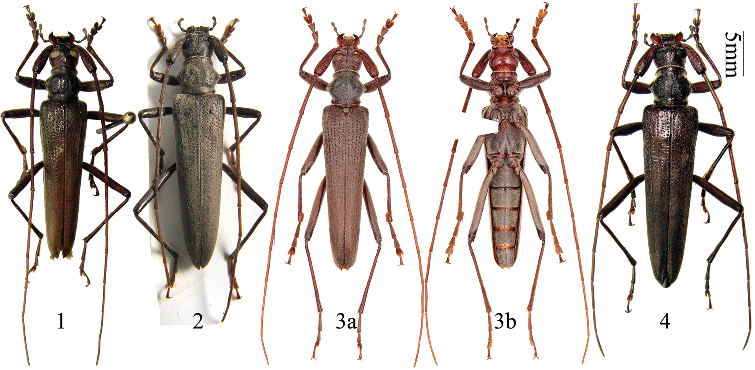
Distenia gracilis (Blessig, 1872). 1 male, from Far East Russia 2 female, from Far East Russia 3 male, from Liaoning, China a dorsal view b ventral view 4 female, from Liaoning, China. Scale 5 mm.
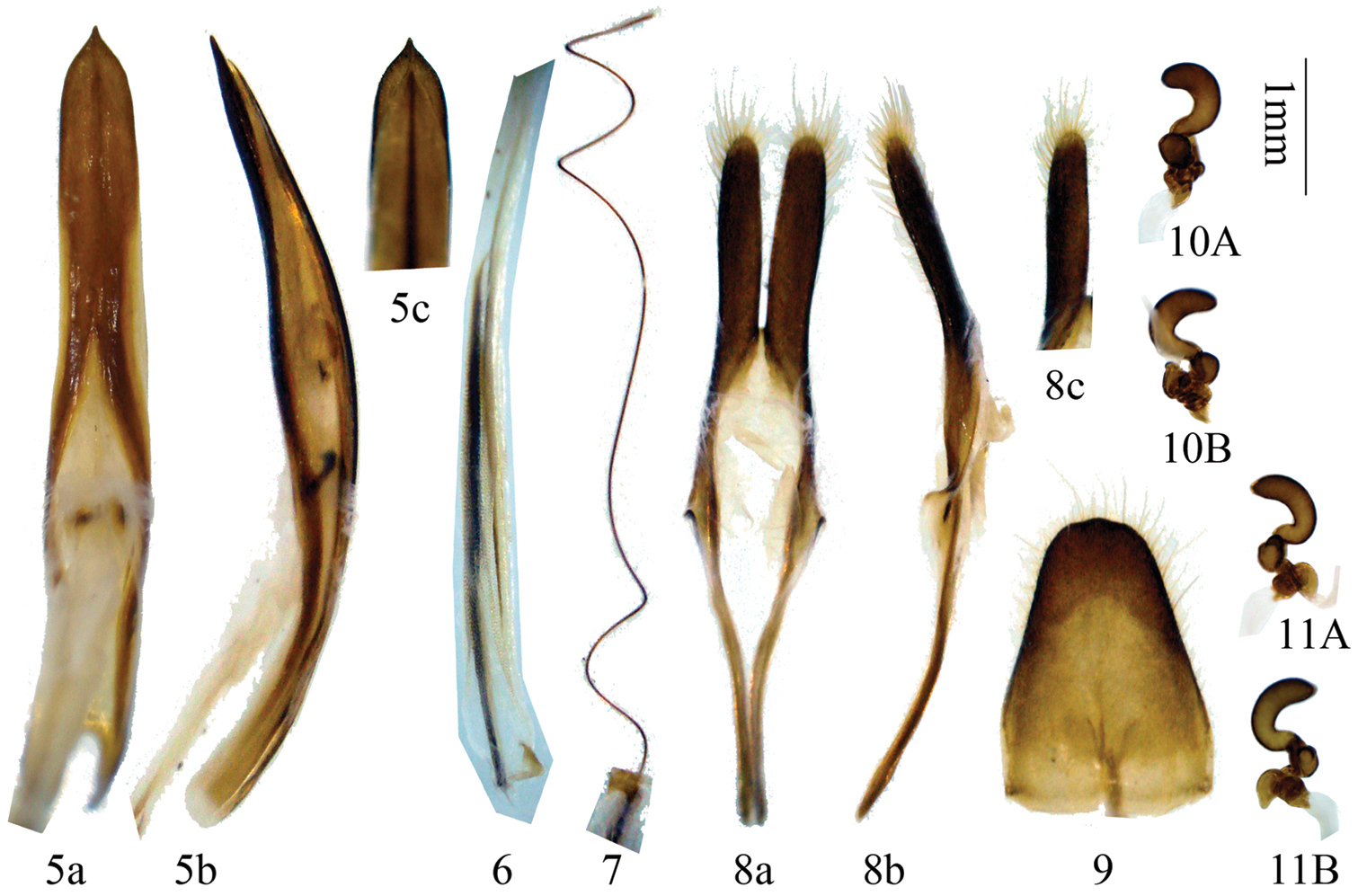
Genitalia of Distenia gracilis (Blessig, 1872). 5–9 male, from Far East Russia 5 median lobe 6 rods of endophallus 7 hair-like thin rod of ejaculatory duct 8 tegmen a ventral view b lateral view. c dorsal view 9 tergite VIII in dorsal view 10–11 female, spermathecal capsule, both from Liaoning, China. A–B from different sides. Scale 1 mm.
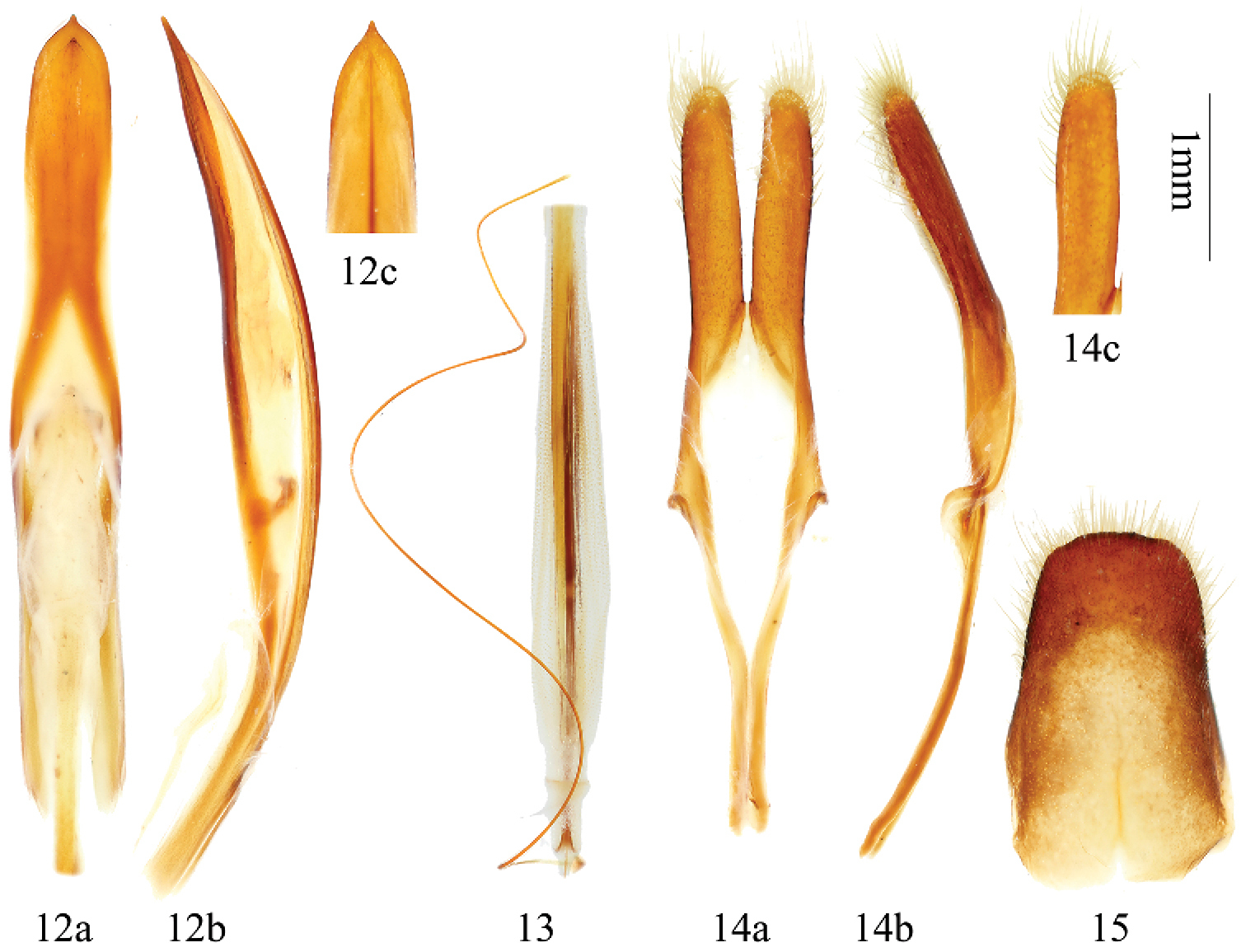
Genitalia of Distenia gracilis (Blessig, 1872), male, from Liaoning, China 12 median lobe 13 rods of endophallus, including hair-like thin rod of ejaculatory duct 14 tegmen a ventral view b lateral view c dorsal view 15 tergite VIII in dorsal view. Scale 1 mm.
http://species-id.net/wiki/Distenia_japonica
Figs 16–24, 39–40It is polyphagous with the following host plants recorded under Distenia gracilis (confused with Distenia japonica): Acer sp. (ACERACEAE), Abies sachalinensis Masters (PINACEAE), Alnus sp. (BETULACEAE), Betula sp. (BETULACEAE), Chosenia sp. (SALICACEAE), Picea sp. (PINACEAE), Pinus sp. (PINACEAE), Quercus sp. (FAGACEAE), Salix sp. (SALICACEAE), Ulmus sp. (ULMACEAE).
According to
This species was first described by
Japan, Russia (Far East, Islands).
Japan: 1 male, syntype, Japan (NHML, ex collection G. Lewis, examined through pictures); 1 male, Japan, Iwate Prefecture, Niisato-mura, Genbeidaira, 1982.VII.31, coll. N. Ohbayashi (CBWX); 1 female, Japon, Iwate Prefecture, Niisato-mura, Genbeidaira, 1982.VII.31, coll. N. Ohbayashi (CBWX); 1 male 1 female, Kyoto, Kibone, 1932.VII.1, coll. S. Yie (IZAS); 1 female, Tokushima, Mt. Tsurugi, 1971.VII.11, coll. H. Toshima (IZAS); 1 female, Tottori Pref., Mt. Hokki-Daisan, 1958.VII.22, coll. H. Toshima (IZAS).
Differences of Distenia gracilis, Distenia japonica and Distenia orientalis sp. n.
| Species / Character | Distenia gracilis | Distenia japonica | Distenia orientalis sp. n. |
|---|---|---|---|
| Antennal segment extending beyond tip of elytra | in male 8th, in female 9th | in male 8th, in female 9th | in male 7th, in female 8th |
| Color of antennae and legs | uniformly black-brown | Uniformly brown | Mostly black-brown, with several orange-red rings |
| Scape in male | With basal grooves, punctures coarser | With basal grooves, punctures finer | Without basal grooves, with rugose punctures |
| Scape length / maximum width | ca.3.0 in male, ca. 2.8 in female | ca.3.5 in male, ca. 3.0 in female | ca.3.1 in male, ca. 3.4 in female |
| Last segment of maxillary palp | Stouter, length / maximum width < 2.5 in male, < 2.6 in female (Figs 37a, 38a) | Stoutest, length / maximum width < 2.1 in male, < 2.4 in female (Figs 39a, 40a) | Slender, length / maximum width > 2.5 in male, > 3.0 in female (Figs 41a, 42a) |
| Pronotum | Without transverse rugae, swelling indistinct (Figs 37c, 38c) | Without transverse rugae, swelling more distinct (Figs 39c, 40c) | With some transverse rugae (Figs 41c, 42c) |
| Mosotibiae of male | Apical protruding lobe very distinct (Fig. 37f ) | Apical protruding lobe distinct (Fig. 39f) | Without apical protruding lobe (Fig. 41f) |
| Punctures on elytra | With distinct longitudinal rows, the row near suture not very dense (Figs 37d, 38d) | With distinct longitudinal rows, the row near suture very dense (Figs 39d, 40d) | Longitudinal rows indistinct, the row near suture very sparse (Figs 41d, 42d) |
| Sternite VII (ventrite V) | Figs 37e, 38e | Figs 39e, 40e | Figs 41e, 42e |
| Median lobe | Figs 5, 12 | Figs 19 | Figs 29 |
| Spermathecal capsule | Figs 10–11 | Figs 24 | Figs 34–36 |
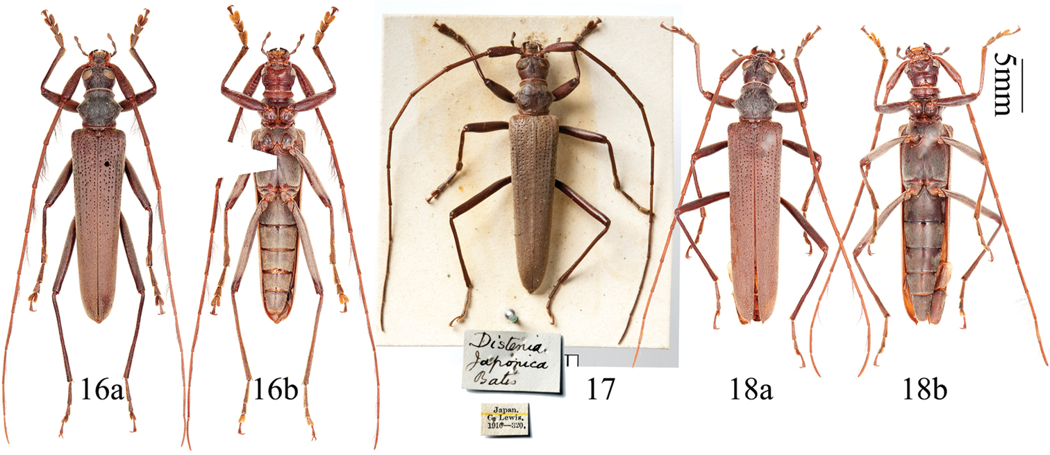
Distenia japonica Bates, 1873. 16 male, from Iwate, Japan 17 syntype, male, from Hyogo, Japan 18 female, from Iwate, Japan a dorsal view b ventral view. Scale 5 mm.
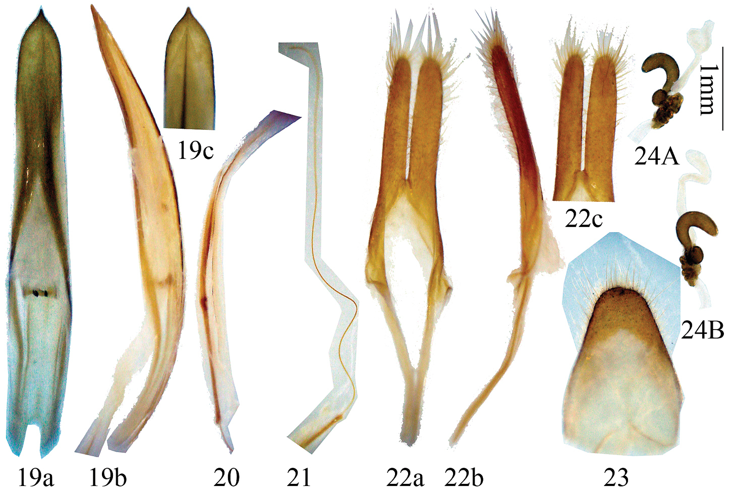
Genitalia of Distenia japonica Bates, 1873. 19–23 male, from Kyoto, Japan 19 median lobe 20 rods of endophallus 21 hair-like thin rod of ejaculatory duct 22 tegmen. a ventral view b lateral view c dorsal view 23 tergite VIII in dorsal view 24 female, spermathecal capsule, from Kyoto, Japan. A–B from different sides. Scale 1 mm.
http://species-id.net/wiki/Distenia_japonica_yakushimana
According to
This subspecies was described based on the female holotype from Japan, Ryukyu island, Mt. Miyanouradake (alt. 1200 m), collected by Hajime Yokoyama on August 3, 1962. It is deposited in Osaka Museum of Natural History. We did not examine the holotype or other specimens but followed
Japan (Yaku-shima).
urn:lsid:zoobank.org:act:14814F4C-97D8-4C2C-9125-7AA5DEDACFA6
http://species-id.net/wiki/Distenia_orientalis
Figs 25–36, 41–42Male: body length 18.7–25.5 mm, width at humeri 4.0–6.0 mm. Female: body length 22.0–26.6 mm, width at humeri 5.0–6.5 mm. Body uniformly black-brown, with rusty tinge (especially in male), except bases of tibiae (about 1/3 to 1/2), tips of antennal segments IV-XI (increasing from IV to XI), and extreme tips of last segments of maxillary and labial palps which are reddish-brown, and ventral side of tarsi and base of mandible being brown.
Body elongate, slender. Head with dense rugose punctures, with mouthparts turned forward and somewhat downward. Last segment of maxillary palp expanded and obliquely truncate apically. Frons between eyes with narrow interrupted longitudinal suture. Antennae long; scape very thick in male and more slender in female, without a groove on basal half, in male with coarse rugose punctures (Fig. 41a), in female not rugose but with finer punctures (Fig. 42a); scape not reaching midlength of pronotum in either sex; pedicel very small; subsequent segments slender; in male 7th, in female 8th segment extends beyond tip of elytra; antennal segments with recumbent long hairs beneath. The relative length of antennal segments, male: 10.6:1:12.9:13.2:13.1:12.5:11.9:11.1:9.7:8.7:8.8 (variable in narrow range); female: 9.9:1:10.2:10.3:10.3:10.1:9.5:8.5:7.4:6.5:6.3 (variable in narrow range).
Pronotum broadest in middle, with acute conical lateral spines, near posterior and anterior margins with slight transverse constriction, with rugae on disc, and with dense minute punctures and dense gray pubescence. Scutellum not longer than width at base, apically rounded, with yellowish pubescence.
Elytra narrow, taper uniformly toward apex, length 3.0–3.4 times the total width at humeri, and anterior half with deep punctures forming several indistinct longitudinal rows. Abdominal ventrite V in female (Figs 26b, 34d) elongate, gently rounded posteriorly; in male (Figs 28b, 33d) distinctly emarginate, with minute tender closely recumbent hairs. Legs long and slender, mesotibiae (of both male and female) without apical protruding lobe.
Male terminalia (Figs 29–33): Tegmen (Fig. 32) approximately 5.0 mm in length; lateral lobes slender, length about 5 times the width, ventral side and apex with short setae; median lobe plus median struts (Fig. 29) slightly curved, longer than tegmen; the median struts less than 1/8 of the whole median lobe in length; apex of ventral plate bluntly pointed; internal sac bearing a basal armature (Fig. 29b) and two median rods of endophallus (Figs 30, 31), of which the strongly sclerotized one (coming from the gonopore) connected to a very long (much longer than the median rods) hair-like rod (inside ejaculatory duct, Fig. 30). Tergite VIII (Fig. 33) longer than broad, narrowed apically from middle, with rounded apex, apical half bearing short dorsal setae.
Female terminalia (Figs 34–36): Paraproct moderate in size, its baculi thick and long, straight and not bifurcate at base; valvifer indistinct; coxite with rough surface, each baculum very thick at base and narrowed towards apex; coxite lobes sclerotized at each inner part, with tactile hairs; stylus articulated to the tip of each coxite lobe (slightly laterally), sclerotized except for apex and bearing tactile hairs; dorsal baculi sinuate and longer than paraproct baculi; proctiger baculi long and almost straight. Spermathecal capsule (Figs 34–36) large, heavily sclerotized and of very intricate structure, its apical part narrow, strongly bent at middle and basally with a protrusion (in shape of a question mark “?”), basal part irregularly twisted and with rather broad protrusion to which attaches the spermathecal gland at the middle part. Tignum much shorter than half of abdomen. In one measured specimen, tignum was 4.4 mm for an adult with 12.0 mm abdomen length in ventral view.
The differences of the three species are shown in Table 1.
The name of the new species refers to its distribution in southeast China, instead of northeast China (which is the distribution of Distenia gracilis).
This species has been misidentified as Distenia gracilis since
It is the 29th recorded species for the Chinese Disteniidae fauna (
One female from Mt. Wutaishan of Shanxi Province shows a strange dot on the distributional map. We believe that the distribution region will be extended after further survey.
China: Zhejiang Prov., Fujian Prov., Guangdong Prov., Jiangxi Prov., Shanxi Prov.
Holotype, male, Zhejiang, Xitianmushan, alt. 1200 m, 2008.VII.2, coll. Hao Huang (SNUC, ex CBWX). Paratypes: China, Zhejiang: 1 male, Xitianmushan, alt. 1300 m, 2009.IV.19 (larva), 2009.V.14 (adult), coll. Wenxuan Bi (CBWX); 1 male, Xitianmushan, alt. 1100 m, 2008.III.1 (larva), 2008.V.27 (adult), coll. Wenxuan Bi (CBWX); 1 female, Tianmushan nature reserve, alt. 1100 m, 2008.VII.30, coll. Yongxiang Wu (CJM); 1 female, China, Chekiang, Tien-mu-shan, 1937.VI.30, coll. E. Surnson (ZMMU); 1 female, Xitianmushan, alt. 1000m, 2012.VII.11, coll. Deyao Zhou (CZDY); 1 female, Tienmushan, 1937.VIII.3 (IZAS, IOZ(E)1859289); 2 males, same data (IZAS, IOZ(E)1859290-91); 2 males, same data but 1937.VIII.4 (IZAS, IOZ(E)1859292-93); 1 male, same data but 1937.VII.21 (IZAS, IOZ(E)1859288); 1 female, Longquan, Fengyangshan, Lu'ao village, alt. 1100 m, 2008.VII.31, coll. Wenxuan Bi (CBWX); Qingyuan county, Baishanzu nature reserve, alt. 1000 m, 2009.VII.25-VIII.5, coll. Zhizhou Yu (CYZZ). China, Fujian: 1 male, Chong’an, Sangang, 1979.VIII.14 (IZAS, IOZ(E)1859287); 1 male, Fujian, Wuyishan nature reserve, 2009.VII.10–15. coll. Ming Jin (CJM). China, Jiangxi: 1 female, Wuyishan nature reserve, Yejiachang station, alt. 900 m, 2004.VIII.2 (CCCC). China, Guangdong: 1 female, Ruyuan county, Nanling nature reserve, 2008-2009, coll. Lei Gao (CCCC).
China, Shanxi: 1 female, Wutaishan, alt. 2000 m, 1996.VII.17, coll. Wenzhu Li (IZAS, IOZ(E)1859062).
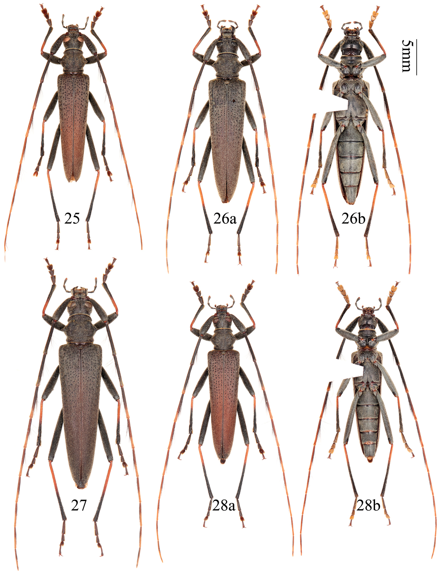
Distenia orientalis sp. n. 25 holotype, male, from Xitianmushan, Zhejiang, China 26 paratype, female, from Tianmushan, Zhejiang, China 27 paratype, female, from Fengyangshan, Zhejiang, China 28 paratype, male, from Wuyishan, Fujian, China a dorsal view b ventral view. Scale 5 mm.
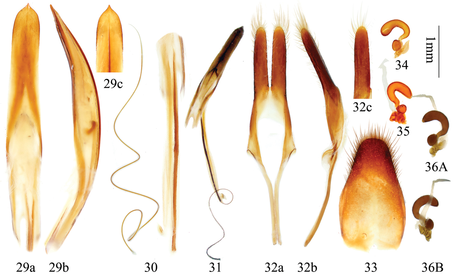
Genitalia of Distenia orientalis sp. n. 29–33 male, from Xitianmushan, Zhejiang, China 29 median lobe 30 rods of endophallus and hair-like thin rod of ejaculatory duct 31 whole median lobe, showing the position of rods of endophallus, not to scale 32 tegmen a ventral view b lateral view c dorsal view 33 tergite VIII in dorsal view 34–36 female, spermathecal capsule 35 from Fengyangshan, Zhejiang, China 34 & 36 from Tianmushan, Zhejiang, China. A & B from different sides. Scale 1 mm.
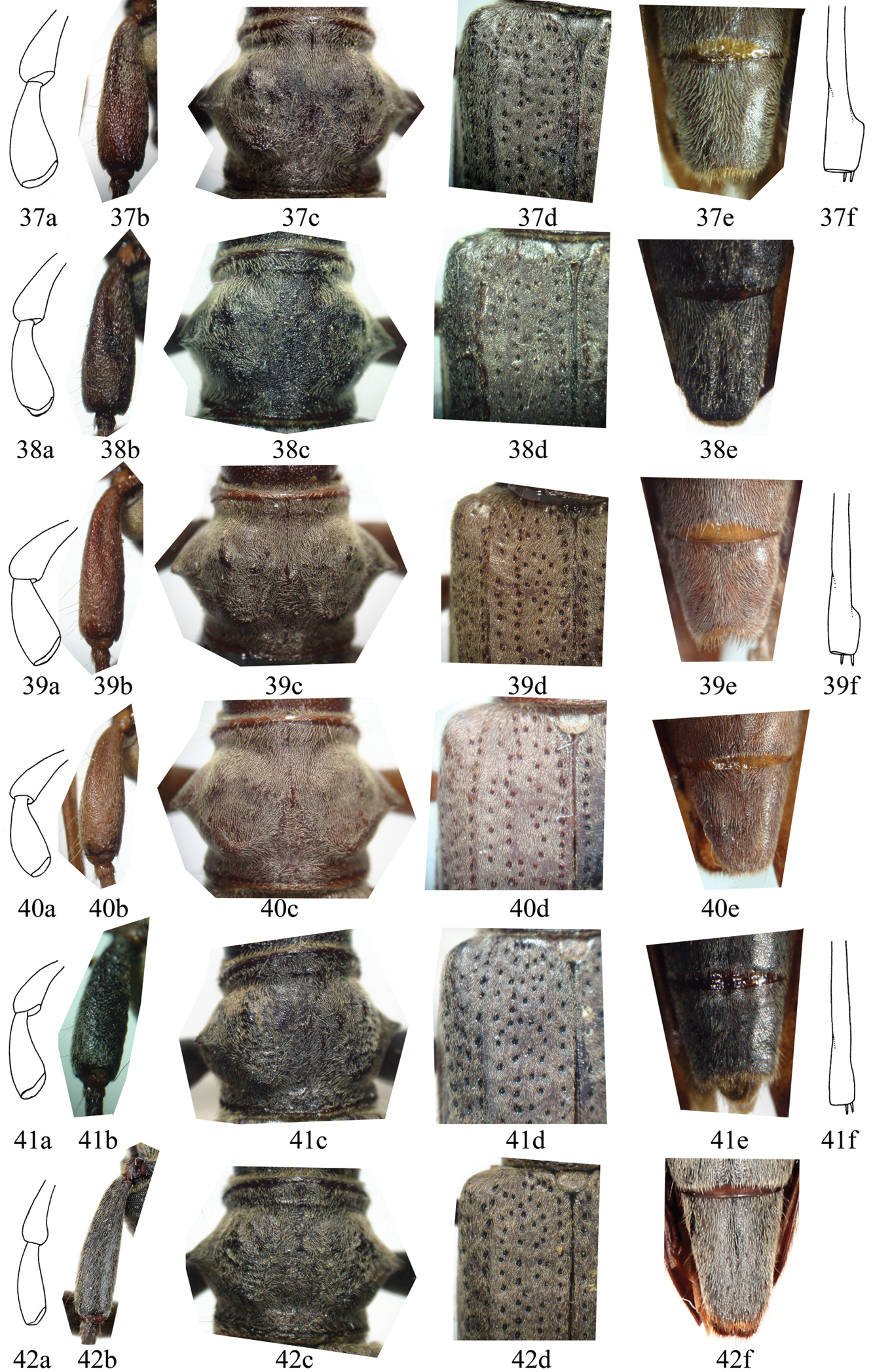
Six important characters of Distenia spp. not to scale. 37–38 Distenia gracilis. 37 male from Far East, Russia 38 female from Liaoning, China 39–40 Distenia japonica 39 male from Kyoto, Japan 40 female from Kyoto, Japan 41–42 Distenia orientalis sp. n. 41 male from Tianmushan, China 42 female from Tianmushan, China a last segment of maxillary palp, showing the tip and the ration of length to width b scape c pronotum d basal part of elytron e ventrite V f mesotibia of male, showing the apical protruding lobe.
We wish to express our sincere thanks to Yulong Lin (Taiwan, China) for the meaningful discussion at the beginning of this research, to Steven W. Lingafelter (National Museum of Natural History, Smithsonian Institution, Washington, USA), Mikhail Danilevsky (IEER) and Michiaki Hasegawa (Toyohashi Museum of Natural History, Aichi, Japan) for improving this manuscript. Our special thanks are due to Alexey Gusakov (ZMMU), Changchin Chen (CCCC), Deyao Zhou (CZDY) and Zhizhou Yu (CYZZ) for giving us the permission to use the related collections and for providing material and information. We thank Paul Hurst and Sharon L. Shute (NHML) for taking the syntype pictures of Distenia japonica Bates for us, and thank Wenzhu Li (IZAS) for making the pictures of maxillary palpi and mesotibiae. Special thanks are due to Jennifer Hammock and Steven W. Lingafelter (Smithsonian Institution, National Museum of Natural History, Washington, USA) for their kindly improving the English. This research was supported by a grant (No. O529YX5105) from the Key Laboratory of the Zoological Systematics and Evolution of the Chinese Academy of Sciences, and by NSFC program J1210002 and 31000967.