

(C) 2013 Si-han Lu. This is an open access article distributed under the terms of the Creative Commons Attribution License (CC BY 4.0), which permits unrestricted use, distribution, and reproduction in any medium, provided the original author and source are credited.
For reference, use of the paginated PDF or printed version of this article is recommended.
Citation: Si-han Lu S-h , Zhang L, Qiao L, Qin D-z (2013) Lumicella, a new genus of the tribe Empoascini (Hemiptera, Cicadellidae, Typhlocybinae) from China. ZooKeys 364: 11–17. doi: 10.3897/zookeys.364.6618
Lumicella rotundata gen. et sp. n. is described based on specimens from Fujian Province, China. Habitus photos and illustrations of male genitalia of this new species are provided. Differences between the new genus and closely related genera are discussed.
Homoptera, Auchenorrhyncha, leafhoppers, taxonomy, distribution
The fauna of Empoascini in China is very rich and diverse, this is associated with China’s high biodiversity. To date, 31 genera of this tribe have been described in Chinese fauna (
The specimens examined in this study are deposited in the Entomological Museum, Northwest A&F University, Yangling, Shaanxi, China (NWAFU). The entire male abdomen of the examined specimens were removed and cleared in 10% NaOH and drawn from preparations preserved in glycerin. External morphology was observed under an Olympus SZX-10 microscope. Photographs of the specimens were made using a Nikon SMZ 1500 microscope with a Retiga 2000R camera (CCD). Images were produced using the software Auto-Montage Pro. The male genitalia were drawn using a Olympus PM-10AD, and wings were drawn with a Leica MZ-12.5 microscope. All the pictures were edited and enhanced using Adobe Photoshop CS7.0 (Adobe Systems). The body measurements are from apex of the vertex to the tip of the forewing.
Morphological terminology predominantly follows
Lumicella rotundata Lu & Qin, sp. n., here designated.
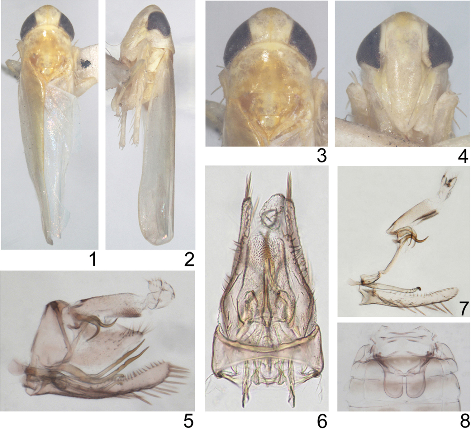
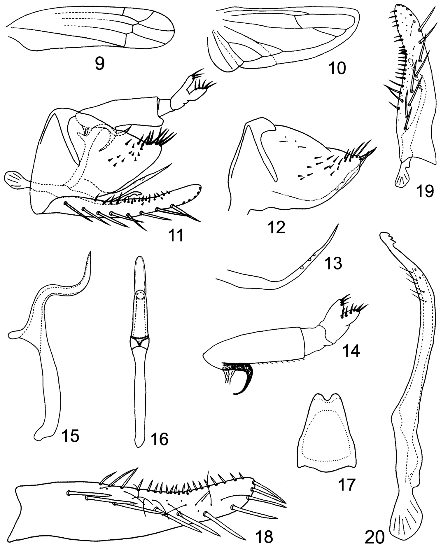
Body small. Head with eyes broader than maximum width of pronotum (Figs 1, 3). Vertex short, rounded anteriorly (Figs 1, 3), profile of transition to face rounded (Fig. 2), coronal suture long (Figs 1, 3). Face narrow and slightly convex in profile, lateral frontal suture present (Figs 2, 4). Forewing narrow, rounded apically, apical cells occupying less than one-third total length, all apical cell with separate bases, 2nd apical cell with margins subparallel but slightly broadened at apex, c and r cells nearly equal in width, narrower than m and cua cells; veins RP, MP’ arise from r cell and MP”+CuA’ from m cell (Fig. 9). Hindwing with bifurcation point of CuA basad of coalescence of CuA with MP” (Fig. 10).
Lumicella rotundata sp. n. 1 male adult (abdomen removed), dorsal view 2 same, left lateral view 3 head and thorax, dorsal view 4 face 5 male genitalia, left lateral view 6 same, dorsal view 7 anal tube and anal styli, aedeagus, connective, paramere and subgenital plate, left lateral view 8 abdominal apodemes.
Lumicella rotundata sp. n. 9 forewing 10 hindwing 11 male genitalia, left lateral view 12, pygofer side and ventral pygofer appendage, left lateral view 13 ventral pygofer appendage, left lateral view 14 anal tube and anal styli, left lateral view 15 aedeagus, left lateral view 16 same, dorsal view 17 connective 18 subgenital plate 19 subgenital plate and paramere, dorsal view 20 paramere.
Male basal abdominal sternal apodemes developed, apically rounded and parallel sided (Fig. 8). Male pygofer elongate, strongly narrowing caudad, terminally with rigid microsetae on each side of lobe, ventral appendage present (Figs 5, 6, 11–13), dorsal bridge short, less sclerotized in middle dorsocaudad (Fig. 6). Subgenital plate much exceeding pygofer side, A-group setae distinct, C-group setae arranged in a single row and reaching apex of plate (Figs 5, 11, 18, 19). Paramere slim, apophysis bearing prominent dentifer and a few slender setae (Figs 5, 7, 11, 19, 20). Connective lamellate (Fig. 17). Aedeagus without dorsal apodeme, preatrium well developed, shaft tubular and curved twice, gonopore apical on ventral side (Figs 15, 16). Anal tube process curved and narrowed terminally (Figs 5, 7, 11, 14).
The generic name is an arbitrary combination of letters, and is regarded as feminine.
In Alebroides Matsumura group, the new genus is similar to Ghauriana Thapa, Membranacea Qin & Zhang, Dattasca Dworakowska, Luvila Dworakowska, Szara Dworakowska, Szuletaia Dworakowska, Luodianasca Qin & Zhang, Nikkotettix Matsumura and Znana Dworakowska in having veins RP, MP’ of forewing arise from r cell and MP”+CuA’ from m cell, all apical cells in fore wing having separate bases (in Nikkotettix and Znana, 3rd apical cell stalked or sessile) and CuA in the hindwing branched apically. However, this new genus differs from Membranacea, Luodianasca, Luvila and Szara in the presence of the ventral pygofer appendage (ventral pygofer appendage absent in these four genera), from Dattasca and Szuletaia in having bifurcation point of CuA basad of coalescence of CuA with MP” (apicad of coalescence of CuA with MP” in Dattasca and Szuletaia), from Znana in having coronal suture not reaching apex of vertex (surpassing apex of vertex and reaching the level of ocelli on face in Znana); from Ghauriana in the subgenital plate having A-group setae (A-group setae undifferentiated in Ghauriana), from Nikkotettix in the absence of ventral process at the base of aedeagal shaft (with ventral process at the base of aedeagal shaft in Nikkotettix). The new genus also differs from Membranacea in the presence of anal tube appendage (anal tube appendage absent in Membranacea) and from Luvila in having the C-group setae of subgenital plate arranged in a single row subbasally (C-group setae arranged in two rows subbasally in Luvila).
China (Fujian).
http://zoobank.org/771BE1DE-E369-4879-A4E0-08276BF30F46
http://species-id.net/wiki/Lumicella_rotundata
Figs 1–20Body length: Male 3.7–3.9mm.
General colour variable: lighter coloured specimens yellow to ochre-yellow. Vertex with borders at eyes creamy-yellowish, semilunar patch mesocaudad of ocelli creamy. Face and basal antennal segments light yellow. Eyes blackish-brown. Disc of pronotum golden-yellow, irregular arch of hypodermal pattern light-yellow in addition to three large creamy patches along anterior margin. Centre of scutellum sordid cream, scutoscutellar sulcus beige. Darker specimens brown to sordid brown, semilunar patch mesocaudad of ocelli, borders at eyes, genae, patches on pronotum and centrally on scutellum, sordid cream.
Male genitalia: Basal sternal abdominal apodemes exceeding half of segment 4 (Fig. 8). Male pygofer with about 16 rigid setae on outer and inner surface of lobe, ventral pygofer appendage slim and bent caudodorsad near base, surpassing caudal margin of lobe, tapering and sculptured with depressions subapically (Figs 5, 11–13). Subgenital plate with nearly same width in basal third, apical 2/3 gradually narrowing towards apex, A-group setae (3–4) rigid, B-group seate (15-17) small, roughly uniseriate along dorsal margin in apical half, C-group setae (13–14) arising near base of plate, sharply terminated, D-group setae roughly bi- or tri-seriate, starting caudad of C-group setae (Figs 5, 11, 18, 19). Paramere sinuate in caudal part, apically bearing 3 big teeth preceded by ca. 6 fine setae and few sensory pits (Figs 5, 11, 19, 20). Connective narrowing to deeply emarginate apex (Fig. 17). Aedeagal shaft tubular, longer than preatrium, in profile its middle part right-angled and curved caudoventrad followed by vertical apical region, gonopore large on ventral side, in ventral view aedeagus with rounded apex (Figs 5, 11, 15, 16). Anal tube process well sclerotized, originating subapically from ventral margin of anal tube, nearly reaching 1/3 height of pygofer (Figs 5, 11, 14).
Holotype. ♂ (NWAFU), China, Fujian Province, Wuyi Mountain, 17 Aug 2008, coll. X. Gao and X. T. Li. Paratypes. 4♂♂(NWAFU), same data as holotype; 1♂(NWAFU), China, Fujian Province, Wuyi Mountain, 17 Sept 1980, coll. T. Chen; 10♂♂ (NWAFU), China, Fujian Province, Wuyi Mountain, 17 Aug 1984, coll. Z. X. Cui.
The name is derived from the Latin word “rotundus” (round), which refers to the rounded apex of the aedeagal shaft.
Known only from the type locality in Fujian Province in southeastern China.
Unknown.
We are grateful to Prof. John Richard Schrock (Emporia State University, Emporia, USA) for reviewing the manuscript. This work was supported by the National Natural Science Foundation of China (No. 31270689).

