


(C) 2012 Qingyuan Zhao. This is an open access article distributed under the terms of the Creative Commons Attribution License 3.0 (CC-BY), which permits unrestricted use, distribution, and reproduction in any medium, provided the original author and source are credited.
For reference, use of the paginated PDF or printed version of this article is recommended.
Two new genera of the spider family Theridiosomatidae, Alaria gen.n. with the type species Alaria chengguanensis sp. n., Menglunia gen.n. with the type species Menglunia inaffecta sp. n., are described from Guizhou and Yunnan, China. Nine more new species from Guangxi, Guizhou, Hainan and Yunnan Provinces of southern China are described: Baalzebub rastrarius sp. n., Baalzebub youyiensis sp. n., Karstia nitidasp. n., Karstia prolata sp. n., Ogulnius hapalus sp. n., Theridiosoma plumaria sp. n., Theridiosoma triumphalis sp. n., Theridiosoma vimineum sp. n., Zoma fascia sp. n. The type specimens are deposited in the Institute of Zoology, Chinese Academy of Sciences in Beijing.
Biodiversity, taxonomy, canopy, cave, leaf litter
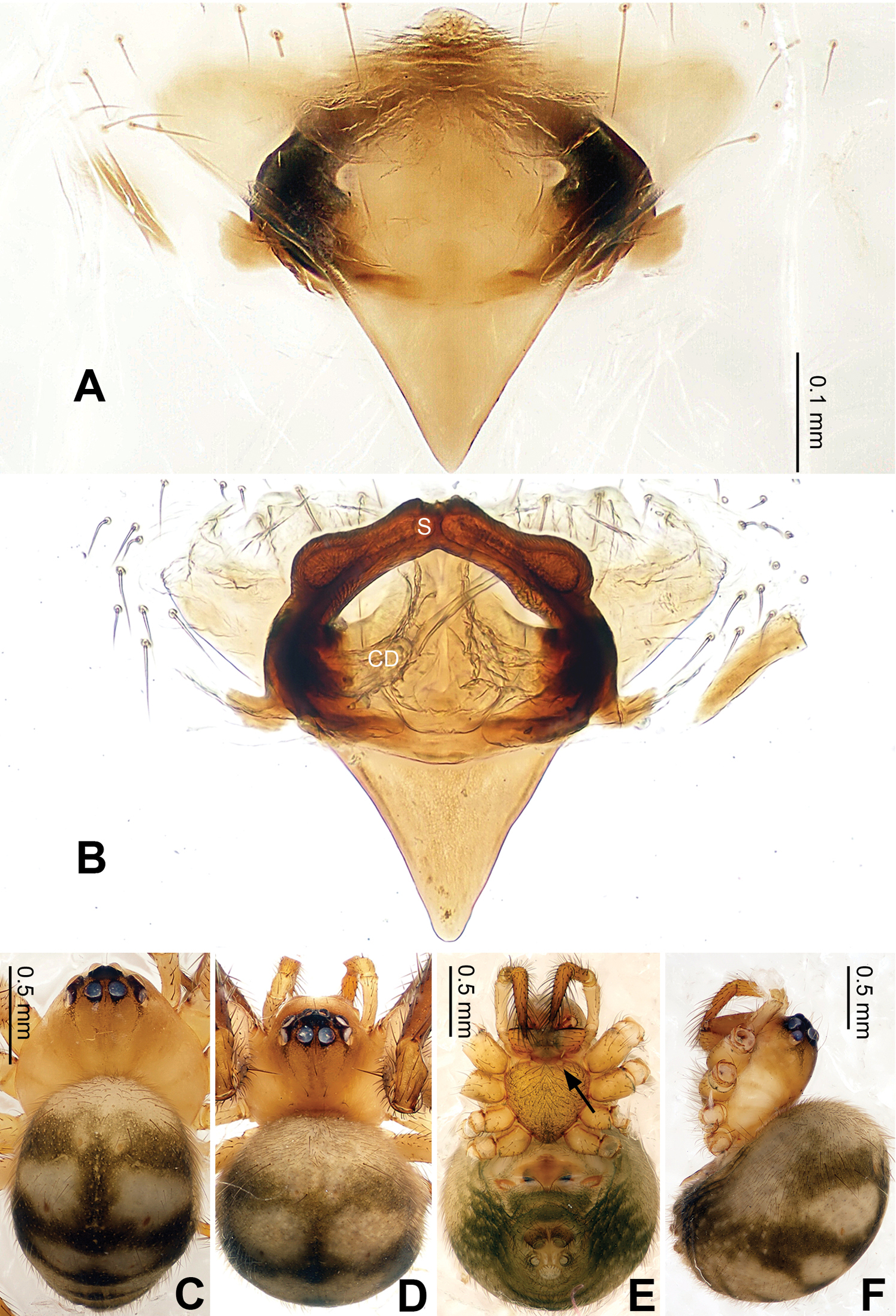
Theridiosomatids are small (usually ≤ 3mm), widely distributed, and cryptozoic spiders, which can be found in damp, dark habitats (litter layer of forest or caves). They can be easily recognized by the presence of a pair of pits on the anterior margin of the sternum near the labial base (Fig. 9D), the disproportionately large globular pedipalps (except for genus Menglunia), and the long dorsal trichobothria on the third and fourth tibia (
The genera of family Theridiosomatidae were reviewed and revised by Coddington in
Theridiosomatids in China are mainly found in the southern provinces: Guangxi, Guizhou, Hainan, Taiwan and Yunnan, which is consistent with the tropical and subtropical preferences typical for this family. Some theridiosomatids are associated with caves, which are characterized by stable humidity and temperature. In this paper, we provide descriptions and distribution data for eleven new species collected in Guangxi, Guizhou, Hainan and Yunnan.
Specimens were examined using a LEICA M205 C stereomicroscope. Further details were studied under an Olympus BX51 compound microscope. All illustrations were made using a camera lucida attached to an Olympus BX51 compound microscope, and then inked on ink jet plotter paper. Male and female genitalia were examined and illustrated after being dissected from the spiders’ bodies. Left pedipalps of male spiders were illustrated, except as otherwise indicated. Vulvae of female were removed and cleared in lactic acid or warm 10% potassium hydroxide (KOH) solution before illustration. All embolic divisions and vulvae were illustrated after being embedded in Arabic gum. Type specimens examined were preserved in 75% ethanol solution. Photos were taken with an Olympus c7070 wide zoom digital camera (7.1 megapixels) mounted on an Olympus SZX12 stereomicroscope. Images from multiple focal planes were combined using Helicon Focus (version 3.10.3) image stacking software. All measurements are given in millimeters. Leg measurements are shown as: total length (femur, patella, tibia, metatarsus, tarsus).
SEM images were taken using the HITACHI S-3000N at the Institute of Genetics and Developmental Biology, Chinese Academy of Sciences. Specimens for SEM examination were critical point dried and sputter coated with gold-palladium. Specimens were mounted on copper pedestal using double-sided adhesive tape.
All type specimens are deposited in the Institute of Zoology, Chinese Academy of Sciences in Beijing.
Chaetotaxy. Macrosetae are marked for the dorsal (d), prolateral (p), retrolateral (r), and ventral (v) surfaces of the legs. Metatarsal trichobothrium (Tm) is given as the ratio of the distance between the proximal margin of the metatarsus and the root of the trichobothrium divided by the total length of the metatarsus (
Abbreviations and conventions. Abbreviations used in the text are given in Table 1. References to figures in cited papers are listed in lowercase type (fig.); Figures of this paper are noted with an initial capital (Fig.).
When extra materials are examined and recorded, and the paratype’s collecting information is the same as holotype’s, it will be implied in brackets as [same data as holotype].
List of abbreviations used in the text and figures.
| Male pedipalp | |
| Co | conductor |
| E | embolus |
| EA | embolic apophysis |
| MA | median apophysis |
| PC | paracymbium |
| T | tegulum |
| Vulva | |
| CD | Copulatory duct |
| S | spermatheca |
| Spinnerets spigot morphology | |
| AC | aciniform gland spigot |
| AG | aggregate gland spigot |
| ALS | anterior lateral spinneret |
| CY | cylindrical gland spigot |
| FL | flagelliform gland spigot |
| MAP | major ampullate gland spigot |
| mAP | minor ampullate gland spigot |
| PI | piriform gland spigot |
| PLS | posterior lateral spinneret |
| PMS | posterior median spinneret |
| t | tartipore |
| Institution | |
| IZCAS | Institute of Zoology, Chinese Academy of Sciences |
(Species known from one sex is marked as ‘m’ or ‘f’ to represent their holotype’s sex ‘male’ or ‘female’; if unmarked, it means they are known from both sexes)
| 1 | Posterior median eyes separated by about their diameter or more (Figs 20C–D) | 2 |
| Posterior median eyes separated by less than 1/2 their diameter or less (Figs 7C–D) | 3 | |
| 2 | The turning made by the copulatory ducts bends outwardly (Fig. 20B). Median apophysis mesally with a projection oriented distoventrally (Fig. 19A). Embolic apophysis long, whip-like (Fig. 19C) | Ogulnius hapalus sp. n. |
| – | The turning made by the copulatory ducts bends inwardly. Median apophysis with apex oriented distodorsally. Embolic apophysis filiform ( |
Ogulnius barbandrewsi |
| 3 | Females | 4 |
| – | Males | 21 |
| 4 | Scape present (Fig. 2B) | 5 |
| – | Scape absent (Fig. 16A) | 14 |
| 5 | Scape protruding from beneath the epigynal plate (Fig. 4A) | 6 |
| – | Scape protruding from epigyne’s posterior rim (Fig. 7A) | 9 |
| 6 | Scape less sclerotized, smaller and partially exposed ( |
7 |
| – | Scape more sclerotized, and utterly exposed. Spermathecae juxtaposed. Copulatory ducts rise and curl up to form a blunt-tipped projection at each side (Figs 2A, 5D) | Alaria chengguanensis sp. n. |
| 7 | Spermathecae juxtaposed, unseparated | 8 |
| – | Spermathecae separated ( |
(f) Wendilgarda assmensis |
| 8 | Epigyne with a deeper median invagination of the posterior margin |
Wendilgarda muji |
| – | Epigyne with a blunt tip and concave posterior margin ( |
Wendilgarda sinensis |
| 9 | Spermathecae distally fused (Fig. 7B) | 10 |
| – | Spermathecae juxtaposed or overlapped (Fig. 11B) | 11 |
| 10 | Scape with a central pit on the epigynal plate. Spermathecae long, narrow, form an arcade-shaped conformation (Figs 7A–B) | Baalzebub rastrarius sp. n. |
| – | Scape with a semi-transparent distal tip. Spermathecae small, elliptical (Figs 9A–B) | (f) Baalzebub youyiensis sp. n. |
| – | Epigyne subtriangular, pointed posteriorly with concave margins so medial part is more acute than lateral part ( |
(f) Baalzebub nemesis |
| 11 | Spermathecae juxtaposed (Fig. 11B) | 12 |
| – | Spermathecae overlapped at the tip ( |
13 |
| 12 | Epigyne small, with a short, distally spherical scape protruding from posterior margin of epigynal plate (Figs 17A, D, E) | (f) Karstia nitidasp. n. |
| – | Epigyne with a long, apiculate scape protruding perpendicularly (slightly tilted) from posterior margin of epigynal plate (Figs 5A, 18E–F) | (f) Karstia prolata sp. n. |
| 13 | Epigyne with an apiculate, approximately triangular scape. Spermathecae potato-like, bulging and stout ( |
Karstia coddingtoni |
| – | Epigyne with a triangular scape protruding from its posterior rim. Spermathecae peanut-shape stout and simple ( |
Karstia upperyangtz |
| 14 | Spermathecae separated by less their diameter or juxtaposed (Fig. 16B) | 15 |
| – | Spermathecae separated by nearly their diameter ( |
(f) Coddingtonia euryopoides |
| 15 | Spermathecae spherical, separated from each other (Fig. 16B) | Menglunia inaffecta sp. n. |
| Spermathecae juxtaposed (Fig. 2A) | 16 | |
| 16 | Abdomen with silver patches forming curved transverse strip (Figs 29C–D) | 17 |
| Abdomen without silver patches forming curved transverse strip | 18 | |
| 17 | The upper rim of the spermathecae is the same height as the copulatory ducts ( |
Zoma didaiyin |
| – | The spermathecae is above the loop made by the copulatory ducts (Fig. 29B) | Zoma fascia sp. n. |
| 18 | Epigyne with a deep atrium, height of opening about one third the width in posterior view ( |
Epeirotypus dalong |
| – | Atrium absent ( |
19 |
| 19 | Epigyne with pair of processes arising from posterolateral margin running toward each other ( |
(f) Theridiosoma shuangbi |
| – | Epigyne without pair of processes arising from posterolateral margin | 20 |
| 20 | Posterior margin of epigyne with median longitudinal slit ( |
Theridiosoma diwang |
| – | Posterior margin of epigyne without median longitudinal slit, but with a median transverse ridge ( |
Theridiosoma taiwanica |
| 21 | Embolic apophysis absent (Fig. 1C) | 22 |
| – | Embolic apophysis present (Fig. 6C) | 24 |
| 22 | Tegulum without tuberculate-textured mesal lobe | 23 |
| – | Tegulum with tuberculate-textured mesal lobe. Median apophysis lightly sclerotized, with fine distoventral projection. Conductor a complex of sclerotized and membranous structure enveloping thick embolus for most of its length ( |
Epeirotypus dalong |
| 23 | Embolus long, whip-like, mostly enveloped in conductor (Figs 1B–D). Median apophysis disproportionately large, orienting and stretching along the longitudinal axis of pedipalp (Figs 1A, 3A) | Alaria chengguanensis sp. n. |
| – | Embolus short, stout (Figs 15B, 17B). Tegulum suboval shaped (Fig. 15D). Median apophysis with a short projection oriented distoventrally (Fig. 15A) | Menglunia inaffecta sp. n. |
| 24 | Embolic division simple with one filiform embolic apophysis (Fig. 28C) | 25 |
| – | Embolic division complex with more than one embolic apophysis (Fig. 24C) | 26 |
| 25 | Exposed embolic apophysis short with a triangular tip, embolus ‘Z’-shaped (Figs 28A–D) | Zoma fascia sp. n. |
| – | Exposed embolic apophysis long; embolic division with a moderate branching ( |
Zoma dibaiyin |
| 26 | The mesal bristle of the embolic apophysis protruding from beneath the conductor and lying long the mesal side of the conductor itself ( |
27 |
| – | The palp without a mesal bristle of apophysis protruding from beneath the conductor and lying long the mesal side of the conductoritself | 28 |
| 27 | Conductor with a spear-shaped apophysis, above median apophysis ( |
Wendilgarda sinensis |
| – | Conductor without a spear-shaped apophysis. Palpa tibia with one trichobothrium. Median apophysis sclerotized with concave dorsal margin ( |
Wendilgarda muji |
| 28 | Embolic apophysis with blunt, spatulate processes, without a mesal bristle ( |
29 |
| – | Embolic apophysis fragments, filiform, with tips protruding out of conductor (Fig. 24B) | 31 |
| 29 | Median apophysis elongated and has a trough of groove along its upper surface. Cymbium with apophysis ( |
30 |
| – | Median apophysis small, triangular and cleft (Figs 6A, D). Embolic apophysis spatulate arching structure with abruptly acuminated distal ends (Fig. 6C) | Baalzebub rastrarius sp. n. |
| 30 | Cymbium apophysis small. Paracymbium with a long spine on the distal end (Chen 2009: fig. 13) | Karstia upperyangze |
| – | Cymbium apophysis big, distally crooked. Paracymbium without a long spine (Chen 2009: fig. 27) | Karstia coddingtoni |
| 31 | Conductor with plumose branching (Fig. 22C). Median apophysis with a curved lobe attenuates distally (Fig. 23A) | (m) Theridiosoma plumaria sp. n. |
| – | Conductor without plumose branching | 32 |
| 32 | Two embolic apophysis fragments form a 'V'-shaped conformation (Figs 24A–B) | (m) Theridiosoma triumphalis sp. n. |
| – | One embolic apophysis fragment forms a beak-shaped conformation (Figs 26A–B). Conductor with a piece of long, pliant apophysis protruding from its ridge and stretching towards median apophysis (Figs 26B, D) | (m) Theridiosoma vimineum sp. n. |
Alaria chengguanensis sp. n.
The generic epithet is derived from the Latin ‘alarius’, meaning ‘of wings’, which refers to the two projections of the copulatory ducts at each side, which resembles a pair of wings. Gender is feminine.
The unique structure of epigyne distinguishes Alaria from other theridiosomatids. Like in Wendilgarda and Chthonopes, the scape in female Alaria protrudes from beneath epigynal plate (
Alaria chengguanensis sp. n.
urn:lsid:zoobank.org:act:5EEE5079-3F6D-48CC-84D1-1A26BAD71F78
http://species-id.net/wiki/Alaria_chengguanensis
Figs 1–5Holotype: CHINA, Guizhou: Bijie City, Chengguan Town, Xiaohe Village, Xiniu Cave, 27°21.231'N, 105°17.186'E, elevation ca 1515 m, 30 April 2007, J. Liu & Y.C. Lin (IZCAS), 1 male.
Paratypes: [same data as holotype] (IZCAS), 13 males, 8 females.
This specific name chéng guān ( ) refers to its type locality; adjective.
See diagnosis for genus.
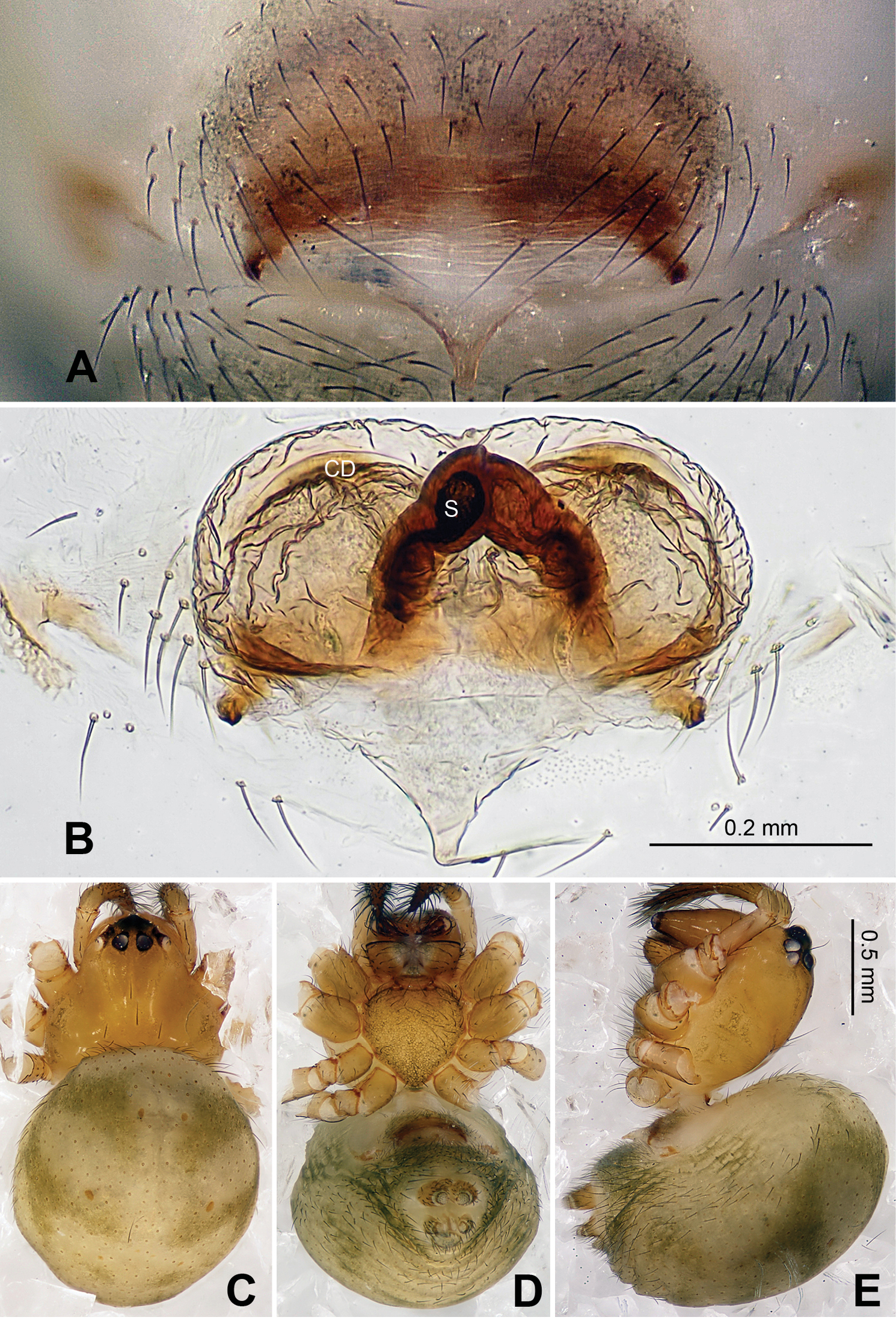
Carapace brownish red, with symmetric dark veins. Sternum yellow with dark brown margins. Legs yellow, dark brown distally at joints, especially tibiae. Abdomen tan with dark grey and silver patches (Figs 2A–B).
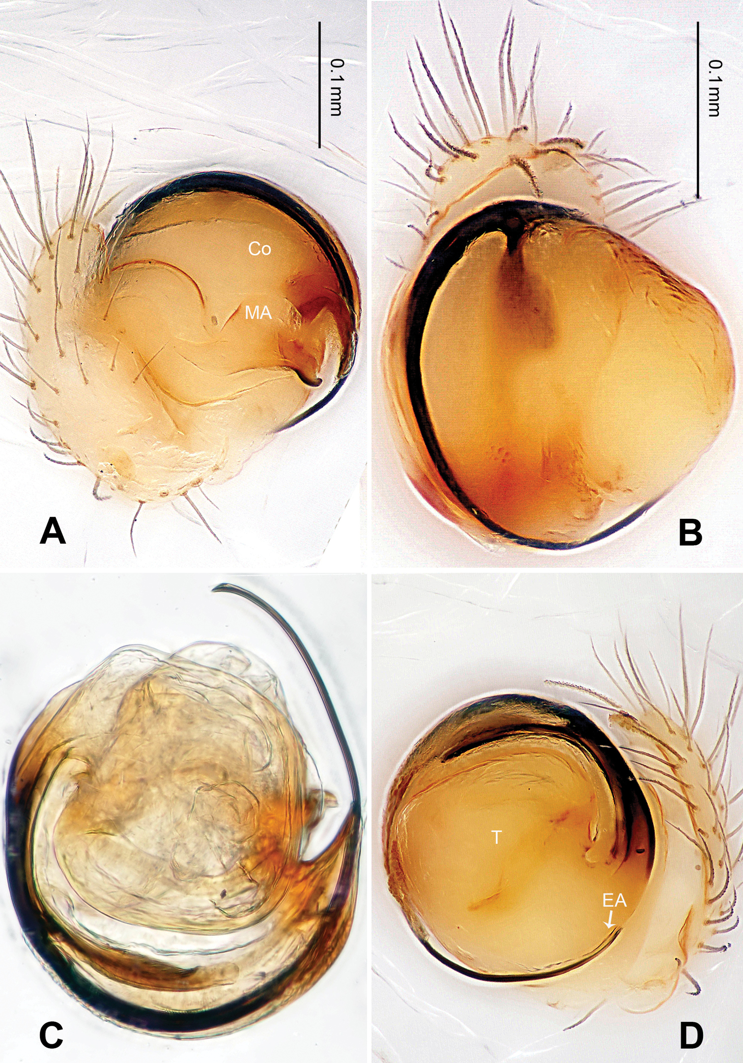
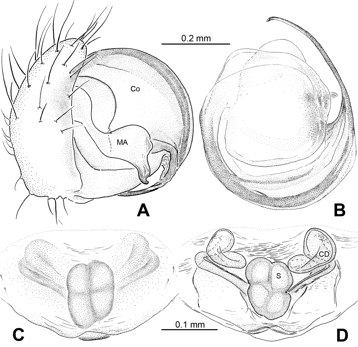
Male pedipalp: Patella with strong sinuous macroseta. Tibia with two trichobothria. Cymbium with small cluster of long setae proximally (Figs 1B, 3D). Paracymbium elongated, curved near base (Figs 1B, 3D). Tegulum smooth. Median apophysis wide, longitudinally grooved, strongly sclerotized, with curved and pointed distal region. Conductor a thick, looped, blade-shaped structure enveloping embolus for most of its length (Figs 1B–D). Embolus long, slim.
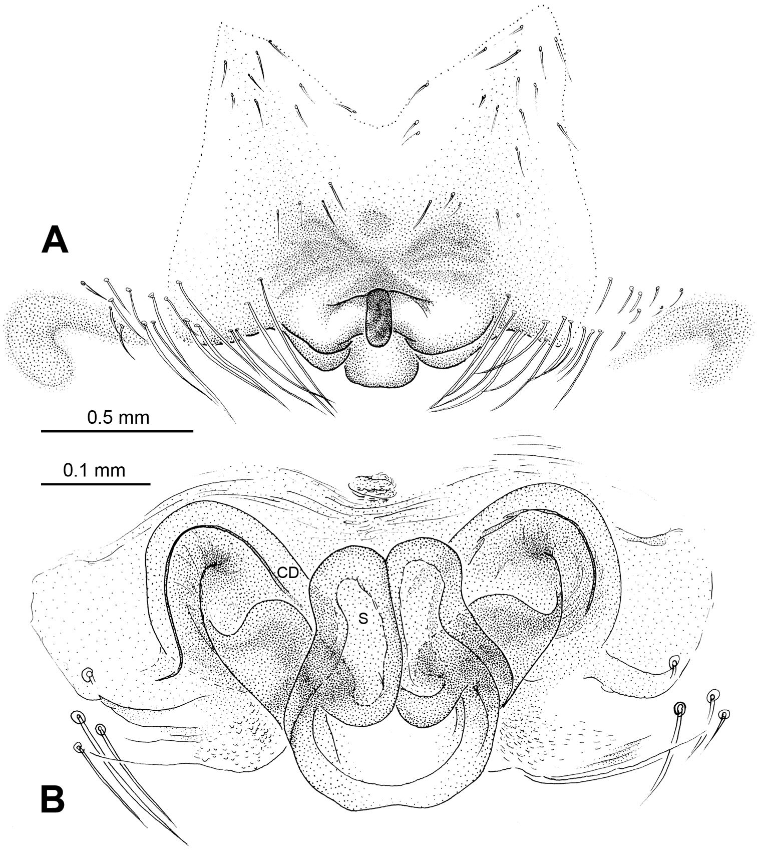
Vulva: Epigyne with long, tongue-shaped scape protruding from beneath its concaved margin. Scape slightly humped with a small transverse opening at its distal end. Spermathecae juxtaposed. Copulatory ducts rise and curl up to form a blunt-tipped projection at each side (Figs 2A, 5D).
Male (holotype): Total length 2.10, carapace 1.24 long, 1.08 wide, clypeus 0.13, sternum 0.6 long, 0.6 wide, coxae IV separated by two thirds their width. Posterior median eyes separated by half their diameter. Macrosetae: Leg I: femur d 1, patella d 1, tibia d 1, p 1, v 2, r 1, metatarsus p 1, v 1; Leg II: femur d 2, patella d 2, tibia d 1, p 1, v 1, r 1, metatarsus p 1, v 1; Leg III: femur d 3, patella d 1, tibia p 1, v 2, metatarsus d 1, p 1, r 1; Leg IV: femur d 1, tibia p 1, v 1, r 1, metatarsus p 1, v 1, r 1. Metatarsal trichobothria: Tm I: 0.27; Tm II: 0.30; Tm III: 0.84; Tm IV: 0.24. Leg measurements: I 3.98 (1.25, 0.50, 0.85, 0.88, 0.50); II 3.09 (1.00, 0.43, 0.63, 0.63, 0.40); III 2.27 (0.68, 0.35, 0.38, 0.48, 0.38); IV 2.95 (0.93, 0.38, 0.63, 0.63, 0.38).
Female (one of paratypes): Total length 2.50, carapace 1.25 long, 1.25 wide, clypeus 0.08, sternum 0.78 long, 0.63 wide, coxae IV separated by 1 time their width. Posterior median eyes separated by half their diameter. Macrosetae as in male. Metatarsal trichobothria: Tm I: 0.21 ; Tm II: 0.25; Tm III: 0.48; Tm IV: 0.27. Leg measurements: I 4.66 (1.05, 0.63, 0.95, 0.95, 0.63); II 3.75 (1.20, 0.55, 0.75, 0.75, 0.50); III 2.48(0.75, 0.33, 0.50, 0.50, 0.40); IV 3.75 (1.25, 0.50, 0.75, 0.75, 0.50).
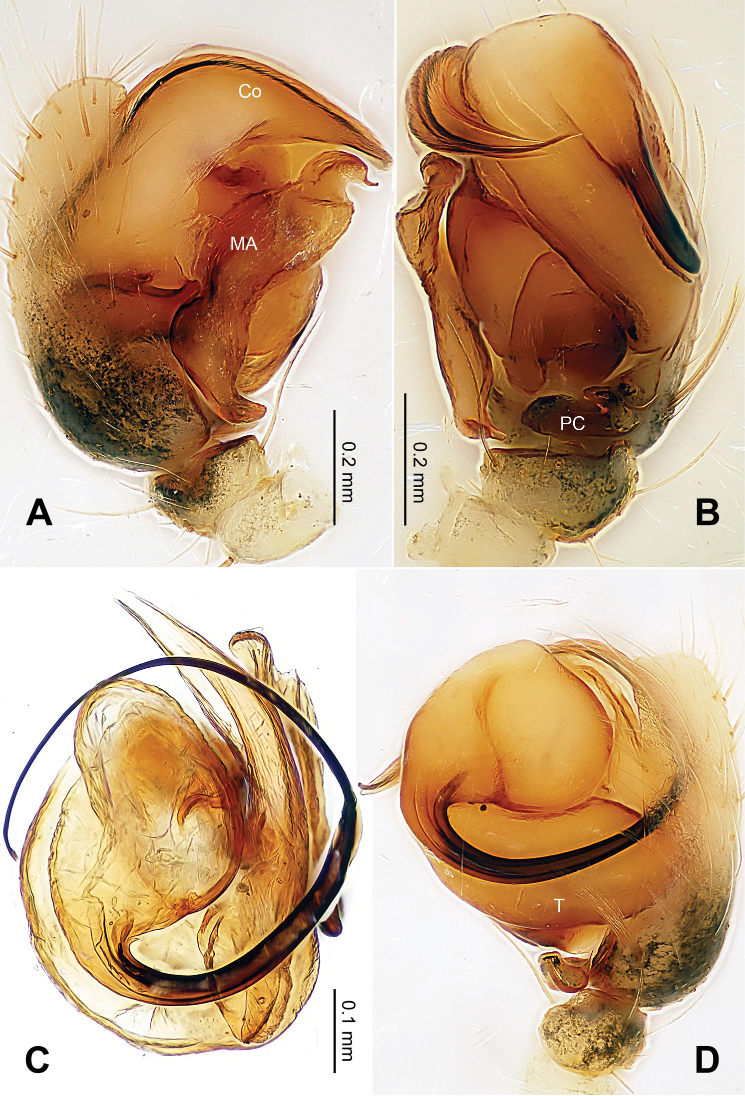
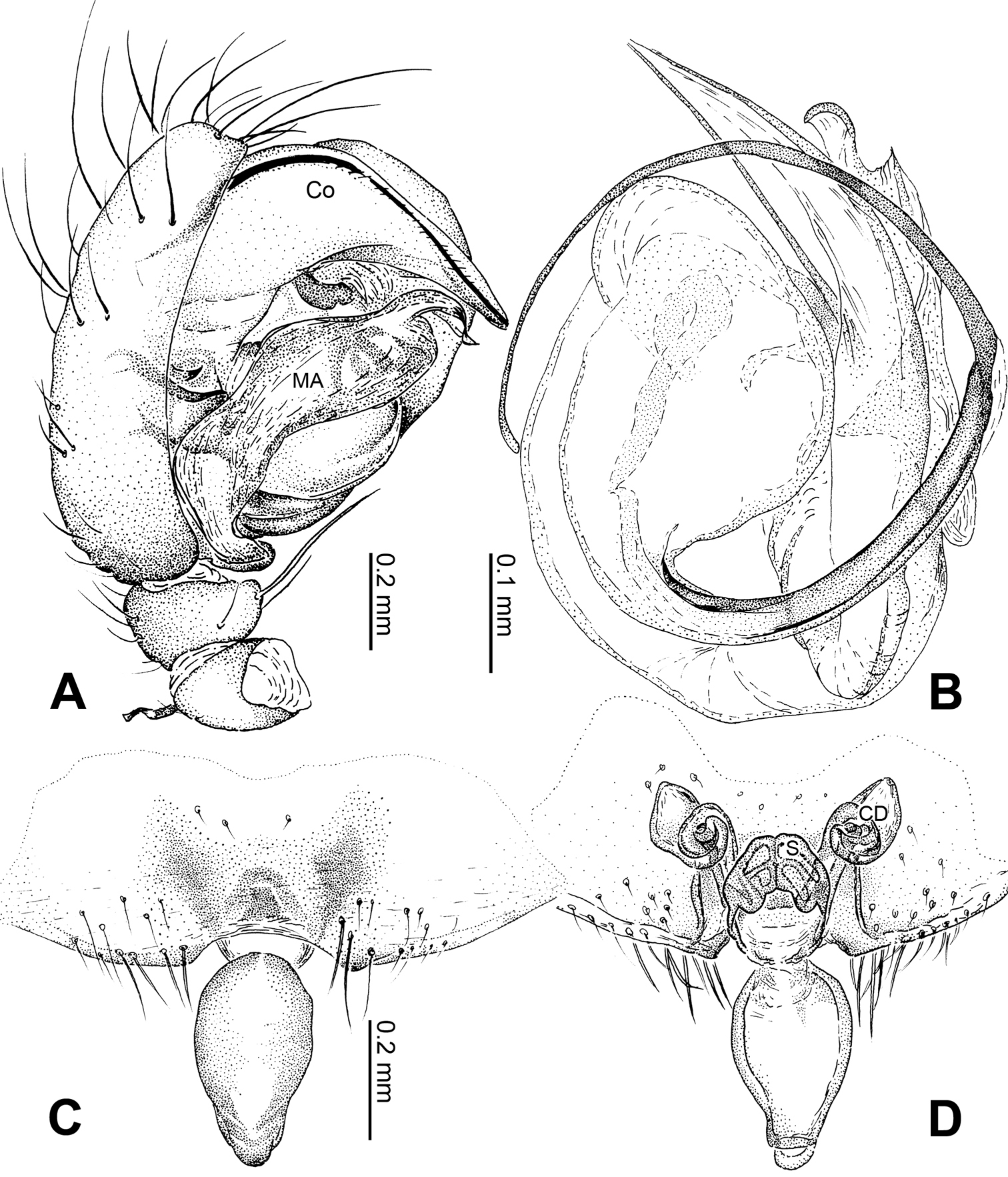
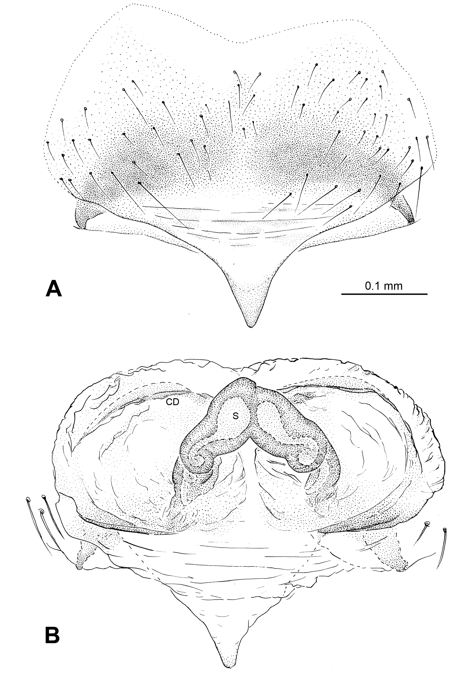
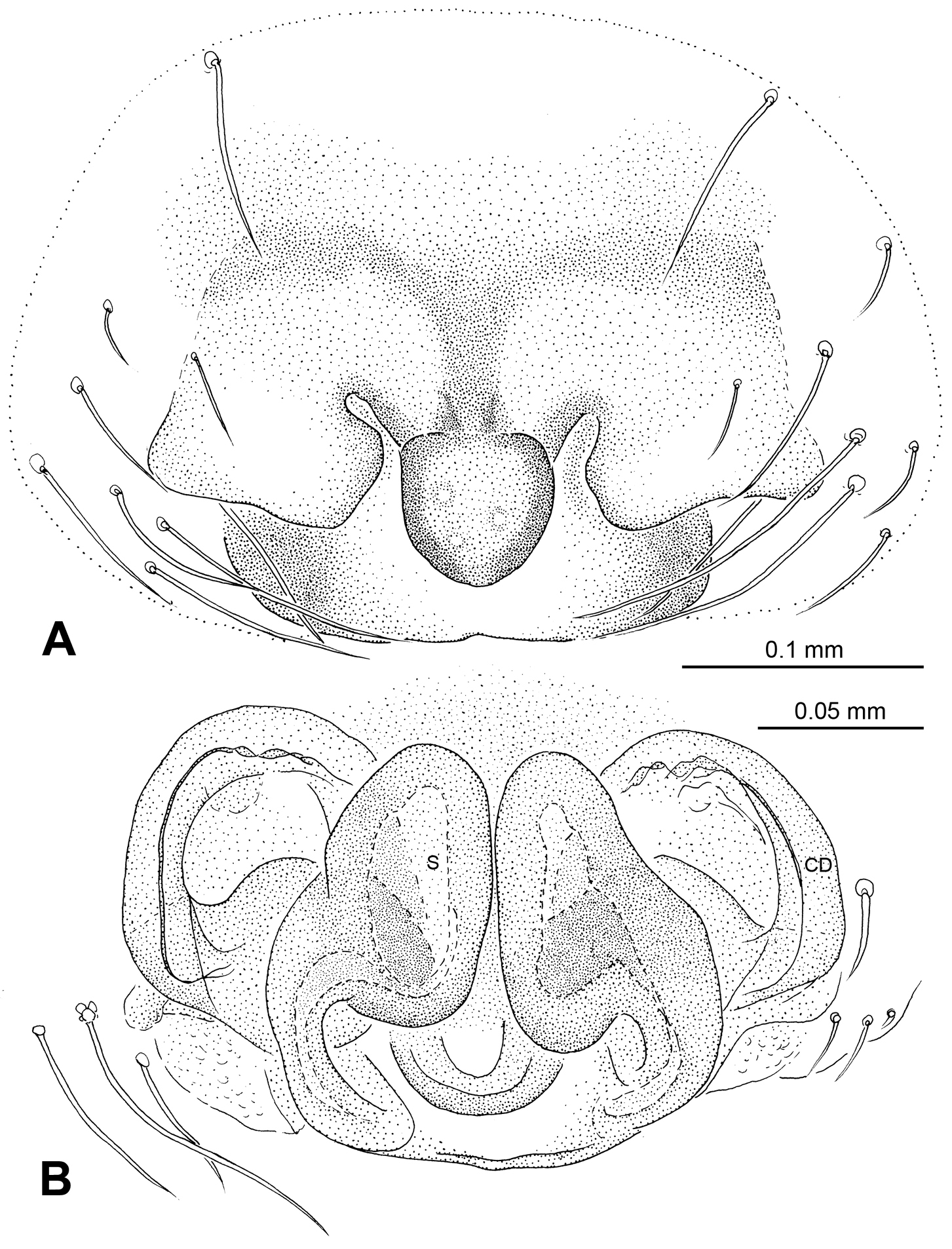
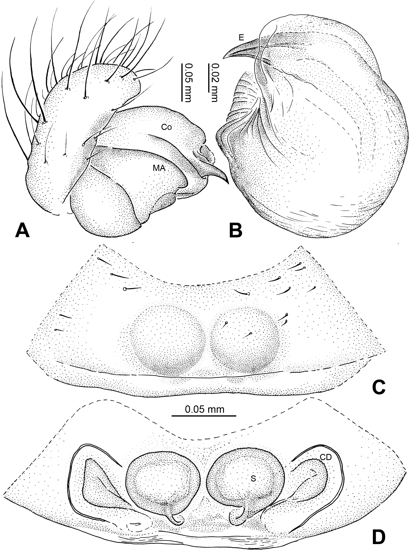
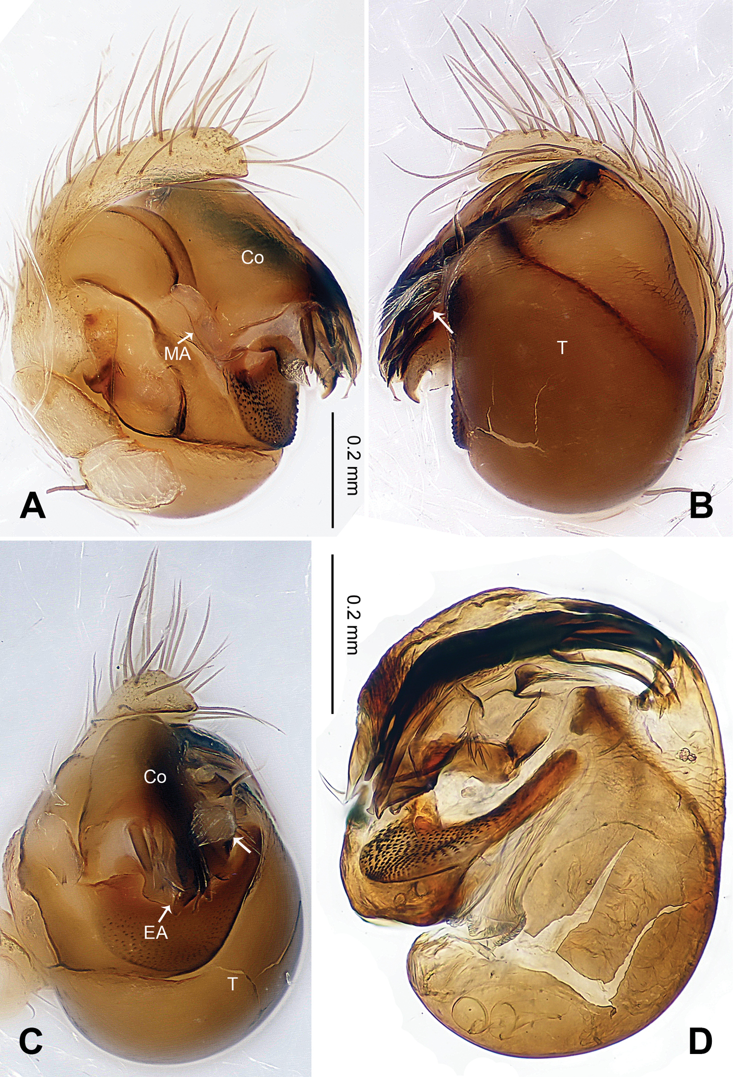
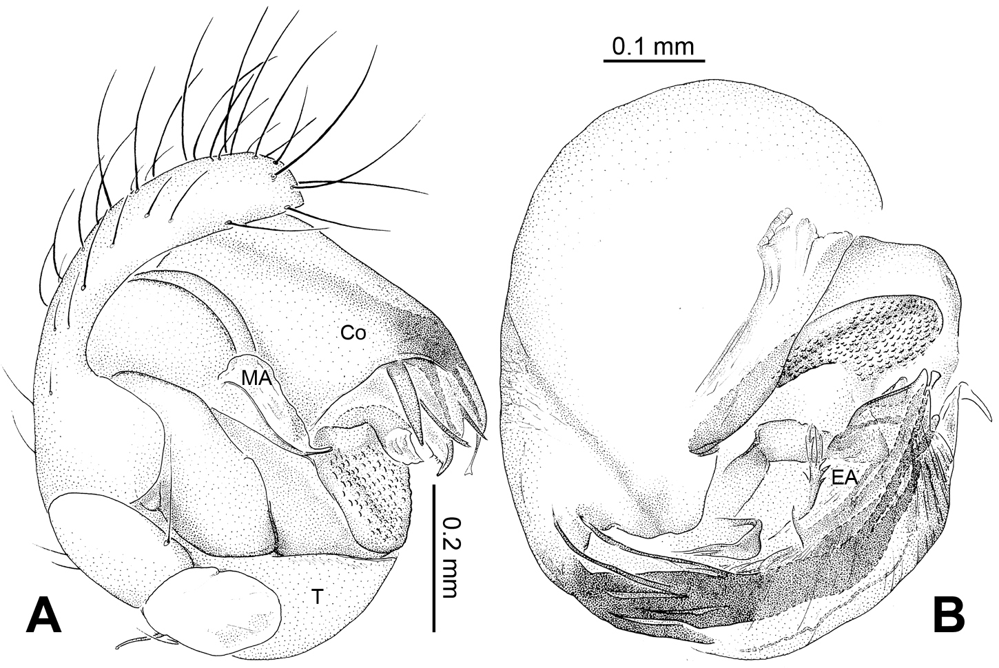
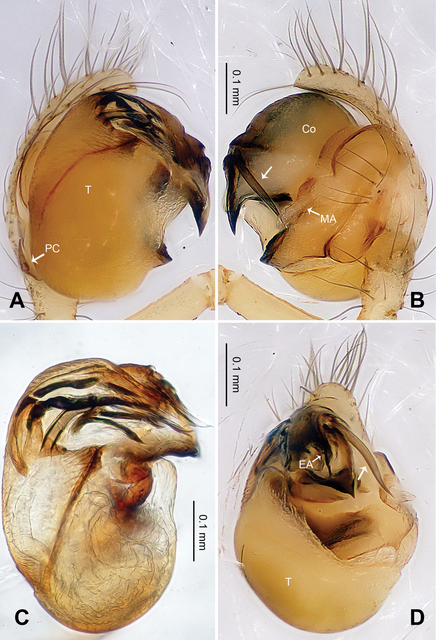
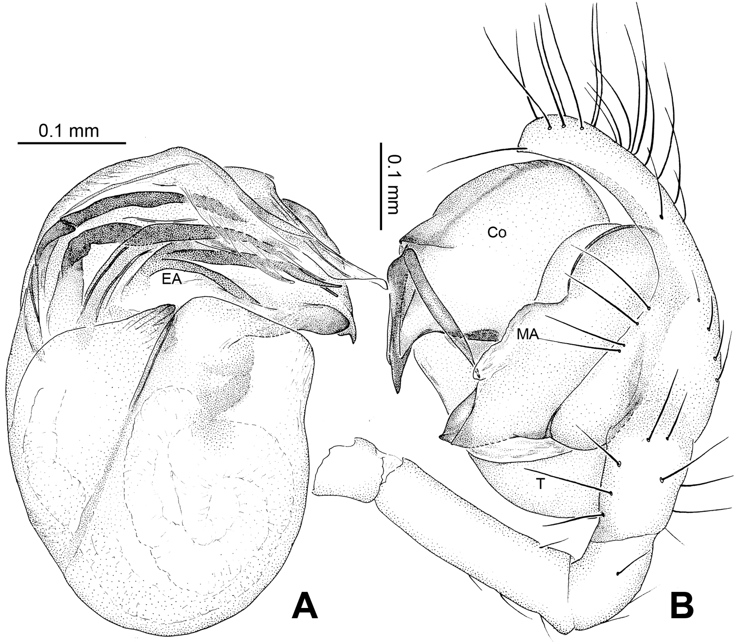
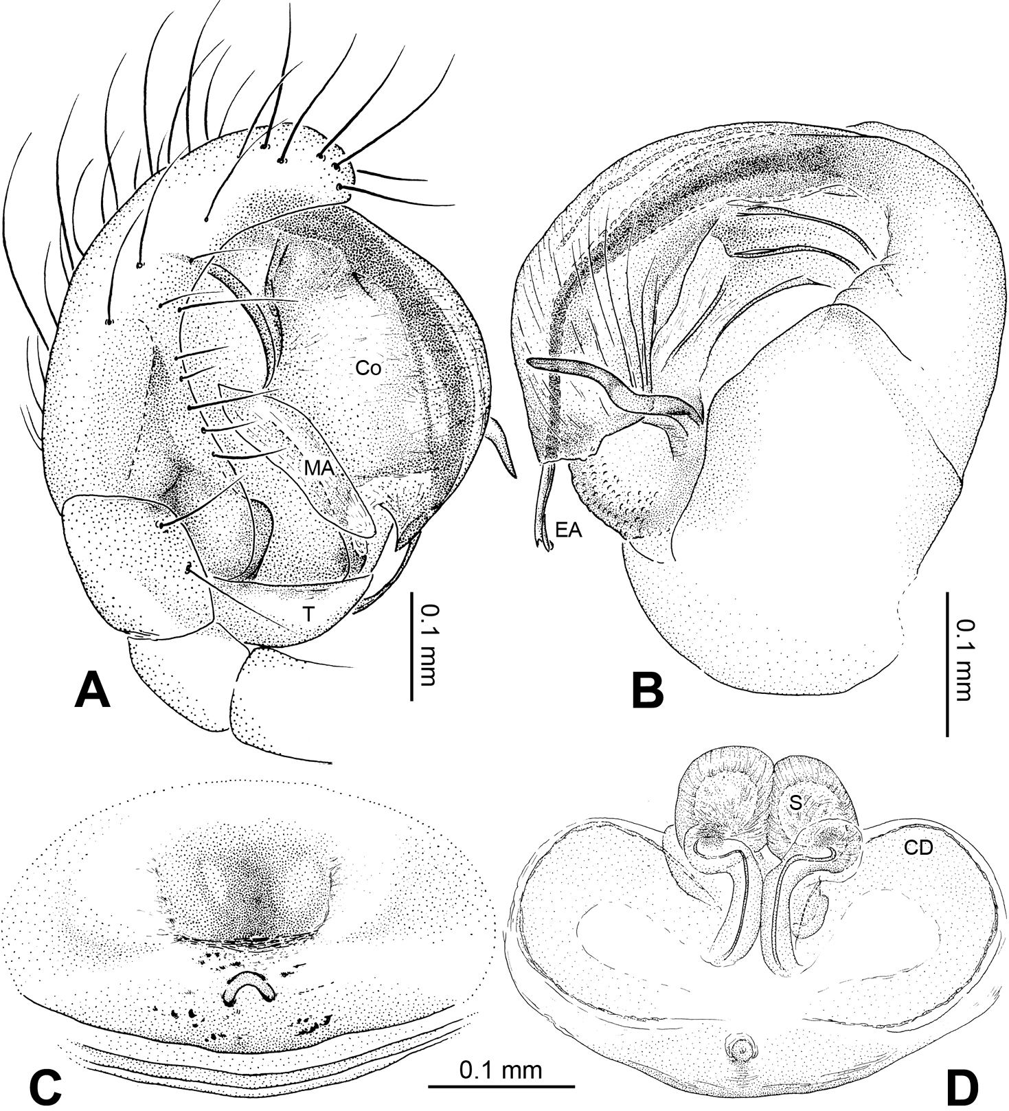
Alaria chengguanensis sp. n., male holotype. A Pedipalp, prolateral view B Pedipalp, ventral view C Embolic division, dorsal view D Pedipalp, retrolateral view. Co conductor; MA median apophysis; PC paracymbium; T tegulum. Scale bars: D as A.
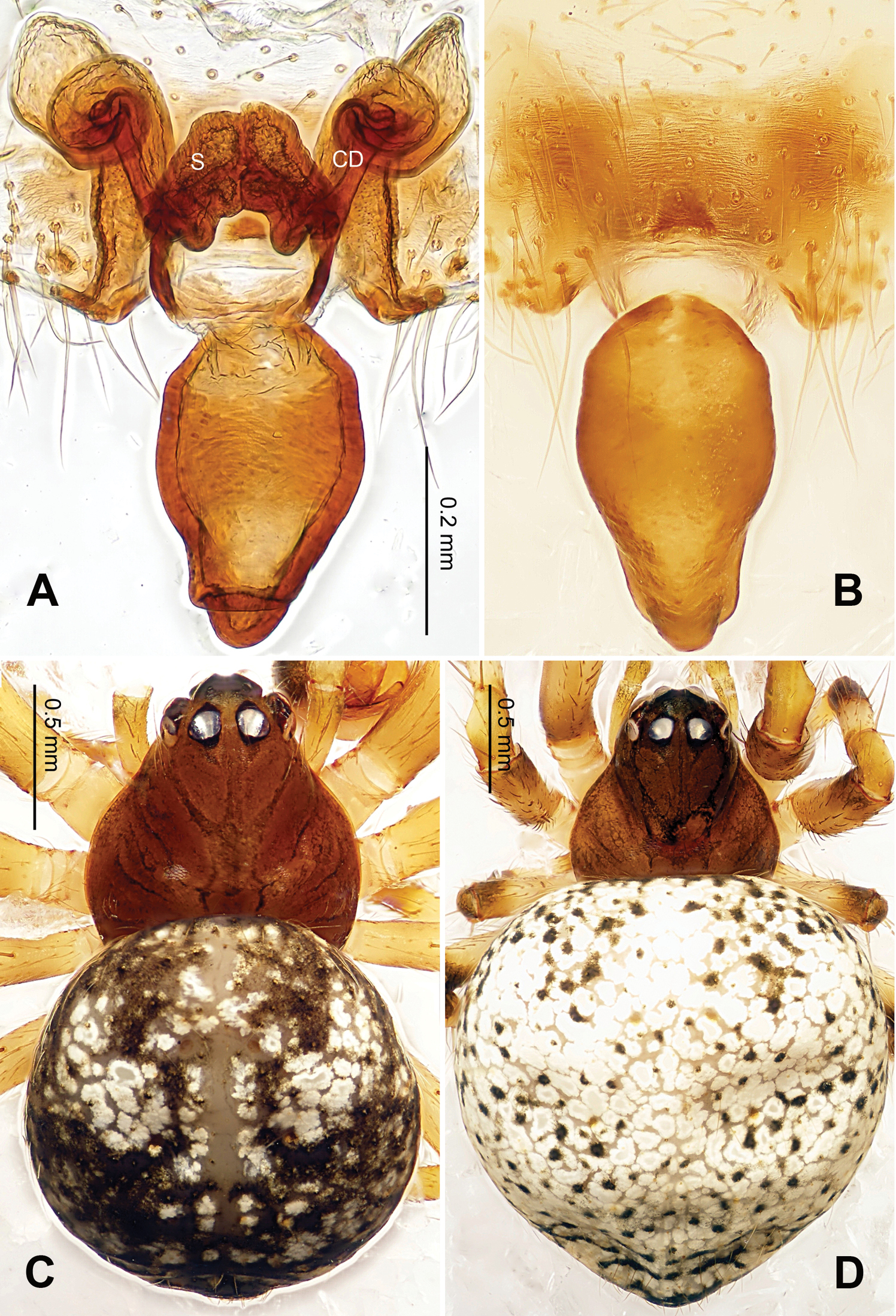
Alaria chengguanensis sp. n., male holotype (C) and female paratype (A–B, D). A Vulva, dorsal view B Epigyne, ventral view C Male, dorsal view D Female, dorsal view. CD copulatory duct; S spermatheca. Scale bars: B as A.
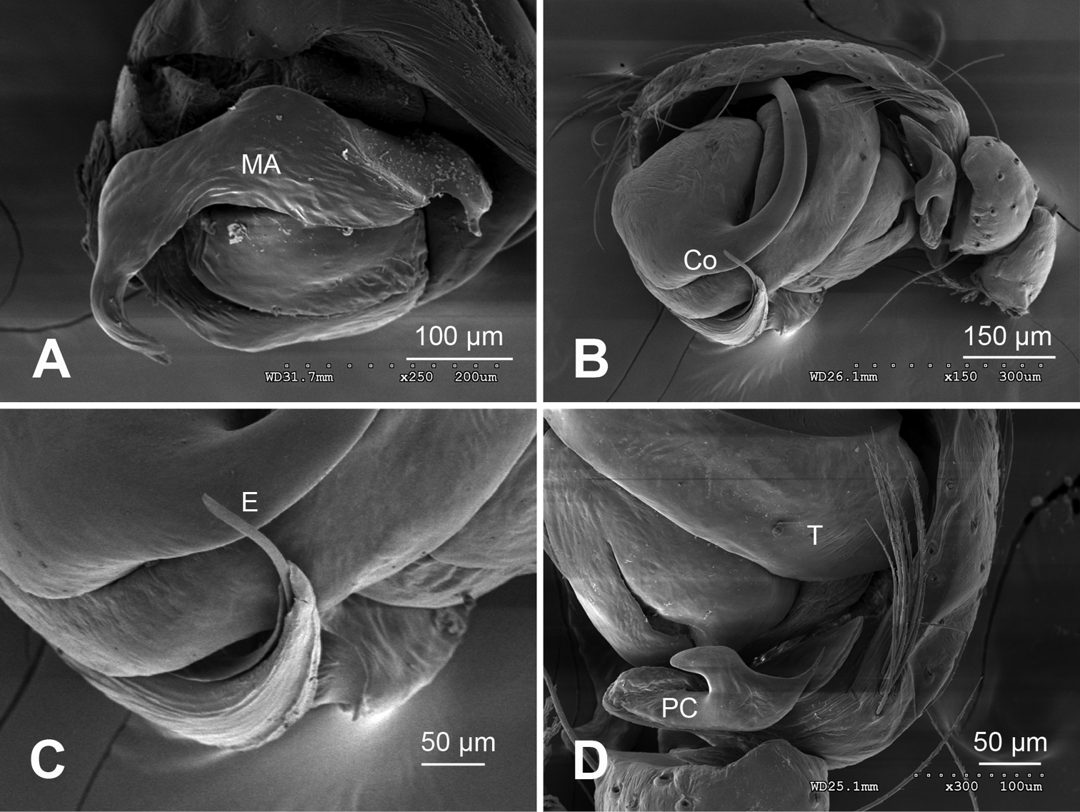
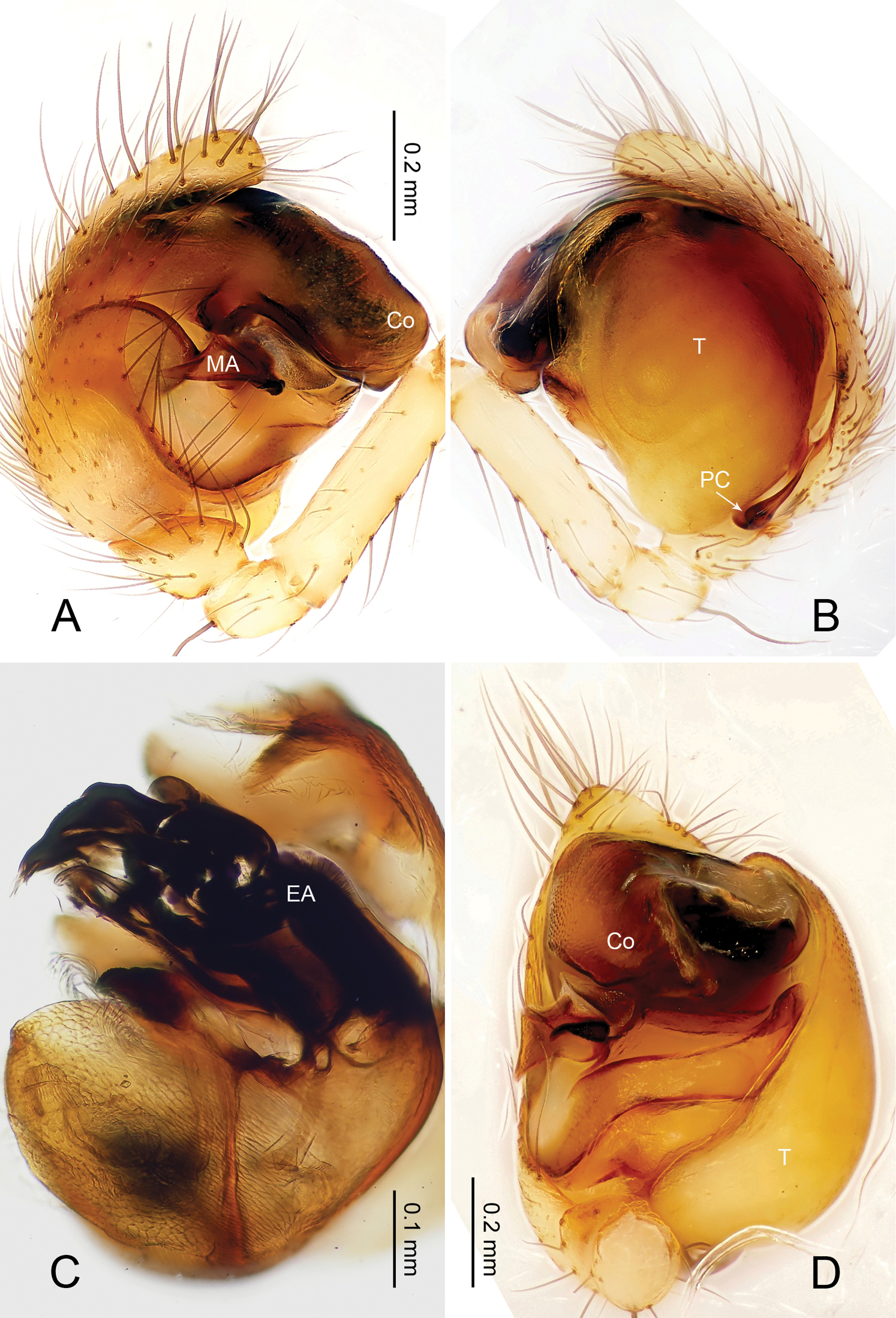
Alaria chengguanensis sp. n., SEM of pedipalp of a male paratype. A Prolateral view, detail showing MA B Retrolateral view C Retrolateral view, detail showingembolus D Retrolateral view, detail showing PC. Co conductor E Embolus; MA median apophysis; PC paracymbium; T tegulum.
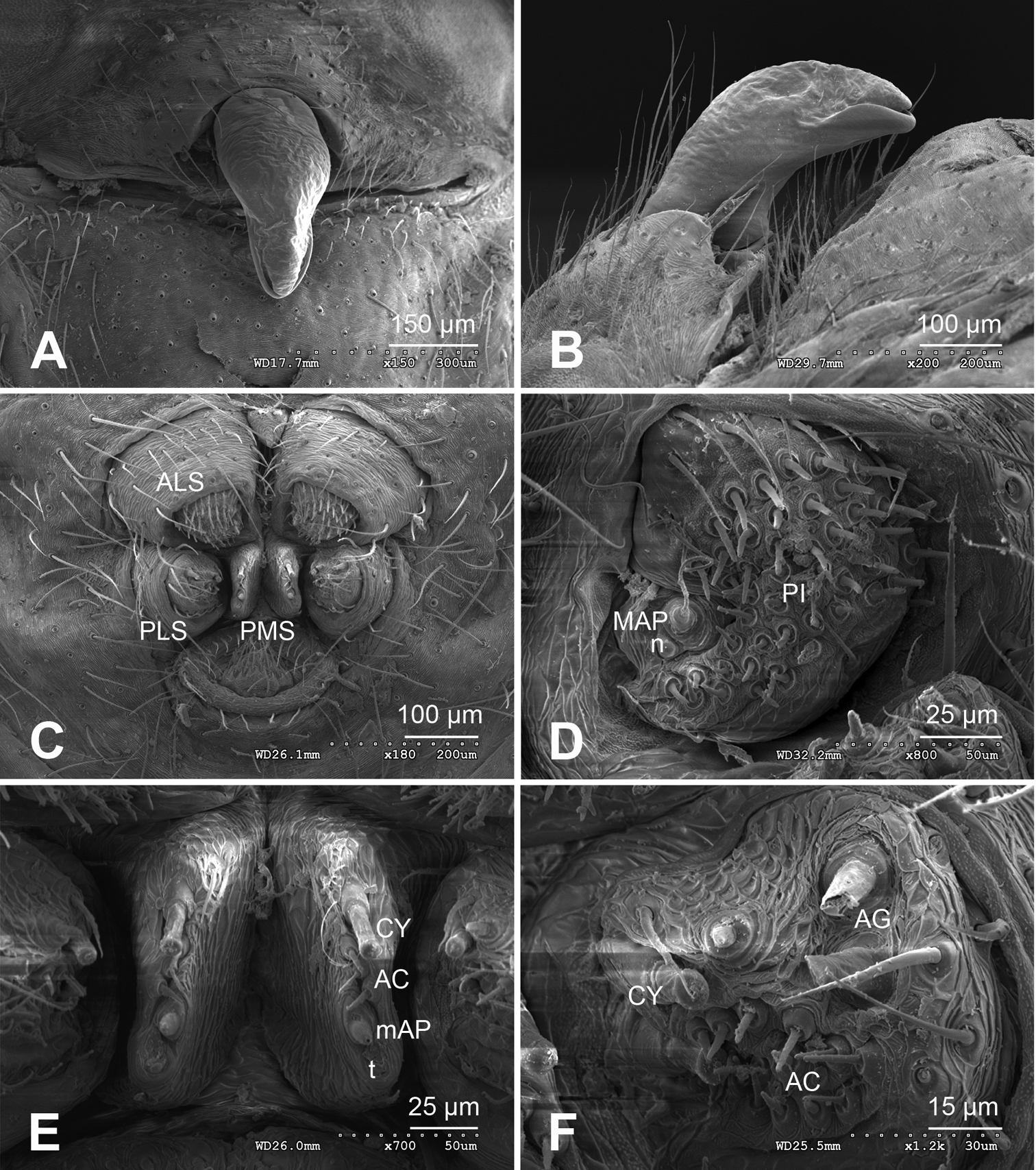
Alaria chengguanensis sp. n., SEM of a female paratype. A Epigyne, ventral view B Epigyne, lateral view C Spinnerets D ALS E PMS F PLS. AC aciniform gland spigot; AG aggregate gland spigot; ALS anterior lateral spinneret; CY cylindrical gland spigot; MAP major ampullate gland spigot; mAP minor ampullate gland spigot; n nubbin; PI piriform gland spigot; PLS posterior lateral spinneret; PMS posterior median spinneret; t tartipore.
Alaria chengguanensis sp. n., male holotype (A–B) and female paratype (C–D). A Pedipalp, prolateral view B Embolic division, dorsal view C Epigyne, ventral view D Vulva, dorsal view. CD copulatory duct; Co conductor; MA median apophysis; S spermatheca. Scale bars: D as C.
Baalzebub Coddington, 1986: 71. Type species Baalzebub baubo Coddington, 1986.
urn:lsid:zoobank.org:act:7E74FF15-FCE5-45FB-807A-12724669D543
http://species-id.net/wiki/Baalzebub_rastrarius
Figs 6–8Holotype:CHINA, Guizhou: Bijie City, Chengguan Town, Xiaohe Village, Xiniu Cave, 27°21.231'N, 105°17.186'E, elevation ca 1515 m, 30 April 2007, J. Liu & Y.C. Lin (IZCAS), 1 male.
Paratypes: [same data as holotype] (IZCAS), 1 male, 7 females. CHINA, Guizhou: Xishui County, Sangmu Town, Tuhe Village, Dongkouwan Cave, 28°15.679'N, 106°18.355'E, elevation ca 1271 m, 16 March 2011, Z.G. Chen & Z.W. Zha (IZCAS), 4 males, 8 females; Dafang County, Wenge Town, Taibai Village, Yelaoda Cave, 27°10.869'N, 105°28.289'E, elevation ca 1398 m, 12 March 2011, Z.G. Chen & Z.W. Zha (IZCAS), 2 males, 2 females.
The epithet comes from Latin word ‘rastrarius’ which means ‘of a hoe’, referring to the hoe-shaped conductor in prolateral view; adjective.
The presence of small, cleft median apophysis and blunt, spatulate processes of embolic apophysis in males, and tip-fused spermathecae in females indicates that this species belongs to the genus Baalzebub. Conductor in males envelopes the entire embolic apophysis (Fig 6A), which is similar to Baalzebub albinotatus, butthe rectangular conductor and the small, pointed cymbium apophysis are different from other described Baalzebub species. Females distinguished by the triangular epigynal plate, similar to Baalzebub baubo (
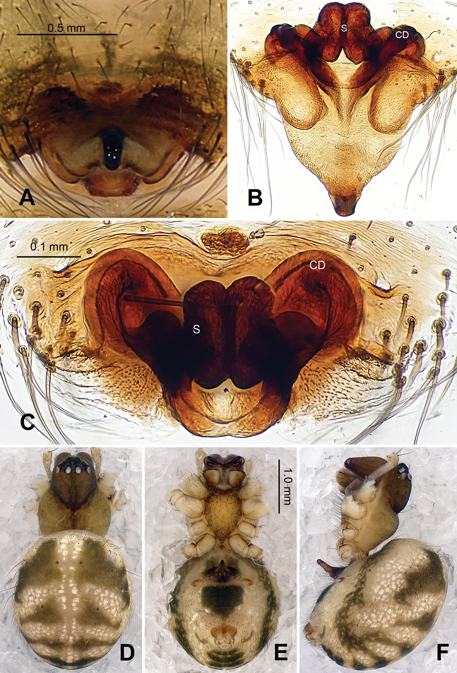
Carapace yellow tan. Sternum yellow with dark orange margins and setae. Legs yellow, dark brown distally at joints, especially tibiae. Abdomen tan with 2–4 rows of dark grey patches, the first two of which are connected by a perpendicular grey patch at midline, and two pairs of small dorsal brown spots (Figs 7C–F).
Male pedipalp: Patella with strong sinuous macroseta. Tibia with two trichobothria. Cymbium with a row of short regular bristles at its junction with cymbial lamella. Paracymbium with a long, pointed distal end (Fig. 6B). Cymbium apophysis small and pointed (Fig. 6B). Median apophysis triangular, strongly sclerotized (Figs 6A, 8A). Conductor a sub-rectangular translucent theca covering the complex embolic division and embolic apophysis. Embolic apophysis a spatulate arching structure with abruptly acuminated distal ends (Fig. 6C).
Vulva: Epigyne with a smooth triangular scape protruding from posterior margin of epigyne plate (Figs 7A–B). A deep pit lies on the epigynal mesal anterior edge. Spermathecae long, narrow, meeting at the tip to form an arcade-shaped structure. Copulatory ducts follow simple curve (Figs 7B, 8D).
Male: Total length 1.75, carapace 0.94 long, 1.09 wide, clypeus 0.25, sternum 0.53 long, 0.56 wide, coxae IV separated by their width. Posterior median eyes separated by less than half their diameter. Macrosetae: Leg I: femur p 1, patella d 1, tibia d 3, r 1; Leg II: femur r 1, patella d 2, tibia d 1, r 1; Leg III: patella d 2, tibia d 1; Leg IV: patella d 2, tibia d 1. Metatarsal trichobothria: Tm I: 0.25; Tm II: 0.29; Tm III: 0.23; Tm IV: 0.26. Leg measurements: I 4.66 (1.50, 0.50, 1.26, 1.00, 0.40); II 3.45 (1.10, 0.40, 0.80, 0.80, 0.35); III 2.50 (0.75, 0.30, 0.50, 0.50, 0.45); IV 3.10 (0.75, 0.60, 0.75, 0.70, 0.30).
Female: Total length 2.24, carapace 0.93 long, 0.93 wide, clypeus 0.11, sternum 0.5 long, 0.5 wide, coxae IV separated by their width. Posterior median eyes separated by less than half their diameter. Macrosetae as in male. Metatarsal trichobothria: Tm I: 0.28; Tm II: 0.25; Tm III: 0.45; Tm IV: 0.27. Leg measurements: I 4.17 (1.38, 0.48, 0.98, 0.88, 0.45); II 3.39 (1.10, 0.43, 0.73, 0.75, 0.38); III 2.50 (0.75, 0.35, 0.50, 0.55, 0.35); IV 3.11 (1.00, 0.33, 0.65, 0.75, 0.38).
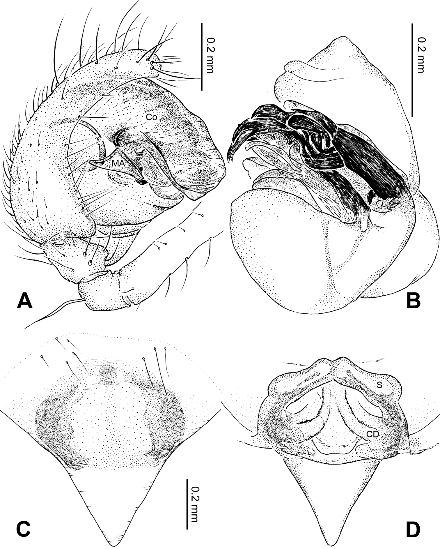
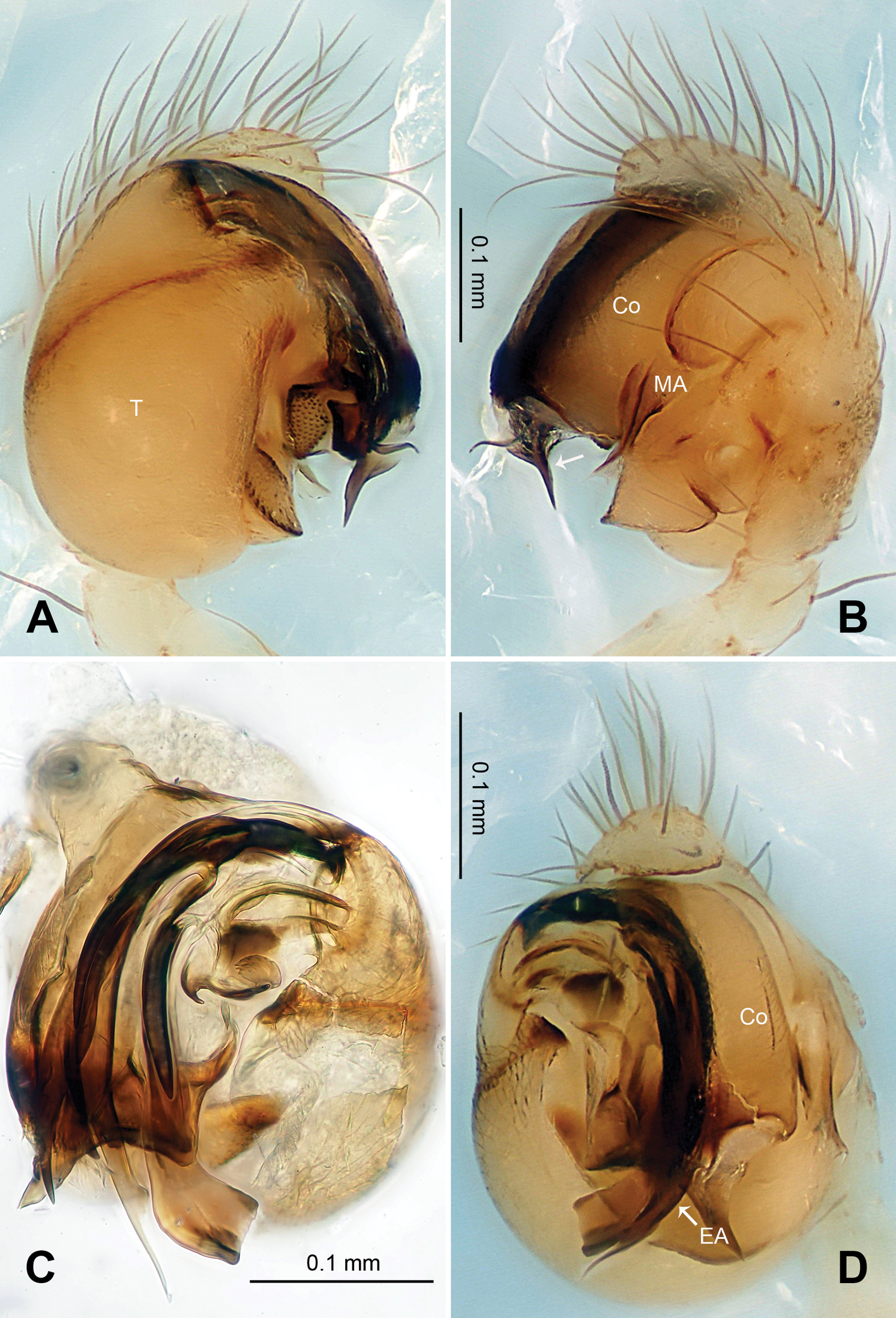
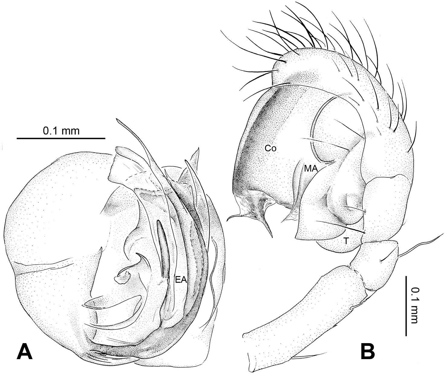
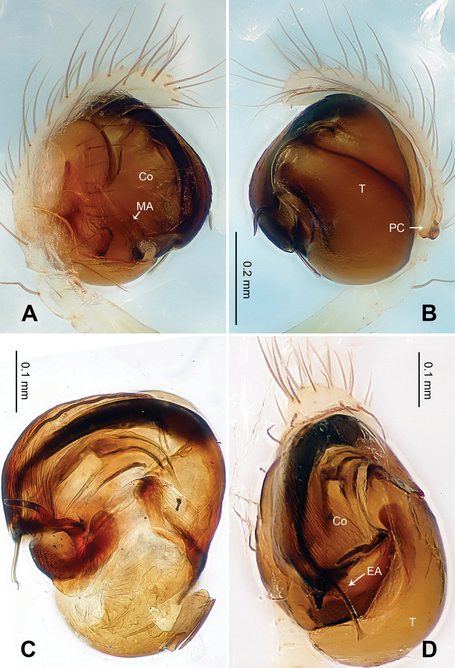
Baalzebub rastrarius sp. n., male holotype. A Pedipalp, prolateral view B Pedipalp, retrolateral view C Embolic division, retrolateral view D Pedipalp, ventral view. Co conductor; EA embolic apophysis; MA median apophysis; PC paracymbium; T tegulum. Scale bars: B as A.
Baalzebub rastrarius sp. n., male holotype (C) and female paratype (A–B, D–F). A Epigyne, ventral view B Vulva, dorsal view C Male, dorsal view D Female, dorsal view E Female, ventral view F Female, lateral view. CD copulatory duct, S spermatheca. Scale bars: B as A, D as E.
Baalzebub rastrarius sp. n., male holotype (A–B) and female paratype (C–D). A Pedipalp, prolateral view B Embolic division, retrolateral view C Epigyne, ventral view D Vulva, dorsal view. CD copulatory duct; Co conductor; MA median apophysis; S spermatheca. Scale bars: D as C.
urn:lsid:zoobank.org:act:791FC4A5-1C1F-4E54-97BA-EEDF7FAFFCA9
http://species-id.net/wiki/Baalzebub_youyiensis
Figs 9–10Holotype: CHINA, Guangxi: Pingxiang City, Youyi County, Bantou Village, Niuyan Cave, 22°05.666'N, 106°45.439'E, elevation ca 251 m, 18 January 2011, Z.G. Chen & Z.W. Zha (IZCAS), 1 female.
Paratypes: [same data as holotype] (IZCAS), 3 females.
This specific name formed from the Chinese words for friendship yŏu yì ( ), which is the name of the county where this species was collected; adjective.
Females distinguished from other described Baalzebub by the shape of the semi-transparent scape, which is proportionately shorter and with a blunt tip (Fig. 9A). Spermathecae, contrasted with narrow, long spermathecae in other Baalzebub species, is relatively shorter and smaller compared to the ovoid loops made by copulatory ducts (Fig. 9B)
Carapace broad, orange. Sternum yellow with dusky margins. Legs yellow, brown from patella to tarsus (except for leg I, which only brown distally at joints). Abdomen beige with irregular light, greenish-grey patches (Figs 9C–E).
Vulva: Epigyne with a short, pointed triangular scape with concave lateral margins protruding from posterior margin of epigyne plate, through which dark orange vulva is visible (Fig. 9A). Scape translucent. Epigyne plate with transverse grooves (Fig. 9A). Spermathecae small, elliptical, joining each other at the tip (Figs 9B, 10B). Copulatory ducts simple, with three coils toward spermathecae.
Female: Total length 2.10, carapace 1.00 long, 1.00 wide, clypeus 0.19, sternum 0.53 long, 0.53 wide, coxae IV separated by 1.00 time their width. Posterior median eyes separated by less than half their diameter. Macrosetae: Leg I: femur d 1, p 1, patella d 1, tibia d 2, p 1, r 1; Leg II: patella d 3, tibia d 2, r 1; Leg III: patella d 2, tibia d 2, r 2; Leg IV: patella d 2, tibia d 1. Metatarsal trichobothria: Tm I: 0.20; Tm II: 0.15; Tm III: 0.19; Tm IV: 0.22. Leg measurements: I 3.76 (1.20, 0.40, 0.88, 0.80, 0.48); II 3.2 (1.00, 0.36, 0.72, 0.64, 0.48); III 2.24 (0.72, 0.32, 0.44, 0.45, 0.31); IV 2.92 (1.00, 0.40, 0.60, 0.52, 0.40).
Male unknown.
Baalzebub youyiensis sp. n., female holotype. A Epigyne, ventral view B Vulva, dorsal view C Female, dorsal view D Female, ventral view E Female, lateral view. CD copulatory duct; S spermatheca. Scale bars: A as B, C, D as E.
Baalzebub youyiensis sp. n., female holotype. A Epigyne, ventral view B Vulva, dorsal view. CD copulatory duct; S spermatheca. Scale bars: B as A.
Karstia Chen, 2010: 2. Type species Karstia upperyangtzica Chen, 2010.
urn:lsid:zoobank.org:act:5ABD3E69-462D-4811-9851-EF178144FE62
http://species-id.net/wiki/Karstia_nitida
Figs 11–12Holotype: CHINA, Guangxi: Hechi City, Hechi County, Laba Village, Shoushui Cave, 24°41.229'N, 107°52.609'E, elevation ca 268 m, 31 March 2011, Z.G. Chen & Z.W. Zha (IZCAS), 1 female.
Paratypes: [same data as holotype] (IZCAS), 14 females.
This specific name comes from the Latin word ‘nitidus’ which means ‘shinning and elegant’, referring to the glossiness of the swollen tip of the epigynal scape; adjective.
Females distinguished by the following combination of characters: the structure of the scape, the stout, overlapped spermathecae (Fig. 11B), and the habitus of this species (Figs 11C–E). Spermathecae oval-shaped with vertically longer diameter, slightly detached from each other along their inner margin. The abdomen large, contrasted with distinctly small epigynal area. The scape structure is quite different from other Karstia or Baalzebub species: the tip of scape is swollen and shimmery, and the lateral margins of the plate extend toward the scape tip to form a armet-shaped conformation (Fig. 11A). Generic placement tentative pending discovery and examination of the male.
Carapace pale yellow with yellow ocular region. Sternum yellow with tan margins. Legs yellow, brown distally at joints. Abdomen dark grey mottled with white patches (Figs 11C–E).
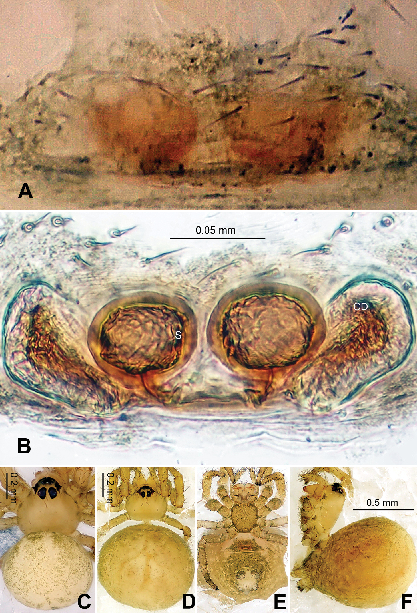
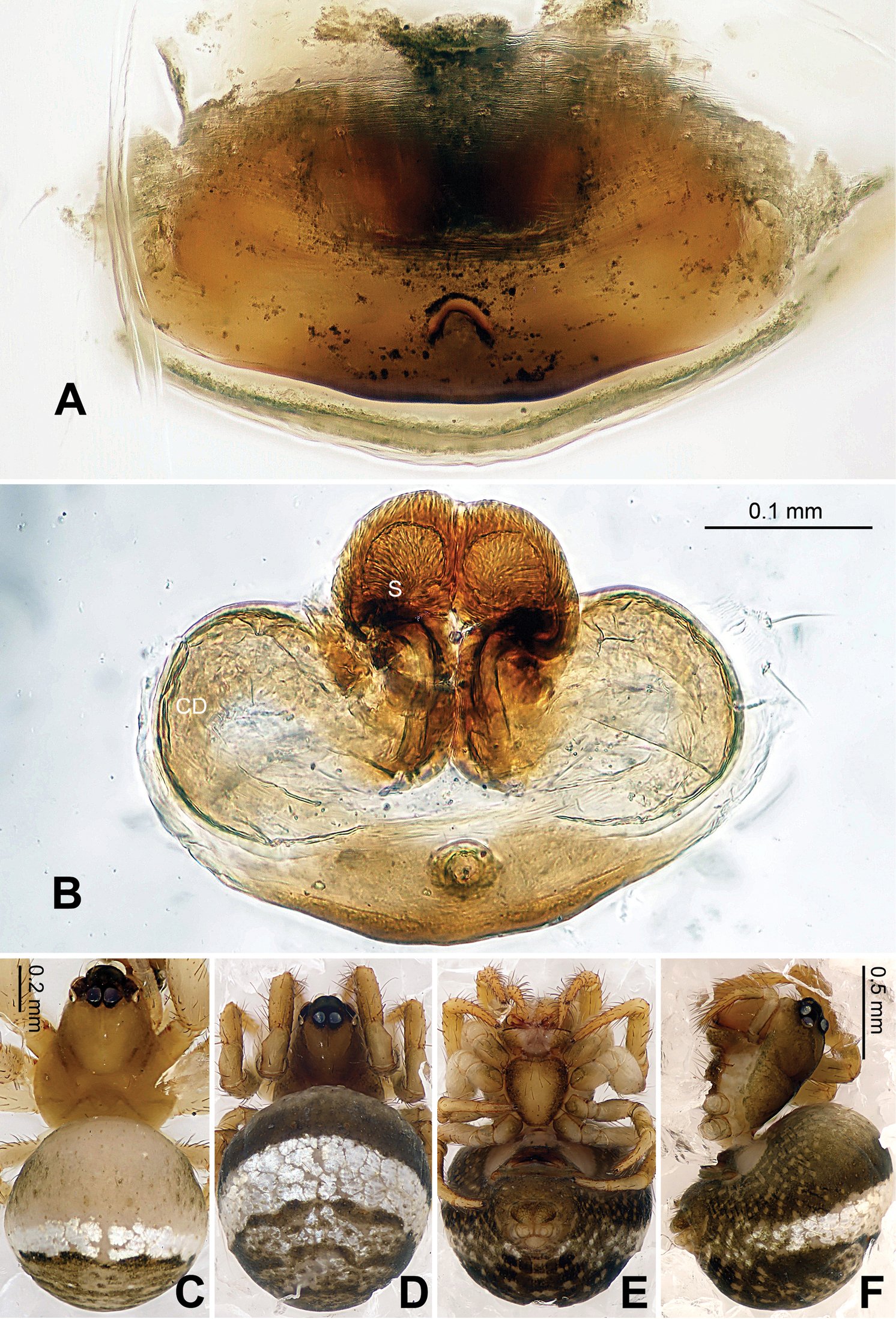
Vulva: Epigyne small, with short, distally spherical scape protruding from posterior margin of epigyne plate. Epigyne plate extends posteriorly, together with scape to form a barbute-shaped conformation (Figs 11A, 12A). End of scape glossy with black purfle (Fig. 11A). Spermathecae peanut-shaped, juxtaposed, slightly detached from each other (Figs 11B, 12B). Copulatory ducts follow simple rout to form small loops and one turning before connected with spermathecae at the bottom (Figs 11B, 12B).
Female: Total length 3.25, carapace 1.38 long, 1.13 wide, clypeus 0.10, sternum 0.70 long, 0.65 wide, coxae IV separated by their width. Posterior median eyes separated by less than half their diameter. Macrosetae: Leg I: femur p 1, r 1, patella d 1, tibia d 2, p 2, v 2, r 1, metatarsus d 1, v 2, r 1; Leg II: femur d 2, patella d 2, tibia d 4, p 1, v2, metatarsus p 1, v 2; Leg III: femur d 1, patella d 2, tibia d 1, v 1, metatarsus d 3; Leg IV: patella d 2, tibia d 1, v 1, r 1, metatarsus d 2. Metatarsal trichobothria: Tm I: 0.23; Tm II: 0.21; Tm III: 0.16; Tm IV 0.22. Leg measurements: I 4.00 (0.56, 0.63, 1.09, 1.09, 0.63); II 4.16 (1.25, 0.50, 0.94, 0.94, 0.53); III 2.98 (0.78, 0.47, 0.63, 0.63, 0.47); IV 3.66 (1.25, 0.38, 0.78, 0.78, 0.47).
Male unknown.

Karstia nitida sp. n., female holotype. A Epigyne, ventral view B Vulva, dorsal view C Habitus, dorsal view D Habitus, ventral view E Habitus, lateral view. CD copulatory duct S spermatheca. Scale bars: C, D as E.
Karstia nitida sp. n., female holotype. A Epigyne, ventral view B Vulva, dorsal view. CD copulatory duct, S spermatheca.
urn:lsid:zoobank.org:act:38E8200D-E27D-4710-ABEE-FBF657944027
http://species-id.net/wiki/Karstia_prolata
Figs 13–14Holotype: CHINA, Guangxi: Pingxiang City, Youyi County, Bantou Village, Niuyan Cave, 22°05.666'N, 106°45.439'E, elevation ca 251 m, 18 January 2011, Z.G. Chen & Z.W. Zha (IZCAS), 1 female.
Paratypes: [same data as holotype] (IZCAS), 9 females.
The specific name is derived from the Latin word ‘prolatus’ meaning elongated’, and refers to the extended epigynal scape; adjective.
Females distinguished by the protruding scape and the overlapped, stout spermathecae. The long acute-angled scape protrudes vertically from the posterior epigynal margin (Figs 13B, F), which is different from other known Karstia species and Baalzebub species.
Relatively large in total body length, compared to other theridiosomatid species. Carapace greenish tan, with brown ocular region. Sternum yellow with dark margins. Femur yellow, brown from patella to tarsus. Abdomen beige with evenly distributed silver spots within dorsal area and 8 or more rows of mesally disrupted greenish grey patches, with a large, wedge-shaped greenish grey patch between epigyne and spinnerets (Figs 13D–E).
Vulva: Epigyne with a long, apiculate scape protruding perpendicularly (slightly tilted) from posterior margin of epigyne plate, flanked by a cluster of long sinuous setae on each side (Figs 13A, E–F). Two deep grooves occur at the posterior base of epigyne which are likely connected to the copulatory openings (Figs 13B, E). Tip of scape glossy black (Fig. 13A). Spermathecae peanut-shaped, juxtaposed (Fig. 13C). Copulatory ducts’ routing simple, half-looped (Figs 13C, 14B).
Female: Total length 3.85, carapace 1.92 long, 1.25 wide, clypeus 0.15, sternum 0.63 long, 0.65 wide, coxae IV separated by 2.00 times their width. Posterior median eyes separated by half their diameter. Macrosetae: Leg I: femur d 2, p 1, patella d 2, tibia d 2, p 2, v 1, r 1, metatarsus p 1, v 3, r 1; Leg II: femur d 2, patella d 2, tibia d 1, p 2, v 1, r 1, metatarsus p 1, v 2, r 1; Leg III: femur d 1, v 1, patella d 1, tibia d 1, p 2, v 2, metatarsus d 3; Leg IV: tibia d 2, p 1, metatarsus d 2. Metatarsal trichobothria: Tm I: 0.13; Tm II: 0.22; Tm III: 0.27; Tm IV: 0.22. Leg measurements: I 6.35 (2.00, 0.75, 1.35, 1.50, 0.75); II 5.25 (1.50, 0.70, 1.10, 1.20, 0.75); III 3.35 (0.95, 0.45, 0.70, 0.75, 0.50); IV 4.60 (1.50, 0.50, 1.00, 1.00, 0.60).
Male unknown.
Karstia prolata sp. n., female holotype. A Epigyne, ventral view B Vulva, posterior view C Vulva, dorsal view D Habitus, dorsal view E Habitus, ventral view F Habitus, lateral view. CD copulatory duct; S spermatheca. Scale bars: B as A, D, F as E.
Karstia prolata sp. n., female holotype. A Epigyne, ventral view B Vulva, dorsal view. CD copulatory duct; S spermatheca.
Menglunia inaffecta sp. n.
The generic epithet refers to the place měng lún ( ) where these specimens were collected. Menglun Town is located at Xishuangbanna in Yunnan province, where tropical rain forest harbors countless species, both vertebrates and invertebrates. Gender is feminine.
Distinguished from other theridiosomatids by the extremely simple, short embolus, and the round, separated spermathecae. The pedipalp in males is an elliptical (slightly rectangular in total), theca-textured, and obscurely circumscribed structure (Figs 15A, D). Conductor is less extensive, and fully covers the embolus (Fig. 18A). The embolus is beak-like, and enveloped in conductor (Fig. 15B). Unlike any other theridiosomatid genus, the median apophysis in Menglunia is merely a small projection, mildly curved without any sharp tip or trough (Fig. 15A). Spermathecae similar to Coddingtonia euryopoides (
Menglunia inaffecta sp. n.
urn:lsid:zoobank.org:act:63DFDE88-8154-412E-B02E-47A71157F286
http://species-id.net/wiki/Menglunia_inaffecta
Figs 15–18Holotype: CHINA, Yunnan: Menglun Town: Xishuangbanna Botanical Garden, 21°55.035'N, 101°16.500'E, elevation ca 558 m, 22 July 2007, primary tropical seasonal rain forest, searching, G. Zheng (IZCAS), 1 male.
Paratypes: [same data as holotype] (IZCAS), 9 males, 9 females.
Its Latin origin ‘inaffectus’ means ‘natural and simple’, which refers to the simplicity of the structure of the male pedipalp; adjective.
See diagnosis for genus.
Carapace yellow tan. Sternum yellow with greenish brown margins and sparse hairs. Legs thick, yellow. Abdomen beige with small, irregularly-distributed greenish brown spots.
Male pedipalp: Patella with erect macroseta. Tibia with one trichobothrium. Paracymbium with a short pointed distal end (Fig. 17A). Tegulum sub-oval. Median apophysis with a small projection oriented distoventrally (Figs 15A, 18A). Conductor kidney-shaped, translucent theca. Embolus beak-shaped, stout (Figs 15A–B, 17B).
Vulva: Epigyne with obscure plate margins (Figs 16A, 17A). Spermathecae round, separated from each other (Fig. 16B). Copulatory duct short and simple (Fig. 16B).
Male (holotype): Total length 1.00, carapace 0.40 long, 0.44 wide, clypeus 0.05, sternum 0.30 long, 0.25 wide, coxae IV separated by 1.5 times their width. Posterior median eyes separated by less than half their diameter. Macrosetae: Leg I: patella d 2, tibia d 2, p 1; Leg II: patella d 1, tibia d 1, p 1, r 1; Leg III: patella d 2. Metatarsal trichobothria: Tm I: 0.27; Tm II: 0.38; Tm III: 0.36. Leg measurements: I 1.10 (0.31, 0.13, 0.25, 0.23, 0.18); II 0.84 (0.25, 0.06, 0.20, 0.20, 0.13); III 0.66 (0.18, 0.11, 0.13, 0.13, 0.11); IV 0.76 (0.19, 0.13, 0.19, 0.14, 0.11).
Female (one of paratypes): Total length 1.00, carapace 0.63 long, 0.48 wide, clypeus 0.05, sternum 0.30 long, 0.30 wide, coxae IV separated by 1.5 times their width. Posterior median eyes separated by less than half their diameter. Macrosetae as in male. Metatarsal trichobothria: Tm I: 0.32; Tm II: 0.38; Tm III: 0.29. Leg measurements: I 1.09 (0.32, 0.20, 0.24, 0.25, 0.18); II 1.02 (0.25, 0.19, 0.23, 0.20, 0.15); III 0.75 (0.19, 0.11, 0.13, 0.18, 0.14); IV 0.91 (0.25, 0.13, 0.21, 0.18, 0.14).
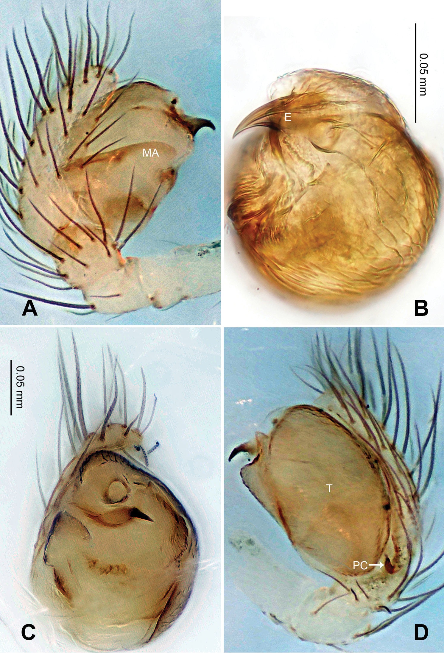
Menglunia inaffecta sp. n., male holotype. A Pedipalp, prolateral view B Embolic division C Pedipalp, ventral view D Pedipalp, retrolateral view. E embolus; MA median apophysis; PC paracymbium; T tegulum. Scale bars: A, D as C.
Menglunia inaffecta sp. n., male holotype (C) and female paratype (A–B, D–F). A Epigyne, ventral view B Vulva, dorsal view C Male habitus, dorsal view D Female habitus , dorsal view E Female habitus, ventral view F Female habitus, lateral view. CD copulatory duct, S spermatheca. Scale bars: A as B, E as D.
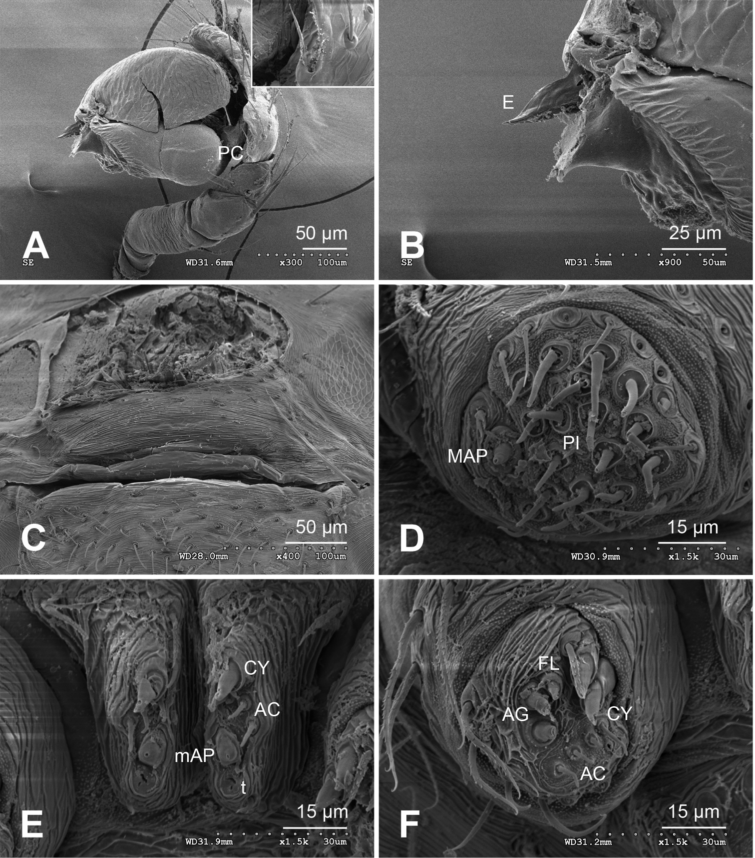
Alaria chengguanensis sp. n., SEM of male paratype (A–B) and female paratype (C–F). A Pedipalp, retrolateral view B Pedipalp, retrolateral view, detail showing embolus C Epigyne D ALS E PMS F PLS. AC aciniform gland spigot; AG aggregate gland spigot; ALS anterior lateral spinneret; CY cylindrical gland spigot E embolus; FL flagelliform gland spigot; MAP major ampullate gland spigot; mAP minor ampullate gland spigot; PC paracymbium; PI piriform gland spigot; PLS posterior lateral spinneret; PMS posterior median spinneret; t tartipore.
Menglunia inaffecta sp. n., male holotype (A–B) and female paratype (C–D). A Pedipalp, prolateral view B Embolic division, retrolateral C Epigyne, ventral view D Vulva, dorsal view. CD copulatory duct; Co conductor; MA median apophysis S spermatheca.
Ogulnius O. Pickard-Cambridge, 1882: 432. Type species Ogulnius obtectus O. Pickard-Cambridge, 1882.
urn:lsid:zoobank.org:act:2CE2BCE0-15BC-488A-A363-872B6BA232F0
http://species-id.net/wiki/Ogulnius_hapalus
Figs 19–21Holotype: CHINA, Yunnan: Menglun Town: Xishuangbanna Botanical Garden, 21°55.035'N, 101°16.500'E, elevation ca 558 m, 22 July 2007, primary tropical seasonal rain forest, fogging, G. Zheng (IZCAS), 1 male.
Paratypes: [same data as holotype] (IZCAS), 2 males, 6 females.
This specific name describes the softness and fragility of this species. The Latin origin is ‘hapalus’ meaning ‘delicate and tender’; adjective.
Though lack of one genetic feature, which is fourth legs are longer than the first legs (subequal in females), other generic characteristics of Ogulnius can be seen in this species: tapering whip-like embolic apophysis in males (Fig. 19C), transverse grooves on epigyne in females, separated and juxtaposed posterior median eyes (
Carapace pale yellow. Sternum ivory. Legs pale yellow, semi-transparent. Abdomen pale with soft, translucent cuticle.
Male pedipalp: Patella with strong sinuous macroseta. Tibia with one trichobothrium. Paracymbium slim with filiform projection distally. Median apophysis curvy, mesally wide, with apex oriented distoventrally (Figs 19A, 21A). Conductor a translucent theca covering about two thirds length of the long filiform embolic apophysis (Figs 19C–D).
Vulva: Epigyne with transverse groove and a concave posterior margin (Fig. 20A). Spermathecae peanut-shaped, juxtaposed (Fig. 20B). Copulatory ducts bend downwardly at top and wide at entrance (Figs 20B, 21B).
Male: Total length 1.00, carapace 0.50 long, 0.40 wide, clypeus 0.09, sternum 0.25 long, 0.25 wide, coxae IV separated by 2.00 times their width. Posterior median eyes separated by 1.5 times their diameter. Macrosetae: Leg I: patella d 1, tibia d 1, p 1; Leg II: patella d 1, tibia d 1; Leg III: patella d 1, tibia d 1; Leg IV: patella d 1, tibia d 1. Metatarsal trichobothria: Tm I: 0.23; Tm II: 0.19; Tm III: 0.27. Leg measurements: I 1.00 (0.30, 0.18, 0.20, 0.20, 0.12); II 0.87 (0.26, 0.12, 0.20, 0.16, 0.13); III 0.57 (0.11, 0.10, 0.11, 0.15, 0.10); IV 0.64 (0.15, 0.10, 0.14, 0.15, 0.10).
Female: Total length 1.41, carapace: 0.50 long, 0.44 wide, clypeus 0.23, sternum 0.30 long, 0.25 wide, coxae IV separated by 2.00 times their width. Posterior median eyes separated 1.5 times their diameter. Macrosetae as in male. Metatarsal trichobothria: Tm I: 0.25; Tm II: 0.21; Tm III: 0.27. Leg measurements: I 0.95 (0.20, 0.25, 0.15, 0.20, 0.15); II 0.82 (0.25, 0.12, 0.15, 0.15, 0.15); III 0.62 (0.12, 0.10, 0.15, 0.15, 0.10); IV 0.90 (0.30, 0.15, 0.15, 0.20, 0.10).
Ogulnius hapalus sp. n., male holotype. A Pedipalp, prolateral view B Pedipalp, ventral view C Embolic division, retrolateral view D Pedipalp, retrolateral view. Co conductor; EA embolic apophysis; MA median apophysis T tegulum. Scale bars: D as A, C as B.
Ogulnius hapalus sp. n., male holotype (C) and female paratype (A–B, D–F). A Epigyne, ventral view B Vulva, dorsal view C Male habitus, dorsal view D Female habitus, dorsal view E Female habitus, ventral view F Female habitus, lateral view. CD copulatory duct; S spermatheca. Scale bars: B as A, D, E as F.
Ogulnius hapalus sp. n., male holotype (A–B) and female paratype (C–D). A Pedipalp, prolateral view B Embolic division, retrolateral view C Epigyne, ventral view D Vulva, dorsal view. CD copulatory duct; Co conductor; MA median apophysis; S spermatheca.
Theridiosoma O. Pickard-Cambridge, 1879: 193. Type species Theridiosoma argenteolum O. Pickard-Cambridge, 1879 (= Theridiosoma gemmosum (L. Koch, 1878)).
urn:lsid:zoobank.org:act:34DDB1DE-B435-4A41-8EF7-A40B43ACC9E0
http://species-id.net/wiki/Theridiosoma_plumaria
Figs 22–23Holotype: CHINA, Hainan: Mt. Jianfengling National Nature Reserve: Tianchi watershed 18°44.383'N, 108°51.062'E, elevation ca 888 m, 19 July 2007, S. Li (IZCAS), 1 male.
Paratype: CHINA: Hainan: Mt. Bawangling National Nature Reserve, 5 kilometers past Dongerjianchazhan, 19°05.186'N, 109°11.802'E, elevation ca 1010 m, 25 July 2007, S. Li (IZCAS), 1 male.
The specific name was taken from the Latin word ‘plumarius’ which means ‘of feathers’. It refers to the plumose branching of its conductor theca (Figs 22B–C); adjective.
Males, similar to Theridiosoma gemmosum (L. Koch) (
Carapace yellow tan. Sternum yellow with brown margins. Legs yellow. Abdomen tan with sparse silver specks and symmetric dark grey patches.
Male pedipalp: Patella with strong macroseta. Tibia with two trichobothria. Paracymbium hooked with pointed distal end. Tegulum large with tuberculate mesal lobe (Figs 22A, D). Median apophysis a curved lobe attenuate distally. Conductor a translucent theca covering fragmented embolic apophysis, with plumose branching and erect tip near tegulum (Fig. 22B). Embolic apophysis fragment tips protruding out of the conductor (Figs 22A–C). Embolic apophysis fragments long, slim (Figs 22D, 23B).
Male: Total length 1.60, carapace 0.80 long, 0.80 wide, clypeus 0.20, sternum 0.40 long, 0.40 wide, coxae IV separated by their width. Posterior median eyes separated by less than half their diameter. Macrosetae: Leg I: femur p 1, patella d 1, tibia d 1, p 1, r 1; Leg II: femur r 1, patella d 1, tibia d 1, p 1, r 1; Leg III: patella d 1, tibia p 1, r 1, metatarsus p 1; Leg IV: patella d 2, tibia d 1. Metatarsal trichobothria: Tm I: 0.0.25; Tm II: 0.19. Leg measurements: I 3.16 (0.93, 0.35, 0.75, 0.75, 0.38); II 2.80 (0.88, 0.28, 0.63, 0.63, 0.38); III 1.66 (0.50, 0.18, 0.35, 0.38, 0.25); IV 1.83 (0.50, 0.25, 0.38, 0.45, 0.25).
Theridiosoma plumaria sp. n., male holotype. A Pedipalp, prolateral view B Pedipalp, retrolateral view C Pedipalp, ventral view D Embolic division, retrolateral view. Co conductor; EA embolic apophysis; MA median apophysis; T tegulum. Scale bars: B, C as A.
Theridiosoma plumaria sp. n., male holotype. A Pedipalp, prolateral view B Embolic division, retrolateral view. Co conductor; EA embolic apophysis; MA median apophysis; T tegulum.
Female unknown
urn:lsid:zoobank.org:act:958EEF84-677E-4E46-B673-AA78B5FF63E6
http://species-id.net/wiki/Theridiosoma_triumphalis
Figs 24–25Holotype: CHINA, Hainan: Mt. Jianfengling National Nature Reserve: Tianchi watershed, 18°44.383'N, 108°51.062'E, elevation ca 888 m, 19 July 2007, S. Li (IZCAS), 1 male.
The specific Latin word ‘triumphalis’ meaning ‘of victory’ refers to the 'V'-shaped conformation made by the two embolic apophysis fragments (Figs 24A, B, 25B); adjective.
Males distinguished from other described Asian Theridiosoma species by the shape of median apophysis: a small, curved projection with an attenuated tip extending distoventrally (Fig. 24B). Embolic apophysis fragmented, similar to Theridiosoma caaguara (
Carapace yellow tan. Sternum yellow with dark margins. Legs yellow. Due to poor preservation condition, abdomen too shriveled to make out its original color pattern.
Male pedipalp: Patella with strong macroseta. Paracymbium elongate with attenuated tip. Tegulum smooth with tuberculate ventral ridge. Median apophysis curved lobe with attenuated distal end. Conductor translucent theca covering most fragmented embolic apophysis, with a small rough ventral area (Fig. 24A). Two embolic apophysis fragment tips protruding out of the conductor tip to form a 'V'-shaped conformation (Fig. 24B). Embolic apophysis with four separate sclerotic fragments (Figs 24C, 25A).
Male: Total length 1.7, carapace 0.50 long, 0.50 wide, clypeus 0.09, sternum 0.31 long, 0.31 wide, coxae IV separated by their width. Posterior median eyes separated by less than half their diameter. Macrosetae: Leg I: femur r 1, patella d 1, tibia d 1, p 1; Leg II: patella d 1, tibia d 2, p 1; Leg III: tibia p 1; Leg IV: tibia d 1. Metatarsal trichobothria: Tm I: 0.21; Tm II: 0.27; Tm III: 0.20. Leg measurements: I 2.01 (0.63, 0.25, 0.50, 0.38, 0.25); II 1.76 (0.50, 0.25, 0.38, 0.38, 0.25); III 1.33 (0.30, 0.13, 0.15, 0.25, 0.15); IV 1.14 (0.38, 0.13, 0.25, 0.25, 0.13).
Theridiosoma triumphalis sp. n., male holotype. A Right pedipalp, prolateral view B Right pedipalp, retrolateral view C Embolic division, retrolateral view D Right pedipalp, ventral view. Co conductor; EA embolic apophysis; MA median apophysis; T tegulum. Scale bars: B as A.
Theridiosoma triumphalis sp. n., male holotype. A Embolic division, retrolateral view B Right pedipalp, prolateral view. Co conductor; EA embolic apophysis; MA median apophysis; T tegulum.
Female unknown
urn:lsid:zoobank.org:act:7E3616F4-8E17-4E5D-9892-98A2860B8AC3
http://species-id.net/wiki/Theridiosoma_vimineum
Figs 26–27Holotype: CHINA, Yunnan: Menglun Town: Xingshuangbanna Botanical Garden, 21°55.428'N, 101°16.441'E, elevation ca 598 m, 19–26 May 2007, primary tropical seasonal rain forest, searching, G. Zheng (IZCAS), 1 male.
The specific name is derived from Latin word ‘vimineus’ meaning ‘pliant’, refers to the long, pliant apophysis that protrudes from the conductor (Fig. 26B); adjective.
Males similar to Theridiosoma semiargentum (Simon) (
Carapace yellow. Sternum tan with dark margins. Legs yellow. Abdomen tan with light grey spots randomly distributed on the dorsal area.
Male pedipalp: Patella with macroseta. Tibia with one trichobothrium. Paracymbium elongate with sharp tip. Tegulum smooth with rough mesal lobe. Median apophysis slightly grooved, semi-transparent at the distal end (Figs 26B, 27B). Conductor translucent theca with a hooked end oriented proximally (Fig. 26A). A piece of long, flat embolic apophysis fragment with a attenuated tip protruding from the ridge of conductor and stretching toward median apophysis (Figs 26B, D). Embolic apophysis fragmented into long, slim bristles (Fig. 26C).
Male: Total length 1.75, carapace 0.95 long, 0.50 wide, clypeus 0.19, sternum 1.54 long, 0.35 wide, coxae IV separated by their width. Posterior median eyes separated by their diameter. Macrosetae: Leg I: femur d 1, patella d 2, tibia d 2, p 1; Leg II: patella d 2, tibia d 2, r 1; Leg III: patella d 1, tibia r 2; Leg IV: tibia d 1. Metatarsal trichobothria: Tm I: 0.23; Tm II: 0.20. Leg measurements: I 2.18 (0.68, 0.25, 0.50, 0.50, 0.25); II 1.86 (0.63, 0.20, 0.38, 0.40, 0.25); III 1.03 (0.35, 0.13, 0.15, 0.25, 0.15); IV 1.31 (0.38, 0.18, 0.30, 0.30, 0.15).
Female unknown
Theridiosoma vimineum sp. n., male holotype. A Right pedipalp, prolateral view B Right pedipalp, retrolateral view C Embolic division of right pedipalp, retrolateral view D Right pedipalp, ventral view. Co conductor; EA embolic apophysis; MA median apophysis; PC paracymbium; T tegulum. Scale bars: A as B.
Theridiosoma vimineum sp. n., male holotype. A Embolic division of right pedipalp, retrolateral view B Right pedipalp, prolateral view. Co conductor; EA embolic apophysis; MA median apophysis; T tegulum.
Zoma Saaristo, 1996: 51. Type species: Zoma zoma Saaristo, 1996.
urn:lsid:zoobank.org:act:4FC78696-B0B2-413F-B8F1-A4FF9D25591F
http://species-id.net/wiki/Zoma_fascia
Figs 28–30Holotype: CHINA: Hainan: Mt. Bawangling National Nature Reserve, 5 kilometers past Dongerjianchazhan, 19°05.186'N, 109°11.802'E, elevation ca 1010 m, 25 July 2007, S. Li (IZCAS), 1 male.
Paratypes: CHINA: Hainan: Mt. Diaoluoshan National Nature Reserve, Diaoluoshan Resort, 18°43.766'N, 109°51.815'E, elevation ca 1010 m, 15 August 2007, S. Li (IZCAS), 1 female; Mt. Jianfengling National Nature Reserve, Huxiaolongyin scenery, on the mountain by the river, 18°45.159'N, 108°54.604'E, elevation ca 900 m, 20 July 2007, S. Li (IZCAS), 1 female; Mt. Limushan Provincial Nature Reserve, Qulinggulinyuan, 19°10.686'N, 109°44.490'E, elevation ca 654 m, 12 August 2007, C.X. Wang (IZCAS), 1 female; [same data as holotype] (IZCAS), 1 female; Mt. Jianfengling National Nature Reserve, east valley of Tianchi, 18°54.691'N, 108°51.588'E, elevation ca 811 m, 17 July 2007, S. Li (IZCAS), 1 female.
The specific name comes from a Latin word ‘fascia’, which refers to the silver band on its abdomen; noun.
Males distinguished by the presence of a filiform embolic apophysis extending beyond the conductor tip and a 'Z'-shaped embolus (Figs 28B, D). Exposed portion of embolic apophysis is smaller compared to Zoma didaiyin (
Female distinguished from Zoma didaiyin by the mildly convex posterior margin of the epigyne and the higher position of the spermathecae relative to the copulatory ducts (Figs 29A, B).
Carapace brownish yellow in males, brown in females. Sternum yellow with dark brown margins. Legs yellow. Abdomen beige in males, dark grey in females with silver pecks forming a transverse belt.
Male pedipalp: Patella with macroseta. Tibia with one trichobothrium. Paracymbium hooked with short, pointed tip (Fig. 28B). Median apophysis lightly sclerotized, similar to Zoma didaiyin (
Vulva: Epigyne a flat orange plate with a low median pit. Spermathecae subspherical, juxtaposed. Copulatory ducts follow simple curve (Fig. 29A, B) .
Male: Total length 1.52, carapace 0.72 long, 0.60 wide, clypeus 0.14, sternum 0.37 long, 0.31 wide, coxae IV separated by their width. Posterior median eyes separated by less than half their diameter. Macrosetae: Leg I: femur p 1, patella d 1, tibia d 1, p 1, r 1; Leg II: patella d 2, tibia d 3, v 2, r 1; Leg III: patella d 1, tibia d 1; Leg IV: patella d, tibia d 1. Metatarsal trichobothria: Tm I: 0.21; Tm II: 0.20; Tm III: 0.27. Leg measurements: I 2.19 (0.70, 0.25, 0.50, 0.47, 0.27); II 1.99 (0.63, 0.23, 0.44, 0.39, 0.30); III 1.16 (0.31, 0.23, 0.22, 0.23, 0.17); IV 1.45 (0.39, 0.21, 0.31, 0.31, 0.23).
Female: Total length 1.80, carapace 0.88 long, 1.00 wide, clypeus 0.18, sternum 0.50 long, 0.30 wide, coxae IV separated by their width. Posterior median eyes separated by less than half their diameter. Macrosetae as in male. Metatarsal trichobothria: Tm I: 0.30; Tm II: 0.19; Tm III: 0.27. Leg measurements: I 2.11 (0.63, 0.31, 0.47, 0.39, 0.31); II 1.97 (0.63, 0.31, 0.39, 0.39, 0.25); III 1.47 (0.47, 0.23, 0.31, 0.25, 0.21); IV 1.16 (0.31, 0.23, 0.23, 0.23, 0.16).
Zoma fascia sp. n., male holotype. A Pedipalp, prolateral view B Pedipalp, retrolateral view C Embolic division, retrolateral view D Pedipalp, ventral view. Co conductor; EA embolic apophysis; MA median apophysis; PC paracymbium; T tegulum. Scale bars: A as B.
Zoma fascia sp. n., male holotype (C) and female paratype (A–B, D–F). A Epigyne, ventral view B Vulva, dorsal view C Male habitus, dorsal view D Female habitus, dorsal view E Female habitus, ventral view F Female habitus, lateral view. CD copulatory duct; S spermatheca. Scale bars: A as B, D, E as F.
Zoma fascia sp. n., male holotype (A–B) and paratype female (C–D). A Pedipalp, prolateral view B Embolic division, retrolateral view C Epigyne, ventral view D Vulva, dorsal view. CD copulatory duct; Co conductor; EA embolic apophysis; MA median apophysis; S spermatheca. T tegulum.
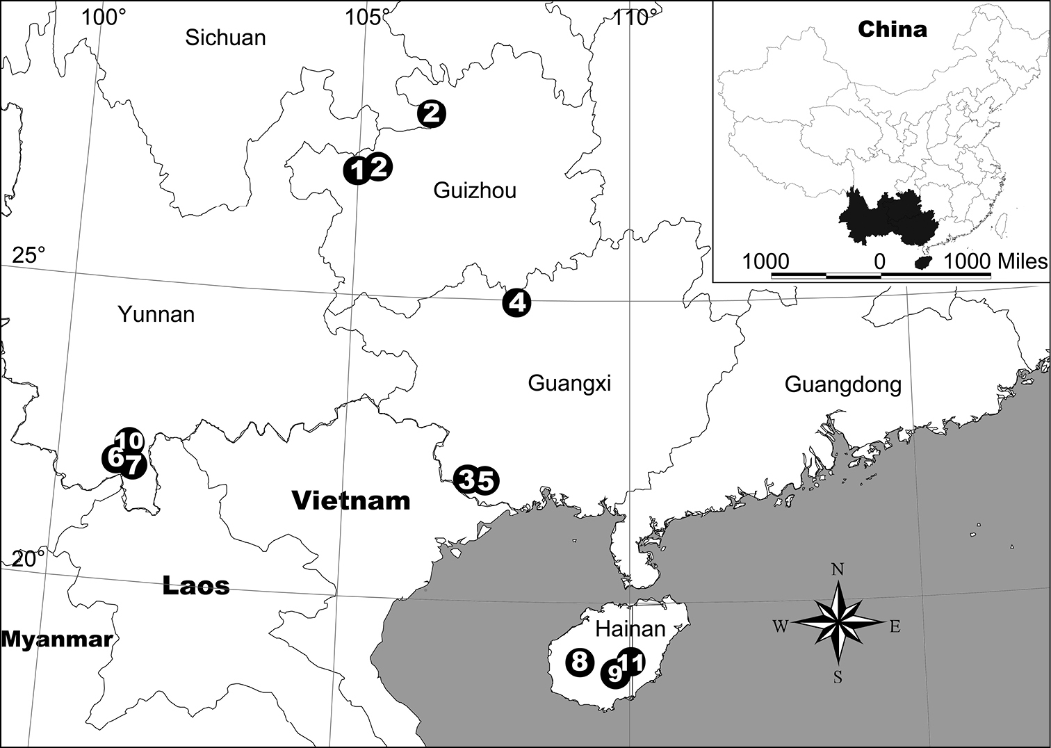
Locality records for eleven theridiosomatid spider species in China. 1 Alaria chengguanensis gen. n., sp. n. 2 Baalzebub rastrarius sp. n. 3 Baalzebub youyiensis sp. n. 4 Karstia nitida sp. n. 5 Karstia prolata sp. n. 6 Menglunia inaffecta gen. n., sp. n. 7 Ogulnius hapalus sp. n. 8 Theridiosoma plumaria sp. n. 9 Theridiosoma triumphalis sp. n. 10 Theridiosoma vimineum sp. n. 11 Zoma fascia sp. n.
The manuscript benefited greatly from comments by Jeremy A. Miller (Naturalis Biodiversity Center) and Michael G. Rix (Western Australian Museum). The study was supported by the National Natural Sciences Foundation of China (China National funds for Distinguished Young Scientists-31025023) and the Knowledge Innovation Program of the Chinese Academy of Sciences (KSCX2-EW-Z-8).