


Citation: Ruan Y, Konstantinov AS, Ge S, Yang X (2014) Revision of the Chaetocnema picipes species-group (Coleoptera, Chrysomelidae, Galerucinae, Alticini) in China, with descriptions of three new species. ZooKeys 387: 11–32. doi: 10.3897/zookeys.387.6672
The Chinese Chaetocnema picipes species-group is revised. It contains 5 species including 3 new species: C. cheni sp. n., C. constricta sp. n. and C. kingpinensis sp. n. The lectotype of C. fortecostata is designated. A key to all known species of this group from China and the illustrations of habitus and genitalia are provided. A distribution map of species is given.
Coleoptera, Alticinae, species group, new species, China, flea beetles
Chaetocnema Stephens, 1831 is a cosmopolitan flea beetle genus with over 400 species known to world (
Two distinct subgenera of Chaetocnema are recognized in the Palearctic. They are separated based on the following characters: relative width of the frontal ridge and density and size of punctures on vertex. Since a distinguishing power of these characters weakens significantly in more southern faunas (
Chaetocnema species of the picipes group are usually found in the field feeding on Rubus, Polygonum and Solanum.
We studied all the specimens in IZCAS previously identified as Chaetocnema concinna (Marsham) from different provinces of China. It turned out that they are indeed Chaetocnema picipes, Chaetocnema fortecostata sp. n. or Chaetocnema constricta sp. n. Chaetocnema concinna is not found in China and all the published records of it in China should be treated as misidentifications.
The female genitalia were dissected and mounted onto slides with Hoyer’s medium, photos were taken with digital camera NIKON 5200D attached to the ZEISS AXIOSTAR PLUS Microscope. The photos of habitus were taken with the 5× lens of the same microscope with extra light source softened by semitransparent paper, so as to observe the real color of these tiny beetles. The photos of aedeagus were taken with the KEYENCE VHX-600 microscope. Scanning electron micrographs were taken with FEI QUANTA 450. A map of species distribution was generated by ARCGIS software. Descriptions of species were initially generated by LUCID software, exported from it and extensively edited.
Morphological terminology follows
Places of distribution of this article are arranged from north to south provinces names in “Materials” paragraphs are in bold font.
Abbreviations: MBL = male body length; MLH = male body length without head; FBL = female body length; FLH = female body length without head; AL/BL = antenna length to body length; MBW = male body width; EL/EW = elytron length (along suture) to width (maximum); PW/PL = Pronotum width (at base) to length; EL/PL = elytron length to pronotum length; EWB/PWB = elytra width at base (in middle of humeral calli) to pronotum width at base; EWM/PWM = maximum width of elytra to maximum width of pronotum.
Abbreviations of collections: BMNH, The Natural History Museum, London, United Kingdom; IZCAS, Institute of Zoology, Chinese Academy of Sciences, Beijing, China; NHRS, Naturhistoriska Riksmuseet, Stockholm, Sweden; USNM, National Museum of Natural History, Washington D.C., USA; ZMAS, Zoological Institute of Russian Academy of Sciences, St. Petersburg, Russia.
Diagnosis. Body small, usually 1.70–2.50 mm. 5–7 punctures on vertex close to each eye. Two short, weakly delineated longitudinal strip without punctures at base of pronotum. All rows of punctures on elytra single and regular, surface between rows smooth and glabrous. Median lobe of aedeagus lacking deep groove or transverse wrinkle on ventral surface, apical dentical weak or absent. Spermatheca pear-shaped or cylindrical, proximal part of spermatheca duct straight. All five species are very similar exteriorly. Color of their bodies and appendages varies between samples collected from different location. The most consistent characteristic to differentiate these five species is the shape of the male genitalia.
Based on the narrow frontal ridge and 5-7 punctures near each eye, species of the picipes group can be placed to the Chaetocnema subgenus.
| 1 | Body broad. First male protarsomere distinctly larger than second, appendages dark in color, anterolateral angles of pronotum round | 2 |
| – | Body narrow. First male protarsomere only slightly larger than second, appendages light in color, anterolateral angles of pronotum obtuse and thickened | 4 |
| 2 | Metatibia proximad to denticle in dorsal view convex, apex of aedeagus subdeltoid, tip of aedeagus broad | Chaetocnema cheni sp. n. |
| – | Metatibia proximad to denticle in dorsal view concave, apex of aedeagus obcordate, tip of aedeagus narrow | 3 |
| 3 | Body bronzish, aedeagus thickened in lateral view | Chaetocnema fortecostata Chen, 1939 |
| – | Body copperish, aedeagus narrow in lateral view | Chaetocnema picipes Stephens, 1831 |
| 4 | Body size in male 1.80-2.54 mm and in female 2.11-2.64 mm, length of antenna to length of body about 0.70, pronotum bronzish and elytra blackish brown | Chaetocnema kingpinensis sp. n. |
| – | Body size in male 1.71–1.80 mm and in female 2.15–2.31mm, length of antenna to length of body about 0.62, pronotum and elytra bronzish | Chaetocnema constricta sp. n. |
http://species-id.net/wiki/Chaetocnema_picipes
Fig. 1Heilongjiang, Liaoning, Inner Mongolia, Beijing, Hebei, Tianjin, Shanxi, Shandong, Gansu, Qinghai, Shaanxi; Europe, North Asia (
Polygonum persicaria Linn. (Polygonaceae), Polygonum aviculare Linn., Brassica rapa Linn. (Cruciferae) (
Chaetocnema picipes very much resembles Chaetocnema cheni sp. n. and Chaetocnema fortecostata sp. n., but it can be reliably separated from them by the shape of the aedeagus (obcordate on the apex in ventral view and narrow in lateral view) and the copperish color of the body.
MBL = 1.67-1.96 mm; MBH = 1.60-1.80 mm; FBL = 2.01-2.27 mm; MBH = 1.90-2.09 mm; AL/BL = 0.60±0.05; MBW = 1.02–1.13 mm; EL/EW = 2.42–2.49; PW/PL = 1.67–1.68; EWB/PWB = 1.10±0.05; EWM/PWM = 1.40–1.41.
Color of elytra, pronotum and head consistently copperish. Antennomere 1 partly dark brown. Antennomeres 2–3 yellow. Antennomere 4 yellow or partly brown. Antennomere 5 partly brown. Remaining antennomeres black. Pro- and mesofemora brown with yellow on the apex. Metafemora brown. Tarsi brown with yellow on base of each tarsomere.
Base of pronotum with two short, obscure longitudinal impressions without punctures near basal margin. Deep row of large punctures at base of pronotum present on sides, lacking in middle. Pronotal base evenly convex. Lateral sides of pronotum slightly convex with maximum width near base. Anterolateral prothoracic callosity protruding laterally forming round angle. Posterolateral prothoracic callosity projects up to lateral margin of pronotum. Diameter of pronotal punctures 2 to 4 times smaller than distance between them.
Elytra with convex sides. Scutellar row of punctures on elytron regular and single. Remaining rows of punctures regular. Elytral humeral calli well developed. Interspaces between rows of punctures smooth and glabrous. Two lines of minute punctures on each interspace.
Head hypognathous. Frontal ridge between antennal sockets narrow and convex. Frontolateral sulcus present. Suprafrontal sulcus shallow and faint or deep laterally, shallow in middle. Suprafrontal sulcus slightly concave. Orbital sulcus (above the antennal socket) deep, but rather narrow. Width of frontal ridge to width of antennal socket: 0.900–1.005. Width of orbital sulcus to width of frontolateral sulcus: 0.611–0.614. Surface of vertex sparsely and unevenly covered with 6–7 punctures near each eye. Numbers of punctures on each orbit: 2–3. Numbers of setae along frontolateral sulcus on each side: 8–10. Numbers of setae on frons (triangular area surrounded by frontolateral sulci and clypeus): 0. Numbers of setae on clypeus: 7. Numbers of setae on labrum: 6. Anterior margin of labrum slightly concave in middle.
First male protarsomere distinctly larger than second one. First male protarsomere, length to width ratio: 1.63–1.67. First and second male protarsomeres, length to length ratio: 2.00–2.03; width to width ratio: 1.55–1.59. First male protarsomere, width at apex to width at base: 2.58–2.64. Length of metatibia to distance between denticle and metatibial apex: 2.50–2.55. Large lateral denticle on metatibia sharp. Metatibial serration proximal to large lateral denticle present, obtuse. Metatibia proximal to denticle in dorsal view concave. First male metatarsomere, length to width ratio: 3.01–3.05. First and second male metatarsomeres, length to length ratio: 1.87–1.89. First and second male metatarsomeres, width to width ratio about 0.98. Third and fourth male metatarsomeres, length to length ratio: 1.64–1.68. Metatibia length to metafemora length: 0.81±0.05. Length of hind leg to length of body: 0.92±0.05.
Median lobe of aedeagus parallel-sided with apical third slightly widening. Apical part of median lobe in ventral view narrowing abruptly. Ventral longitudinal groove of median lobe absent in apical part and poorly developed in middle and basal part. Apical denticle of aedeagus in ventral view poorly differentiated, straight in lateral view. Minute transverse wrinkles on ventral side of median lobe absent. Median lobe in lateral view narrow and evenly curved. Width (in middle) to length of median lobe (in ventral view) about 0.15.
Spermathecal receptacle pear-shaped. Spermathecal pump much shorter than receptacle. Apex of spermathecal pump cylindrical. Spermathecal pump attached to middle of receptacle top. Maximum width of receptacle situated basally. Basal part of receptacle wider than apical. Posterior sclerotization of tignum spoon-shaped, wider than mid section. Anterior sclerotization of tignum wider than mid section. Apex of vaginal palpus subdeltoid, with lateral side slightly arching. Sides of middle part of vaginal palpus (before apex) narrowing from base, slightly widening towards apex. Anterior sclerotization of vaginal palpus slightly widening anteriorly. Anterior sclerotization of vaginal palpus slightly and evenly curved along length. Anterior end of anterior sclerotization broadly rounded. Length of posterior sclerotization greater than width. Posterior sclerotization about as wide as anterior sclerotization.
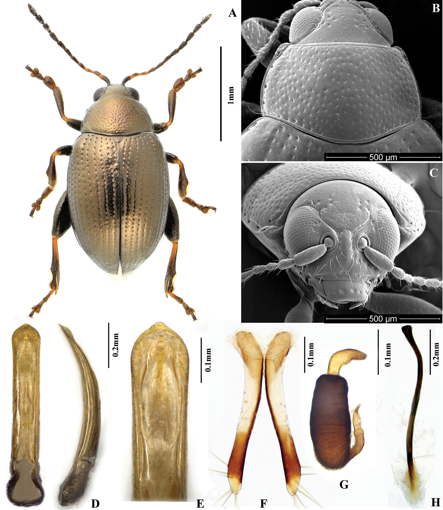
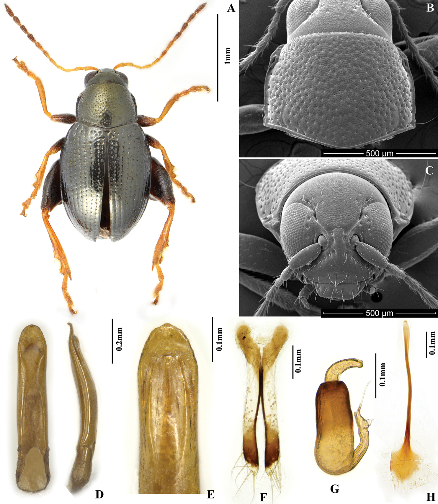
Chaetocnema picipes, (Qinling Mountain, Shaanxi, China). A Male habitus B Pronotum C Head D Aedeagus, ventral and lateral view E Apical part of aedeagus, dorsal view F Vaginal palpi G Spermatheca H Tignum.
(all the materials preserved in IZCAS): 1, Harbin, Heilongjiang, 11.VI.1965, leg. P. M. Hammond; 2♀1♂, Fujin, Heilongjiang, 16.VIII.1970; 15♀3♂, Mishan, Heilongjiang, 11-21.VIII.1970; 10♀2♂, Molida, Daxinganling Mountains, Heilongjiang, VII-VIII.1970; 1♂, Lingyuan, Liaoning; 1♀1♂, Chifeng, Inner Mongolia, 8.VIII.1956; 1♂, Fangshan, Beijing, leg. Cong; 5, Beijing, 5.VII.1980, leg. Subai Liao; 4, Beijing, 28.VI.1980, leg. Subai Liao; 14, Zhongguancun, Beijing, 8.VI.1962, leg. Shuyong Wang; 15, Yanqing, Beijing, 1.VII.1990, leg. Shuyong Wang; 1♀2♂, Shan-hai-Kwan, Hebei, 1.IX.1906, leg. F. M. Thomson; 1♀1♂, Xinglong, Hebei, 10.VII.1963, leg. Shengqiao Jiang; 1♂, Tianjin, 26.IX.1929; 1♀, Tianjing, 11.IV.1955; 5♀4♂, Tianjing, leg. F. M. Thomson, 1904; 3♀5♂, Lishan National Reserve, Shanxi, 112.016°E, 35.420°N, alt.1560m, 26.VII.2012, leg. Yongying Ruan & Zhengzhong Huang, feed on Polygonum sp.; 1♀1♂, Long-tong, Tsinanfou (Jinan), Shandong; 13♀4♂, Qiujiaba, Wenxian, Gansu, alt.2200-2350m, 29.VI.1998, leg. Shuyong Wang; 1♀, Datong, Qinghai, V.1956; 13♀28♂, Niubeiliang National Reserve, Qinling Mountain, Shaanxi, alt.1690m, 30.VI.2013, leg. Yuanyuan Lu; 4, Niubeiliang National Reserve, Qinling Mountain, Shaanxi, alt.1800m, 11.VI.2013, leg.Yongying Ruan; 5♀11♂, Haopingsi National Reserve, Qinling Mountain, Shaanxi, 34.095°N, 107.707°E, alt.1200m, 23.VIII.2013, leg. Yongying Ruan; 3♀9♂, Fengxian, Qinling Mountain, Shaanxi, 34.2352°N, 106.9572°E, alt.1500m, 21.VIII.2013, leg. Yongying Ruan.
This species was recently revised by
We did not find any Chaetocnema picipes specimens from South China, it seems that Chaetocnema picipes is distributed only in the Palaearctic part of China. The southern boundary of the distribution of Chaetocnema picipes is the Qingling Mountain which is also a southern boundary of many other Palaearctic faunistic elements (
http://species-id.net/wiki/Chaetocnema_fortecostata
Fig. 2Shaanxi, Hubei, Chongqing, Sichuan, Zhejiang, Hunan, Jiangxi, Fujian, Yunnan, Guangxi.
Polygonum sp. (Polygonaceae).
Chaetocnema fortecostata sp. n. is similar to Chaetocnema picipes and Chaetocnema cheni sp. n. But the aedeagus in lateral view is robust in Chaetocnema fortecostata and slender in Chaetocnema picipes and Chaetocnema cheni. Chaetocnema fortecostata have bronzish dorsal surface of its body, while Chaetocnema picipes and Chaetocnema cheni are copperish.
MBL = 1.75–1.90 mm; MBH = 1.60–1.79 mm; FBL = 2.03–2.12 mm; FBH = 1.89–2.03 mm; AL/BL = 0.64±0.05; MBW = 0.95–1.08 mm; EL/EW = 2.64±0.05; PW/PL =1.62±0.05; EL/PL = 2.98±0.05; EWB/PWB = 1.07–1.19; EWM/PWM = 1.34±0.05.
Color of dorsal side of body bronzish throughout, including head. Antennomere 1 partly dark brown. Antennomeres 2–3 yellow. Antennomere 4 yellow or partly brown. Antennomere 5 partly brown. Remaining antennomeres black. Pro- and mesofemora brown with yellow on apex. Metafemora brown. Tarsi brown with yellow on base of each tarsomere.
Head hypognathous. Frontal ridge between antennal sockets narrow and convex. Frontolateral sulcus present. Suprafrontal sulcus shallow and faint or deep laterally, shallow in middle. Suprafrontal sulcus slightly concave. Orbital sulcus (above antennal socket) obscure and narrow. Width of frontal ridge to width of antennal socket: 0.56-0.66. Width of orbital sulcus to width of frontolateral sulcus: 0.71±0.05. Surface of vertex sparsely and unevenly covered with 5-6 punctures near each eye. Numbers of punctures on orbit: 1-2 on each side. Numbers of setae along frontolateral sulcus: 5-6 on each side. Numbers of setae on frons (triangular area surrounded by frontolateral sulcus and clypeus): 0. Numbers of setae on clypeus: 5. Numbers of setae on labrum: 6. Anterior margin of labrum slightly convex in middle.
Base of pronotum with two short, obscure longitudinal impressions without punctures near basal margin. Deep row of large punctures at base of pronotum present on sides, lacking in middle. Shape of pronotal base evenly convex. Lateral sides of pronotum slightly convex with maximum width near base. Anterolateral prothoracic callosity protruding laterally forming strong round angle. Posterolateral prothoracic callosity projects up to lateral margin of pronotum. Diameter of pronotal punctures 2 to 4 times smaller than distance between them.
Elytra with convex sides. Scutellar row of punctures regular and single. Remaining rows regular. Elytral humeral calli well developed. Interspace smooth and glabrous. 2 lines of minute punctures on each interspace.
First male protarsomere, length to width ratio: 1.65±0.05. First and second male protarsomeres, length to length ratio: 1.94–2.24, width to width ratio: 1.42–1.45. First male protarsomere, width at apex to width at base: 1.60–1.75. Length of metatibia to distance between denticle and metatibial apex: 2.80–2.90. Large lateral denticle on metatibia sharp. Metatibial serration proximal to large lateral denticle present, obtuse. Metatibia proximad to denticle in dorsal view concave. First male metatarsomere, length to width ratio: 3.60–3.67. First and second male metatarsomeres, length to length ratio: 1.87–1.93, width to width ratio: 0.83. Third and fourth male metatarsomeres, length to length ratio: 0.60–0.65. Metatibia length to metafemora length: 0.76±0.05. Length of hind leg to length of body: 0.87±0.05.
Median lobe of aedeagus quite robust and thickened in lateral view. Apical third of median lobe widening evenly. Apical part of median lobe in ventral view narrowing abruptly. Ventral longitudinal groove of median lobe absent in apical part and poorly developed in middle and basal part. Apical denticle of aedeagus in ventral view poorly differentiated, curved ventrally in lateral view. Minute transverse wrinkles on ventral side of median lobe absent. Median lobe in lateral view slightly sinuous near apex. Maximal curvature of median lobe in lateral view situated medially. Width (in middle) to length of median lobe (in ventral view) about: 0.15.
Spermathecal receptacle pear-shaped. Spermathecal pump much shorter than receptacle. Apex of spermathecal pump cylindrical. Spermathecal pump attached to middle of receptacle top. Maximum width of receptacle situated basally. Basal part of receptacle wider than apical. Posterior sclerotization of tignum spoon-shaped, wider than mid section. Anterior sclerotization of tignum narrower than mid section. Apex of vaginal palpus subdeltoid, with lateral side slightly arching. Sides of mid part of vaginal palpus (before apex) narrowing from base, widening towards apex. Anterior sclerotization of vaginal palpus slightly widening anteriorly. Anterior sclerotization of vaginal palpus slightly and evenly curved along length. Anterior end of anterior sclerotization broadly rounded. Length of posterior sclerotization greater than width. Posterior sclerotization about as wide as anterior.
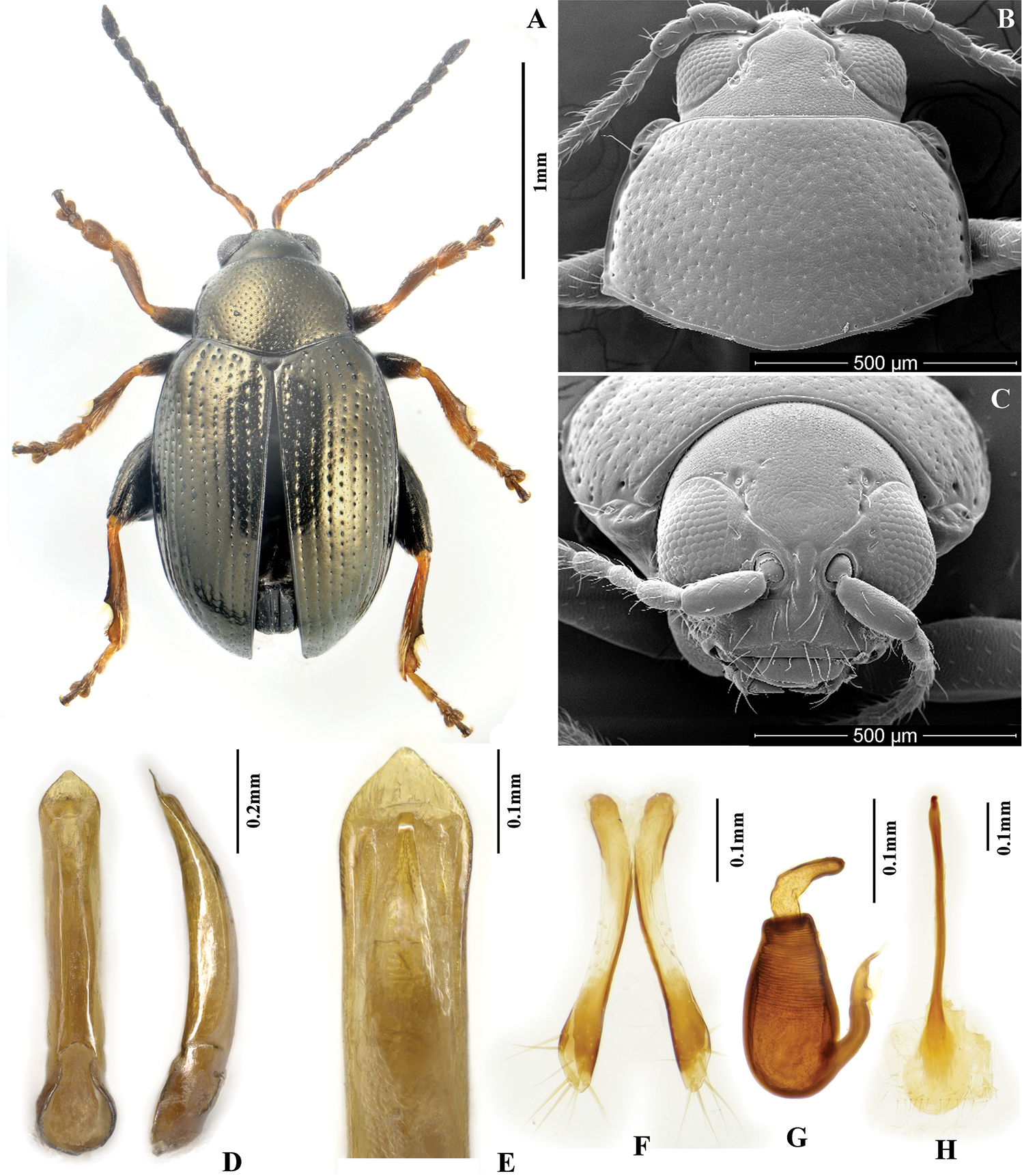
Chaetocnema fortecostata. A Male habitus B Pronotum C Head D Aedeagus, ventral and lateral view E Apical part of aedeagus, dorsal view F Vaginal palpi G Spermatheca H Tignum.
(preserved in IZCAS). Lectotype (designated here): 1♂, (1) Yangshuo, 21.VIII.1938, (2) Lectotype, Chaetocnema fortecostata Chen, 1939, des. Yongying Ruan et al.
Paralectotypes (designated here): 2♂5♀, (1) Yangshuo, 21.VIII.1938, (2) Paralectotype, Chaetocnema fortecostata Chen, 1939, des. Yongying Ruan et al.
(all the materials preserved in IZCAS). 19♀14♂, Huoditang, Qinling Mountain, Shaanxi, alt.1600m, 6.VI.2013, leg. Yongying Ruan, feed on Polygonum sp.; 3, Maoping, Yangxian, Qinling Mountain, Shaanxi, alt.701m, 10.VI.2013, leg. Yongying Ruan; 2♂, Longmen River, Xingshan, Hubei, alt.1300m, leg. Shimei Song; 6, Longmen River, Xingshan, Hubei, 8.IX.1994, alt.1300m, leg. Jian Yao, feed on Polygonum sp.; 1♀2♂, Sanxia Linchang, Badong, Hubei, 26.VI.1994, alt.130m, leg. Jian Yao; 2♀1♂, Beibei, Chongqing, 17.V.1941; 3♂, Longchi, Sichuan, IX.29; 1♀2♂, Fengdu, Sichuan, alt.200m, 29.IX.1994, leg. Shimei Song; 56, Wangerbao, Wangxian, Sichuan, alt.1200m, 4.X.1994, leg. Jian Yao; 1♀1♂, Chudian, Emei Mountain, Sichuan, 28.VI.1957, leg. Fuxing Zhu; 1♀, Tienmo Shan, Zhejiang, 20.IX.1953; 24, Shanmuhe, Hunan, alt.600, 14.VIII.1988, leg. Shuyong Wang; 7♀4♂, Xingzi, Jiangxi, 1932; 1♀1♂, Shuyang, Fuan, Fujian, IV.3013, leg. Yongying Ruan, feed on Polygonum sp.; 5♀2♂, Sangxiang, Xingcun, Chongan, Fujian, alt.740m, 7.VI.1960, leg. Yong Zuo; 187, Baijixun, Weixi, Yunnan, alt.1780m, leg. Shuyong Wang, feed on Polygonum sp.; 14♀3♂, Xiaomengyang, Yunnan, alt.900m, III-IV.1957, leg. Shuyong Wang; 1♂, Damenglong, Xishuangbanna, Yunnan, alt.650m, 6.X.1958, leg. Zhizhi Chen.
There is no holotype or paratype in the IZCAS. But we found eight specimens belonging to what looks like a type series of this species labeled as “Chaetocnema fortecostata sp. n.” with Chen’s handwriting. The locality on the label corresponds with the original description. Therefore we consider these eight specimens as the syntypes. Here we designate one male as the lectotype and the remaining seven as paralectotypes.
This species only occurs in the Oriental China while the northernmost specimens were found on the southern slopes of Qinling Mountain, which is considered as a border between Palearctic and Oriental Regions within China (
http://zoobank.org/3584CDFD-3ACE-4FED-9114-BC746DFEC8B6
http://species-id.net/wiki/Chaetocnema_cheni
Fig. 3We dedicate this species to SH Chen, who originally designated it as new, but left it unpublished. Professor Chen was a classic Chinese entomologist, he laid the foundation for studies of leaf beetles in China.
Hunan, Jiangxi, Sichuan, Yunnan.
Solanum tuberosum Linn. (Solanaceae).
Chaetocnema cheni sp. n. can be differentiated from Chaetocnema kingpinensis sp. n. and Chaetocnema constricta sp. n. by the following characters: first male protarsomere clearly larger than second, appendages darker in color, anterolateral angles of pronotum round. Chaetocnema cheni can be differentiated from Chaetocnema picipes and Chaetocnema fortecostata based on the following characters: metatibia proximad to denticle in dorsal view convex, apex of aedeagus subdeltoid, tip of aedeagus widely rounded.
MBL = 1.85–2.05 mm; MBH = 1.79–1.93 mm; FBL = 2.10±0.05 mm; FBH = 2.05±0.05 mm; AL/BL = 0.60–0.61; MBW = 1.04–1.06; EL/EW = 1.29; PW/PL =1.47±0.05; EL/PL = 2.91±0.05; EWB/PWB = 1.17±0.05; EWM/PWM = 1.53±0.05.
Color of elytra usually same with or slightly different from pronotum. Color of elytra copperish, sometimes bluish black. Color of pronotum copperish, sometimes bronzish. Head dorsally copperish, sometimes bluish black. Antennomere 1 partly dark brown. Antennomeres 2–3 yellow. Antennomeres 4–5 partly brown. Remaining antennomeres black. Pro- and mesofemora brown with yellow apex. Metafemora brown. Tarsi brown with yellow on base of each tarsomere.
Head hypognathous. Frontal ridge between antennal sockets narrow and convex. Frontolateral sulcus present. Suprafrontal sulcus shallow and faint or deep laterally, shallow in middle. Suprafrontal sulcus slightly concave. Orbital sulcus (above antennal socket) deep. Width of frontal ridge to width of antennal socket: 1.19±0.05. Width of orbital sulcus (above antennal socket) to width of frontolateral sulcus: 0.64–0.67. Surface of vertex sparsely and unevenly covered with 6–7 punctures close to each eye. Numbers of punctures on orbit on each side: 1. Numbers of setae along frontolateral sulcus on each side: 9–10. Numbers of setae on frons (triangular area surrounded by frontolateral sulcus and clypeus): 0. Numbers of setae on clypeus: 7. Numbers of setae on labrum: 6. Anterior margin of labrum slightly concave in middle.
Base of pronotum with two short, obscure longitudinal impressions near basal margin. Longitudinal impressions lack punctures. Deep row of large punctures at base of pronotum present on sides, lacking in middle. Shape of pronotal base evenly convex. Anterolateral prothoracic callosity protruding laterally but poorly developed. Posterolateral prothoracic callosity projects up to lateral margin of pronotum. Diameter of pronotal punctures 2 to 4 times smaller than distance between them.
Elytra with convex sides. All rows of punctures on elytron regular and single. Elytral humeral calli well developed. Interspaces of puncture rows smooth and glabrous. Numbers of minute punctures lines on each interspace: 2.
First male protarsomere distinctly larger than second. First male protarsomere, length to width ratio: 1.50±0.05. First and second male protarsomeres, length to length ratio: 1.69±0.05, width to width ratio: 1.23±0.05. First male protarsomere, width at apex to width at base: 1.87–2.00. Length of metatibia to distance between denticle and metatibial apex: 2.34–2.47. Large lateral denticle on metatibia sharp. Metatibial serration proximal to large lateral denticle present, obtuse. Metatibia proximad to denticle in dorsal view convex. First male metatarsomere, length to width ratio: 2.47–2.68. First and second male metatarsomeres, length to length ratio: 1.58–1.62. First and second male metatarsomeres, width to width ratio: 0.92–1.00. Third and fourth male metatarsomeres, length to length ratio: 0.71±0.05. Metatibia length to the metafemora length: 0.76±0.05.
Median lobe of aedeagus widening gradually towards apex. Apical part of median lobe in ventral view narrowing abruptly forming a subdeltoid apex. Ventral surface of median lobe lateral to median groove apically convex. Ventral longitudinal groove absent in apical and middle part, shallow in basal. Apical denticle of aedeagus in ventral view absent. Apical part of aedeagus in lateral view slightly curved ventrally. Minute transverse wrinkles on ventral side of median lobe absent. Median lobe in lateral view slightly sinusoidal near apex. Median lobe narrow in lateral view. Maximal curvature of median lobe in lateral view situated medially. Width (in middle) to length of median lobe (in ventral view) about: 0.14.
Spermathecal receptacle pear-shaped. Spermathecal pump much shorter than receptacle. Apex of spermathecal pump cylindrical. Spermathecal pump attached to middle of receptacle top. Maximum width of receptacle situated basally. Basal part of receptacle wider than apical. Posterior sclerotization of tignum spoon-shaped, wider than mid section. Anterior sclerotization of tignum wider than mid section. Apex of vaginal palpus subdeltoid, with lateral side slightly arching. Sides of mid part of vaginal palpus (before apex) narrowing from base, slightly widening towards apex. Anterior sclerotization of vaginal palpus slightly widening anteriorly, slightly and evenly curved along length. Anterior end of anterior sclerotization nearly flat. Length of posterior sclerotization greater than width. Width of posterior sclerotization greater to anterior sclerotization.
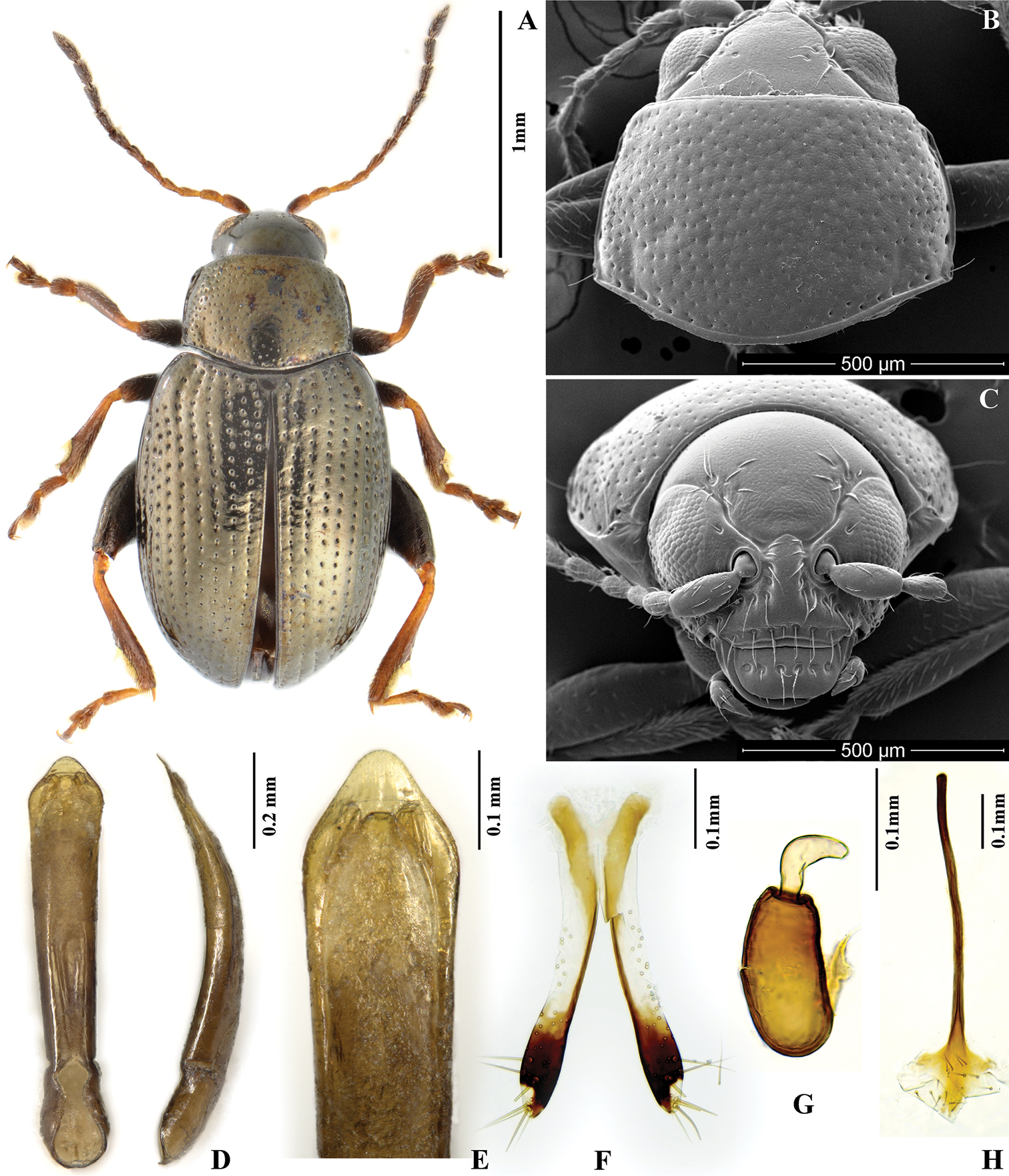
Chaetocnema cheni. A Holotype, habitus B Pronotum C Head D Aedeagus, ventral and lateral view E Apical part of aedeagus, dorsal view F Vaginal palpi G Spermatheca H Tignum.
(all the materials preserved in IZCAS): Holotype: 1♂ (Fig. 3: A), Longling, Yunnan, alt.1600m, 1955.V.20, leg. В. Попов (B. Popov). Paratypes: 2♀1♂, Tianping Mountain, Sangzhi, Hunan, alt.1370, 1988.VIII.15, leg. Shuyong Wang; 3♀1♂, Jiujiang, Jiangxi, 1958.VII-VIII; 2♀, Jiujiang, Jiangxi, 1948.VII; 2♀4♂, Jinfou Mountain, Sichuang, 1945.VIII.16. leg. Shuyong Wang; 20, Liziping, Wushan, Sichuan, alt.1850m, 1993.V.18-19, leg. Youwei Zhang; 4♂5♀, Liziping, Wushan, Sichuan, alt.1850m, 1993.VIII.5-6, leg. Xingke Yang, feed on Solanum tuberosum Linn.; 1♂, Jinpinghe, Yunnan, alt.1700m, 1956V.14, leg. Keren Huang.
There is a noticeable variability in body color among studied specimens. The holotype collected from Longling, Yunnan province is copperish in color, but the paratypes from Jinfou Mountain, Sichuang Province have greenish-bronzish pronotum and blue-blackish elytra.
http://zoobank.org/22CDA31D-F5B5-4207-895A-DCC97EEDE3DF
http://species-id.net/wiki/Chaetocnema_constricta
Fig. 4The name of this species is based on a tiny and tight beetle body.
Anhui, Sichuan, Chongqing, Guizhou, Zhejiang, Jiangsu, Jiangxi, Fujian, Yunnan, Guangxi.
Rubus corchorifolius Linn. f. (Rosaceae), Rubus fruticosus Linn., Polygonum sp. (Polygonaceae).
Body of Chaetocnema constricta sp. n. usually tiny and narrow. It can be differentiated from Chaetocnema picipes, Chaetocnema fortecostata sp. n. and Chaetocnema cheni sp. n. by the following characters: first male protarsomere only slightly larger than second, appendages light in color, anterolateral angles of pronotum obtuse and thickened. Exteriorly this species resembles Chaetocnema kingpinensis. But Chaetocnema kingpinensis is larger in body size, having longer appendages and pronotum (relative to body length). If viewed under a soft light, Chaetocnema constricta’s body is entirely bronzish, while Chaetocnema kingpinensis has usually bronzish pronotum and blackish brown elytra.
MBL = 1.71–1.80 mm; MBH = 1.52–1.65 mm; FBL = 2.15–2.31 mm; FBH = 1.88–2.16 mm; AL/BL = 0.61–0.62; MBW = 0.90–0.94; EL/EW = 1.28±0.05; PW/PL = 1.44±0.05; EL/PL = 2.55±0.05; EWB/PWB = 1.13±0.05; EWM/PWM = 1.37±0.05.
Elytra bronzish, exactly same color as pronotum. Head dorsally bronzish. Antennomere 1 partly dark brown. Antennomeres 2-4 yellow. Antennomeres 5-6 yellow with brown apex. Remaining antennomeres brown with yellow base. Pro- and meso- femora brown with yellow apex. Metafemora brown. Tibia mostly yellow, dark at distal half. Tarsi yellow.
Head hypognathous. Frontal ridge between antennal sockets narrow and convex. Frontolateral sulcus present. Suprafrontal sulcus shallow and faint or deep laterally, shallow in middle. Suprafrontal sulcus slightly concave. Orbital sulcus (above the antennal socket) very deep. Orbital sulcus forming an obvious narrow deep concave above orbit. Width of frontal ridge to width of antennal socket: 0.70–0.75. Width of orbital sulcus (above antennal socket) to width of frontolateral sulcus: 1.20–1.45. Surface of vertex sparsely and unevenly covered with 5–6 punctures on each side close to eye. Numbers of punctures on orbit on each side: 1–2. Numbers of setae along frontolateral sulcus on each side: 9–10. Numbers of setae on frons (triangular area surrounded by frontolateral sulci and clypeus): 0. Numbers of setae on clypeus: 4. Numbers of setae on labrum: 6. Anterior margin of labrum slightly concave in middle.
Base of pronotum with two short longitudinal impressions visible only near basal margin. Longitudinal impressions lack punctures. Deep row of large punctures at base of pronotum present on sides, lacking in middle. Pronotal base evenly convex. Anterolateral prothoracic callosity has finely developed blunt angle protruding antero-laterally. Posterolateral prothoracic callosity projects beyond lateral margin of pronotum. Diameter of pronotal punctures subequal to distance between them.
Elytra with convex sides. Scutellar row of punctures regular and single. Remaining rows regular. Elytral humeral calli well developed. Interspaces of rows of punctures smooth and glabrous. Two lines of minute punctures on each interspace.
First male protarsomere slightly larger than second. First male protarsomere, length to width ratio: 1.90–2.00. First and second male protarsomeres, length to length ratio: 1.60–1.80, width to width ratio: 1.05–1.13. First male protarsomere, width at apex to width at base: 1.70–1.88. Length of metatibia to distance between denticle and metatibial apex: 2.88–3.04. Large lateral denticle on metatibia sharp. Metatibial serration proximal to large lateral denticle present, obtuse. Metatibia proximate to denticle in dorsal view concave. First male metatarsomere, length to width ratio: 1.86–1.91. First and second male metatarsomeres, length to length ratio: 1.91–1.93, width to width ratio: 0.95–1.07. Third and fourth male metatarsomeres, length to length ratio: 0.59–0.76. Metatibia length to metafemora length: 0.82±0.05. Length of hind leg to length of body: 0.91±0.05.
Apical third of median lobe of aedeagus parallel-sided. Apical part of median lobe in ventral view narrowed abruptly and forms big cap on top. Ventral longitudinal groove of median lobe poorly developed, with obtuse margins. Apical part of longitudinal groove as wide as basal. Middle part of longitudinal groove narrower than basal. Apical denticle of aedeagus in ventral view absent. Minute transverse wrinkles on ventral side of median lobe absent. Median lobe in lateral view sinusoidal near apex. Maximal curvature of median lobe in lateral view situated medially. Median lobe thickened in lateral view. Width (in middle) to length of median lobe (ventral view): 0.17.
Spermathecal receptacle pear-shaped and cylindrical. Spermathecal pump much shorter than receptacle. Apex of spermathecal pump cylindrical. Spermathecal pump attached to middle of receptacle top. Basal part of receptacle about as wide as middle and apical parts separately. Posterior sclerotization of tignum spoon-shaped, wider than mid section. Apex of vaginal palpus subdeltoid, with lateral side slightly arching. Sides of mid part of vaginal palpus slightly narrowing from base, and slightly widening towards apex. Anterior sclerotization of vaginal palpus slightly narrowing anteriorly. Anterior end of anterior sclerotization narrowlly rounded. Length of posterior sclerotization greater than width. Posterior sclerotization about as wide as anterior sclerotization.
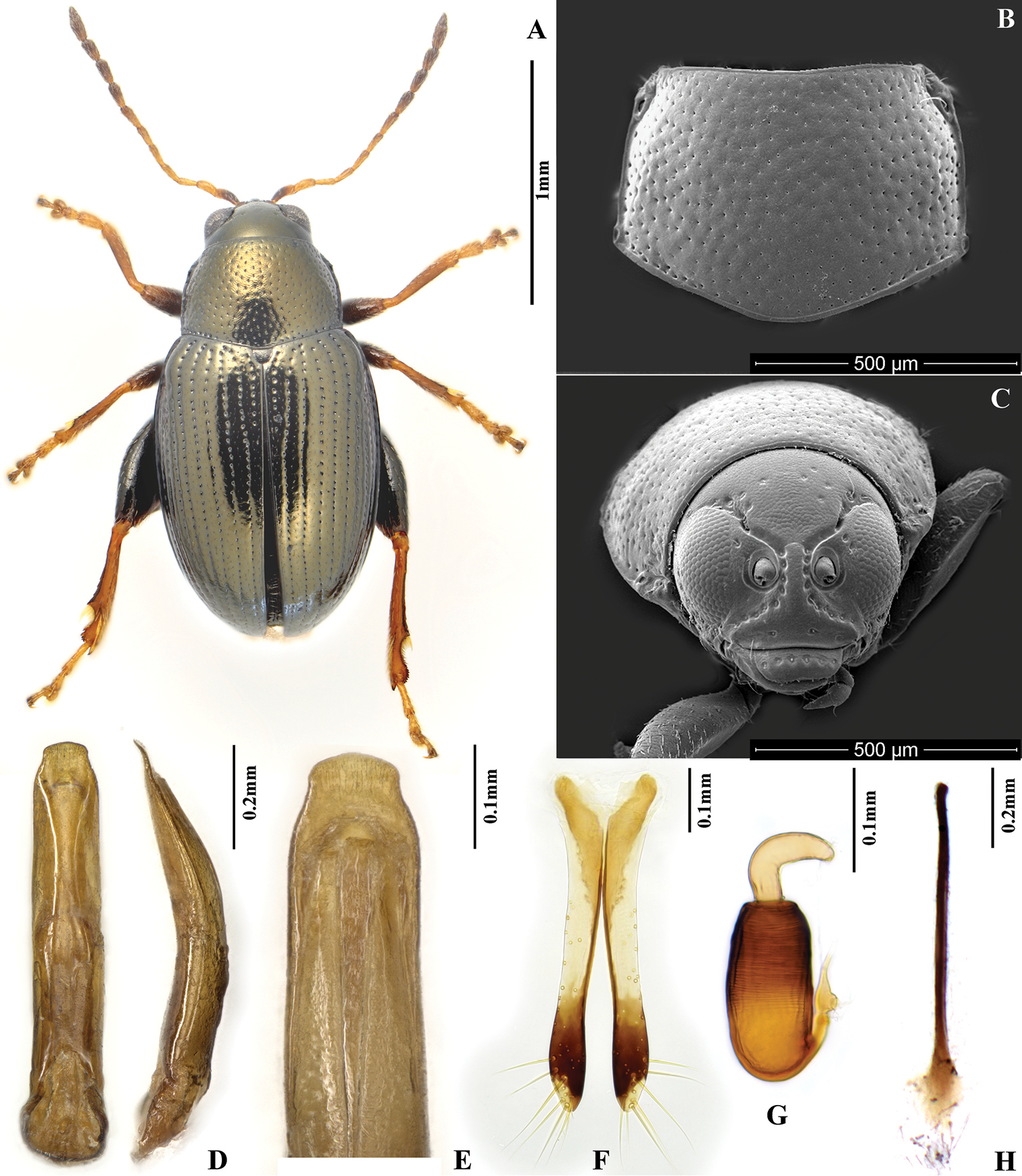
Chaetocnema constricta. A Holotype, habitus B Pronotum C Head D Aedeagus, ventral and lateral view E Apical part of aedeagus, dorsal view F Vaginal palpi G Spermatheca H Tignum.
(all the materials preserved in IZCAS). Holotype: 1♂ (Fig. 4: A), Shuyang, Fuan, Fujian, alt.200m, 2013.VIII.12, leg. Yongying Ruan. Paratypes: 30♀20♂, Huangshan, Anhui, alt.630m, 18.VIII.1978, leg. Shuyong Wang; 6♀7♂, Shaping, Sichuan, 29.XI; 6♀6♂, Ebian, Sichuan, X; 1♀, Beibei, Chongqing, 11.VI.1940; 6♀3♂, Huaxi Guizhou, 8.VI.1980; 6♀1♂, Sanmuping, Tianmu Mountain, Zhejiang, 30.VII.1998, leg. Hong Wu; 2♀, Tianmu Mountain, Zhejiang, 6.VI.1999, leg. Mingyuan Gao; 3♀1♂, Longwang Mountain, Anji, Zhejiang, 1995-1996, leg. Hong Wu; 1♂, Nanjing, Jiangsu, 1994, leg. Miao Hu; 20♀8♂, Jiulianshan, Jiangxi, 20-23.IX.1978, leg. Peiyu Yu, feed on Rubus sp.; 1♂2♀, Dazhulan, Fujian, 15-20.VI.1948; 5♀, Wuyi Mountain, Fujian, alt.500-1100m, V.1997, leg. Jiashe Wang; 1♀, Wuyi Mountain, Fujian, alt.1200m, 1997.VII, leg. Jiashe Wang; 83♀30♂, Wuyi Mountain, Fujian, 5-26.V.1997, leg. Jiashe Wang; 1♀2♂, Nanping, Fujian, 22.VII.1957, leg. Jiashe Wang; 1♂, Aotou, Huangkeng, Jianyang, Fujian, alt.750–950m, 3.VI.1997, leg.Yong Zuo; 1♀, Longling, Yunnan, alt.1600m, 20.V.1995, leg. Zifeng Xue; 1♂, Fangcheng, Guangxi, alt.650m, 14.III.1998, leg. Gexia Qiao; 10♀8♂, Jinxiu, Guangxi, alt.600m, V.1999, leg. Mingyuan Gao; 5♀2♂, Yanshan, Guilin, Guangxi, 15.VI.1963. leg. Shuyong Wang; 1♂, Tianping Mountain, Longsheng, Guangxi, 9.VI.1963, leg. Shuyong Wang; 17♀10♂, Yaoshan, Xiuren, Guangxi, 6.V.1938.
http://zoobank.org/2CD2550A-3AE9-4FA8-8658-10E855D21461
http://species-id.net/wiki/Chaetocnema_kingpinensis
Fig. 5We named this species after a place called “Kingpin” in Yunnan province where some specimens of this species were collected.
Jiangxi, Yunnan, Guangxi.
Rubus sp. (Rosaceae).
Body of Chaetocnema kingpinensis sp. n. quite narrow. It can be differentiated from Chaetocnema picipes, Chaetocnema fortecostata sp. n. and Chaetocnema cheni sp. n. by the following characters: first male protarsomere only slightly larger than second, appendages light in color, anterolateral angles of pronotum obtuse and thickened. This species resembles Chaetocnema constricta exteriorly. But Chaetocnema kingpinensis is lager in body size, with longer appendages and pronotum (relative to body length). If viewed under a soft light, Chaetocnema constricta appears entirely bronze, while Chaetocnema kingpinensis usually has bronzish pronotum and blackish brown elytra.
MBL = 1.80–2.54 mm; MBH = 1.66–2.40 mm; FBL = 2.11–2.64 mm; FBH = 1.80–2.45 mm; AL/BL = 0.70; MBW = 0.89–1.04; EL/EW = 1.29–1.29; PW/PL= 1.32; EL/PL = 1.87; EWB/PWB = 1.14; EWM/PWM = 1.45–1.45.
Color of elytra usually differs from color of pronotum. Elytra often brown to black, sometimes bronzish. Pronotum bronzish. Head dorsally dark bronzish. Antennomere 1 yellow but darker than antennomeres 2–5. Antennomeres 2–5 yellow. Antennomeres 6–7 partly brown. Antennomeres 8–11 brown with yellow at base. Tibiae yellow, tasomeres yellow with claw segment brown at apex. Pro- and mesofemora light brown with yellow apex. Metafemora brown.
Head hypognathous. Frontal ridge between antennal sockets narrow and convex. Frontolateral sulcus present. Suprafrontal sulcus shallow and faint or deep laterally, shallow in middle. Suprafrontal sulcus slightly concave. Orbital sulcus (above antennal socket) deep. Width of frontal ridge to width of antennal socket: 0.84–0.88. Width of orbital sulcus (above antennal socket) to width of frontolateral sulcus: 0.93–1.16. Surface of vertex sparsely and unevenly covered with 5–6 punctures close to each eye. Numbers of punctures on orbit on each side: 3–5. Numbers of setae along frontolateral sulcus on each side: 8–10. Numbers of setae on frons (triangular area surrounded by frontolateral sulcus and clypeus): 0. Numbers of setae on clypeus: 7. Numbers of setae on labrum: 6. Anterior margin of labrum slightly concave in middle.
Base of pronotum with two short longitudinal impressions without punctures visible only near basal margin. Deep row of large punctures at base of pronotum present on sides, lacking in middle. Pronotal base evenly convex. Lateral sides of pronotum thickened, only slightly convex with maximum width near base. Pronotum quite convex from lateral view. Anterolateral prothoracic callosity protruding antero-laterally, forms strong obtuse angle. Posterolateral prothoracic callosity projects beyond lateral margin of pronotum. Setae on each callosity long, exceeding half of pronotal length. Wrinkles between punctures on pronotum well developed. Diameter of pronotal punctures subequal to distance between them.
Elytra with convex sides. Scutellar row of punctures regular and single. All other rows of punctures regular. Elytral humeral calli well developed. Interspaces between rows of punctures on elytra smooth and glabrous. Numbers of minute punctures lines on each interspace: 2.
First male protarsomere only slightly larger than second. First male protarsomere, length to width ratio: 1.95–2.03. First and second male protarsomeres, length to length ratio: 1.43–1.52, width to width ratio: 0.89–0.91. First male protarsomere, width at apex to width at base: 1.45–1.55. Length of metatibia to distance between denticle and metatibial apex: 2.73–2.95. Large lateral denticle on metatibia sharp. Metatibial serration proximal to large lateral denticle present, obtuse. Metatibia proximad to denticle in dorsal view concave. First male metatarsomere, length to width ratio: 2.78–2.85. First and second male metatarsomeres, length to length ratio: 1.80–1.90, width to width ratio: 0.92–0.96. Third and fourth male metatarsomeres, length to length ratio: 0.62–0.71. Metatibia length to metafemora length about: 0.89. Length of hind leg to length of body about: 1.04.
Apical third of median lobe of aedeagus parallel-sided. Apical part of median lobe in ventral view narrowing abruptly. Ventral longitudinal groove of median lobe poorly developed in apical and basal part, narrow or absent in middle part. Apical part of longitudinal groove as wide as basal. Apical denticle of aedeagus in ventral view poorly differentiated. Apical denticle of aedeagus in lateral view strongly curved ventrally. Minute transverse wrinkles absent on ventral side of median lobe. Median lobe in lateral view slightly sinusoidal near apex. Maximal curvature of median lobe in lateral view situated medially. Width (in middle) to length of median lobe (in ventral view) about: 0.18. Median lobe narrow in lateral view.
Spermathecal receptacle pear-shaped, slightly narrow in middle. Spermathecal pump much shorter than receptacle. Apex of spermathecal pump cylindrical. Spermathecal pump attached to middle of receptacle top. Maximum width of receptacle situated basally. Basal part of receptacle wider than apical. Posterior sclerotization of tignum spoon-shaped, wider than mid section. Mid section of tignum nearly straight. Anterior sclerotization of tignum wider than mid section. Apex of vaginal palpus subdeltoid, with lateral side slightly arching. Sides of mid part of vaginal palpus (before apex) narrowing from base, slightly widening towards apex. Anterior sclerotization of vaginal palpus slightly narrowing anteriorly. Anterior sclerotization of vaginal palpus slightly and evenly curved along length. Anterior end of anterior sclerotization broadly rounded. Length of posterior sclerotization greater than width. Posterior sclerotization about as wide as anterior sclerotization.
Chaetocnema kingpinensis. A Holotype, habitus B Pronotum C Head D Aedeagus, ventral and lateral view E Apical part of aedeagus, dorsal view F Vaginal palpi G Spermatheca H Tignum.
(all the materials preserved in IZCAS). Holotype: 1♂ (Fig. 5: A), Lushui, Yunnan, alt.1900m, 8.VI.1981, leg. Shuyong Wang, feed on Rubus sp. 2♀1♂, Jiulianshan national reserve, Jiangxi, 8.IX.1978, leg. Youjiao Liu; Paratypes: 16♀11♂, Lushui, Yunnan, alt.1900m, 8.VI.1981, leg. Shuyong Wang, feed on Rubus sp.; 8♀4♂, Changpotou, Jingping, Yunnan, 22.V.1952, leg. Keren Huang et al., feed on Rubus sp.; 1♂, Hetouzhai, Jinping, Yunnan, alt.2000m, 22.V.1952, leg. Keren Huang et al.; 1♂, Baoshan, Yunnan, alt.1600m, 13.V.1955, leg. Bu-xi-ke & Le Wu.; 2♂, Menghun, Menghai, Xishuangbanna, Yunnan, alt.1200-1400m, 20-23.V.1958; 1♂, Tiantanshan, Jinxiu, Guangxi, alt.600m, 11.V.1999, leg. Mingyuan Gao; 4♀4♂, Tianping Mountain, Longsheng, Guangxi, 9.VI.1963, leg. Shuyong Wang; 1♂, Tianping Mountain, Longsheng, Guangxi, 740m, feed on Rubus sp. 4♀4♂, Tianping Mountain, Longsheng, Guangxi, 9.VI.1963, leg. Shuyong Wang; 1♂, Tianping Mountain, Longsheng, Guangxi, 740m, feed on Rubus sp.
This species was originally recognized as new by SH Chen. A series of paratypes were found in the IZCAS collection, but we did not find the holotype. The species was briefly mentioned by
The specimens of this species collected from Tianping Mountain are extremely large. One male from Guanxi, is 2.54 mm long, and female can be as long as 2.64 mm.
Qinling Mountain, considered as a border between Palearctic and Oriental Regions within China (
Chaetocnema cheni sp. n. seems to be a species in the transition area between Oriental and Palaearctic Region, however Chaetocnema fortecostata, Chaetocnema constricta and Chaetocnema kingpinensis are the Oriental ones.
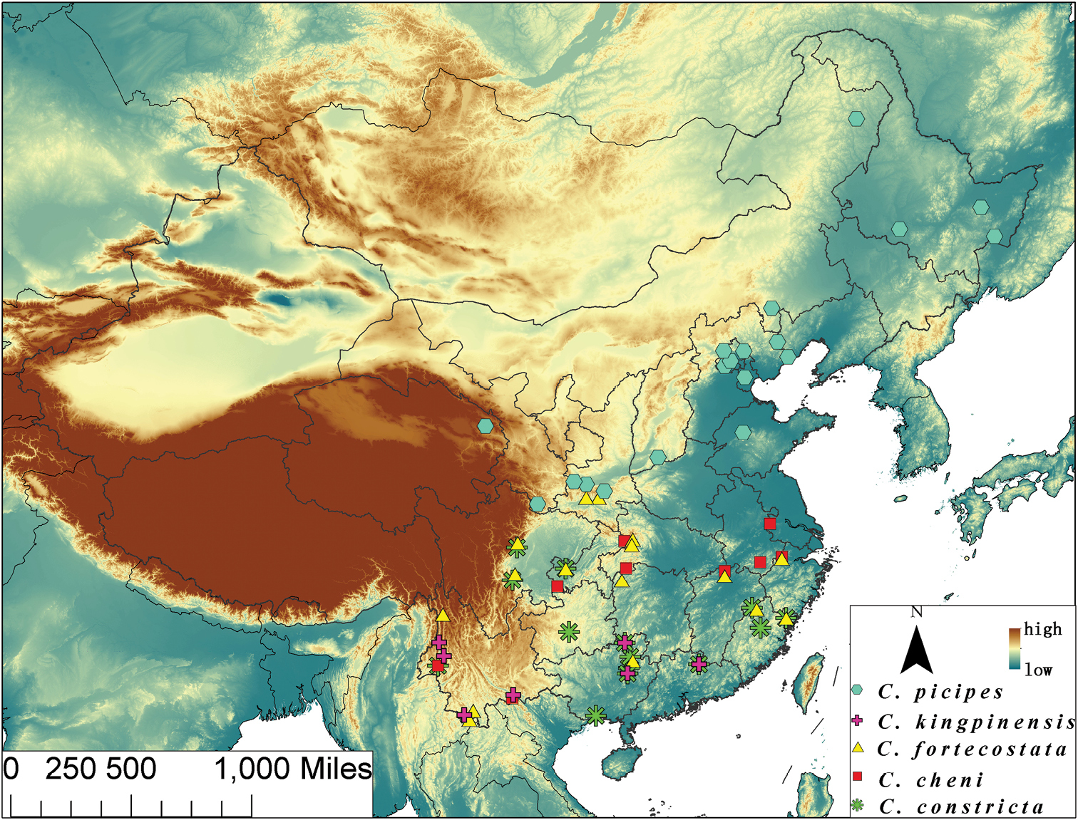
Map of continental China, illustrating localities for distribution of species. Chaetocnema picipes = blue hexagons; Chaetocnema kingpinensis = purple crosses; Chaetocnema fortecostata = yellow triangles; Chaetocnema cheni = red squares; Chaetocnema constricta = green stars.
We express our thanks to Dr. Ganyan Yang (IZCAS), for suggestions on earlier version of this manuscript. This research was supported by grants from the National Science Foundation of China to Xingke Yang (PI, Grant No. 3010300101 and Grant No. 31372239) and the National Science Fund for Fostering Talents in Basic Research (Special Subjects in Animal Taxonomy, NSFC-J1210002).