


(C) 2013 Dan Quan. This is an open access article distributed under the terms of the Creative Commons Attribution License 3.0 (CC-BY), which permits unrestricted use, distribution, and reproduction in any medium, provided the original author and source are credited.
For reference, use of the paginated PDF or printed version of this article is recommended.
Citation: Quan D, Chen J, Jie Liu J (2013) First description of the female of Sinopoda serrata (Wang, 1990) (Araneae, Sparassidae). ZooKeys 321: 89–96. doi: 10.3897/zookeys.321.5752
The female of Sinopoda serrata (Wang, 1990) is described for the first time from Tiantangzhai National Forest Park, Hubei province, China. This species has been recorded from the region of Central China. Morphological descriptions and illustrations of this species are given.
Taxonomy, biodiversity, systematics, huntsman spiders
The spider genus Sinopoda Jäger, 1999 is distributed in East Asia and northern parts of South East Asia with 49 species described so far, 32 of which are known from China (Jäger 2012,
The species Sinopoda serrata (Wang, 1990) was first described in Heteropoda Latreille, 1804, based on male specimens only from Mt. Lushan (Jiangxi Province, China) and Mt. Huangshan (Anhui Province, China) (
Specimens were examined with an Olympus SZX16 stereomicroscope; details were further investigated with an Olympus BX51 compound microscope. All illustrations were made using an Olympus drawing tube. Male palp and epigyne were examined and illustrated after dissection from the spider bodies. Photos were made with a Canon G10 digital camera (14.7 megapixels) mounted on an Olympus SZX16 stereomicroscope. The digital images depicting the habitus and genital morphology were a composite of multiple images taken at different focal planes along the Z axis and assembled using the software package Helicon Focus 3.10. Most hairs and macrosetae were usually not depicted in the palp and epigyne drawings.
Leg measurements are shown as: total length (femur, patella, tibia, metatarsus, tarsus). Number of spines are listed for each segment in the following order: prolateral, dorsal, retrolateral, ventral (in femora and patellae ventral spines are absent and fourth digit is omitted in the spination formula).
Abbreviations: ALE — anterior lateral eyes, AME — anterior median eyes, C — conductor, E — embolus, EA — embolic apophysis, FD — fertilization duct, GA — glandular appendage, LF — lateral furrow, LL — lateral lobes, LS — lobal septum, MSu — membranous sac unexpanded, PLE — posterior lateral eyes, PME — posterior median eyes, PP — posterior part of spermathecae, RTA — retrolateral tibial apophysis, T — tegulum. I, II, III, IV — legs I to IV. Collections: HBU — Hubei University, Wuhan, China; HNU — Hunan Normal University, Changsha, China.
http://species-id.net/wiki/Sinopoda_serrata
Figs 1–171 ♂ (holotype, HNU), Mt. Lushan, Jiangxi Province, China, 15 June 1987, Xianjing Peng leg.; 1 ♂ (paratype, HNU), Mt. Huangshan, Anhui Province, China, October 1979, Jiafu Wang leg.
2 ♂, 7 ♀ (HBU), Tiantangzhai National Forest Park (30°24'01.37"N, 115°18'19.31"E), Hubei, China, 8 September 2012, Fengxiang Liu, Jie Liu and Dan Quan leg.
Male of Sinopoda serrata is similar to Sinopoda albofasciata Jäger & Ono, 2002 in having the unbifurcated RTA, the slightly bent tip of embolus, but can be distinguished from the latter by the following characters: 1. RTA massive, but small in Sinopoda albofasciata (
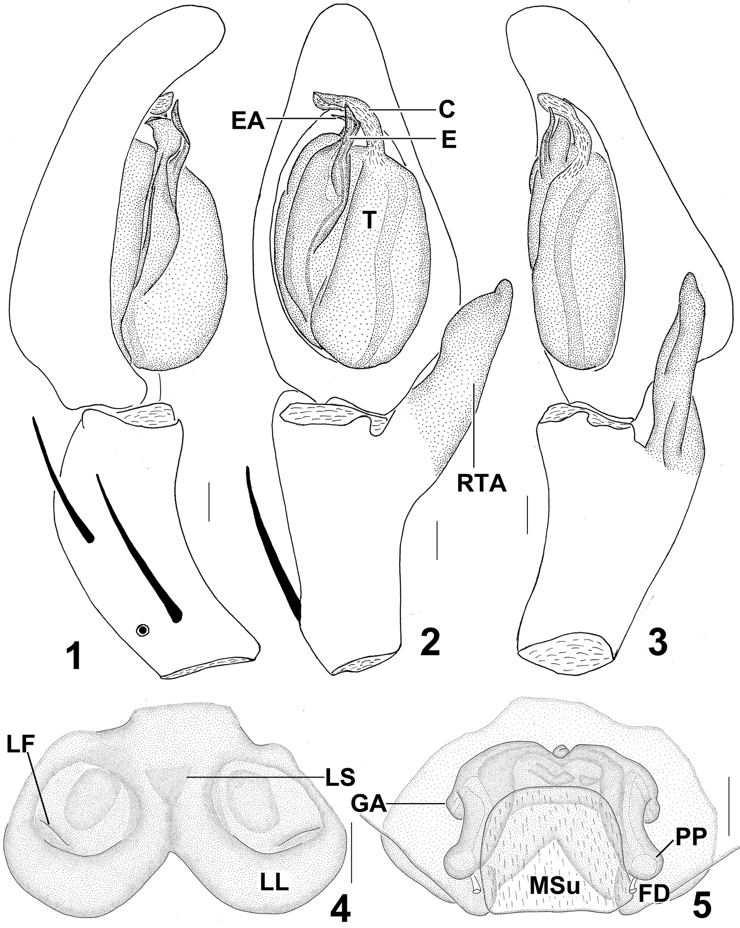
Sinopoda serrata (Wang, 1990), from Tiantangzhai National Forest Park (Hubei Province, China). 1 Left male palp, prolateral view 2 Left male palp, ventral view 3 Left male palp, retrolateral view 4 Epigyne, ventral view 5 Vulva, dorsal view. Scales = 0.2 mm. C conductor, E embolus, EA embolic apophysis, FD fertilization duct, GA glandular appendage, LF lateral furrow, LL lateral lobes, LS lobal septum, MSu membranous sac unexpanded, RTA retrolateral tibial apophysis, PP posterior part of spermathecae, T tegulum.
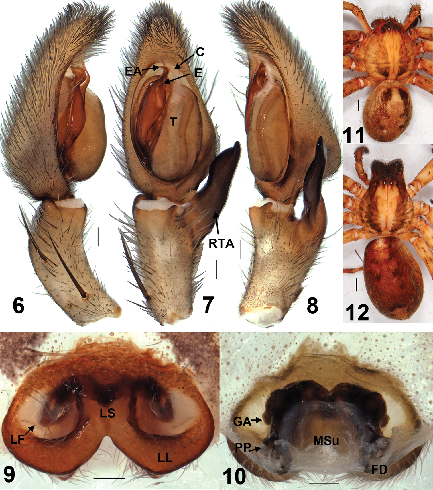
Sinopoda serrata (Wang, 1990), from Tiantangzhai National Forest Park (Hubei Province, China). 6 Left male palp, prolateral view 7 Left male palp, ventral view 8 Left male palp, retrolateral view 9 Epigyne, ventral view 10 Vulva, dorsal view11 Male habitus, dorsal view12 Female habitus, dorsal view. Scales = 0.2 mm (6–10), scales = 1 mm (11–12). C conductor, E embolus, EA embolic apophysis, FD fertilization duct, GA glandular appendage, LF lateral furrow, LL lateral lobes, LS lobal septum, MSu membranous sac unexpanded, RTA retrolateral tibial apophysis, PP posterior part of spermathecae, T tegulum.
Male: Measurements: Prosoma length 4.97, width 4.19, anterior width 2.32, height 2.17; opisthosoma length 5.04, width 2.94. Eyes: AME 0.17, ALE 0.33, PME 0.23, PLE 0.29, AME–AME 0.24, AME–ALE 0.10, PME–PME 0.30, PME–PLE 0.37, AME–PME 0.41, ALE–PLE 0.36, clypeus height at AME 0.33, clypeus height at ALE 0.28. Leg and palp measurements: Palp 7.33 (2.36, 1.34, 1.44, -, 2.19), I 20.38 (4.73, 1.97, 6.76, 5.04, 1.88), II 21.99 (5.96, 2.24, 5.61, 6.12, 2.06), III 16.22 (4.73, 1.63, 4.16, 4.10, 1.60), IV 18.16 (5.01, 1.73, 4.40, 5.03, 1.99). Leg formula: II-I-IV-III. Spination: palp 131, 101, 1021; femur I–III 323, IV 321; patella I–IV 101; tibia I–II, III 2126, IV 2326; metatarsus I–II 1014, III–IV 3036. Chelicerae yellowish-brown. Furrow with 3 anterior teeth, 4 or 6 posterior teeth, and with ca.80 denticles in elongated patch close to anterior teeth. Margins of fang base with one bristle. Palpal claw with 6 or 7 teeth. Sternum, ventral coxae and femora, distal legs as well as frontal chelicerae with long setae, otherwise with shorter setae.
Embolus (E) tip short, slender, slightly curved prolaterally, proximal part of embolus fully visible in the ventral view. Embolic apophysis (EA) short, lamellar, strongly curved prolaterally. Sperm duct (SD) curved in ventral view. RTA large, not bifurcate, arising distally from tibia. Cymbium slightly longer than tibia (Figs 1–3, 6–8).
Colouration in ethanol (Fig. 11): Yellowish- to slightly yellowish-brown. Dorsal prosoma yellowish-brown with petaline patterns, which are divided by the bright yellowish region between posterior eye row and posterior margin of carapace. Sternum, ventral coxae and femora, gnathocoxae, and labium pale yellowish-brown, gnathocoxae and labium proximally reddish-brown. Chelicerae yellowish-brown. Legs pale yellowish-brown with distal parts slightly darker, dorsal femora with dark pattern. Dorsal opisthosoma with two pairs of spots situated in the median part, with a pale yellow inverted triangle-shaped pattern near the spinnerets. Lateral parts of opisthosoma reddish-brown, Ventral opisthosoma with little dark patterns.
Female: Measurements: Prosoma length 4.94, width 4.52, anterior width 2.49, height 2.15; opisthosoma length 7.33, width 4.86. Eyes: AME 0.22, ALE 0.31, PME 0.22, PLE 0.32, AME–AME 0.22, AME–ALE 0.11, PME–PME 0.32, PME–PLE 0.52, AME–PME 0.43, ALE–PLE 0.40, clypeus height at AME 0.32, clypeus height at ALE 0.33. Leg and palp measurements: Palp 6.23 (1.98, 0.94, 1.34, -, 1.97), I 14.37 (4.13, 1.82, 3.80, 3.51, 1.47), II 16.42 (4.61, 2.18, 4.26, 3.91, 1.46), III 13.42 (4.05, 1.68, 3.24, 3.15, 1.30), IV 15.64 (4.46, 1.54, 3.72, 4.16, 1.76). Leg formula: II-IV-I-III. Spination: palp 131, 100, 2121, 1014; femur I–III 323, IV 321; patella I 101, II–IV 001; tibia I–III 2026, IV 2326; metatarsus I–II 1014, III–IV 3036. Chelicerae yellowish-brown. Furrow with 3 anterior teeth, 4 posterior teeth, and with ca.80 denticles in elongated patch close to anterior teeth. Margins of fang base with one bristle. Palpal claw with 7 teeth. Sternum, ventral coxae and femora, distal legs as well as frontal chelicerae with long setae, otherwise with shorter setae.
Epigynal field wider than long. Lateral lobes (LL) fused, posteriorly with median incision. Epigynal pockets running from latero-posterior to medio-anterior, where copulatory openings are situated. Lateral furrows (LF) distinct, running to lateral margins of lateral lobes. Lobal septum (LS) wide, significantly short. Internal duct system significantly wider than long, its left part widely separated from right part. Glandular appendages (GA) small, extending not in posterior half of internal duct system. Posterior part of spermathecae (PP) strongly short, bulging slightly laterally. Fertilization ducts (FD) arising posterio-laterally. Membranous sac between fertilisation ducts unexpanded, almost square-shaped (Figs 4–5, 9–10).
Colouration in ethanol (Fig. 12) as in male.
There is a small difference between the holotype male and the new collected materials: the middle part of RTA slightly covered the cymbium from the ventral view in the new collected materials, but not in the holotype (Figs 2, 7, 14).
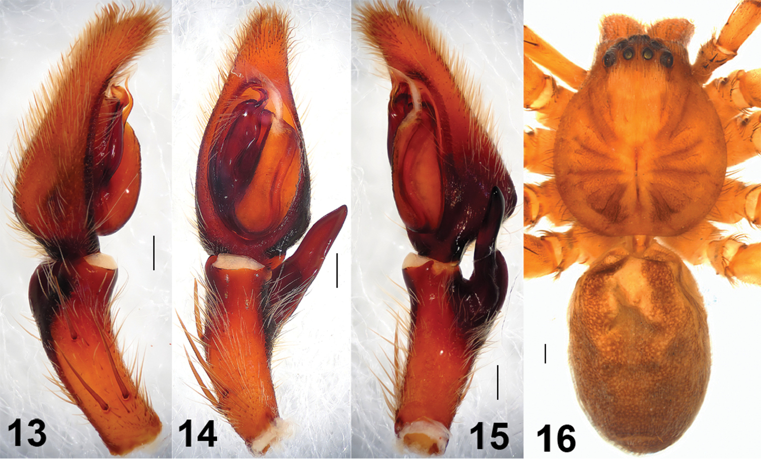
Sinopoda serrata (Wang, 1990), holotype, from Mt. Lushan (Jiangxi Province, China). 13 Left male palp, prolateral view 14 Left male palp, ventral view 15 Left male palp, retrolateral view 16 Male habitus, dorsal view. Scales = 0.2 mm (13–15), scale = 1 mm (16).
China (Hubei, Jiangxi, Anhui) (Fig. 17).
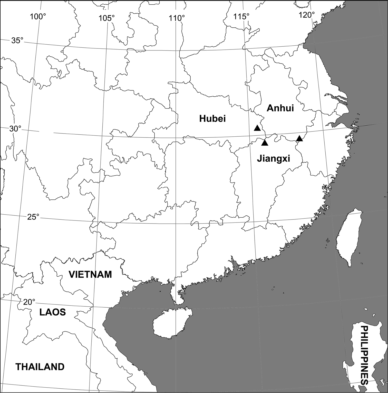
Collection localities of Sinopoda serrata (Wang, 1990) in China.
We thank Mr Fengxiang Liu (College of Life Sciences, Hubei University) for collecting Sparassidae specimens, also thank Prof. Xiang Xu (College of Life Sciences, Hunan Normal University) for checking and taking photos of the type specimens. The manuscript benefited from comments by Dr Peter Jäger (Senckenberg Forschungsinstitut, Germany) and Jeremy Miller (Department of Terrestrial Zoology, Naturalis Biodiversity Center, The Netherlands). We are grateful to one anonymous referee for his comments on the manuscript. This study was financially supported by the National Natural Sciences Foundation of China (NSFC-31272268/31172113).