ZooKeys 243: 1–14, doi: 10.3897/zookeys.243.3668
Redescription of two Pennellids (Copepoda, Siphonostomatoida) from Korea with a key to species of Peniculus von Nordmann, 1832
B. A. Venmathi Maran 1, Seong Yong Moon 2, Sung-Yong Oh 1, Ho Young Soh 2,  Jung-Goo Myoung 1
1 Marine Ecosystem Research Division, Korea Institute of Ocean Science & Technology, P. O. Box 29, Seoul 425-600, Republic of Korea
2 Faculty of Marine Technology, Chonnam National University, Yeosu, Jeollanamdo 550-749, Republic of Korea

Corresponding author: B. A. Venmathi Maran (bavmaran@gmail.com, bavmaran@kiost.ac)

Academic editor: D. Defaye

received 11 July 2012 | accepted 12 November 2012 | Published 16 November 2012


(C) 2012 B. A. Venmathi Maran. This is an open access article distributed under the terms of the Creative Commons Attribution License 3.0 (CC-BY), which permits unrestricted use, distribution, and reproduction in any medium, provided the original author and source are credited.


For reference, use of the paginated PDF or printed version of this article is recommended.

Abstract

Redescriptions of two pennellid copepods, Peniculus minuticaudae Shiino, 1956 and Peniculus truncatus Shiino, 1956, are provided, based on postmetamorphic adult females collected from marine ranched fishes captured at Tongyeong marine living resources research & conservation center, Korea. Peniculus minuticaudae was collected from the soft fin rays of black scraper Thamnaconus modestus. It can be distinguished from the other two closely related congeners Peniculus ostraciontis Yamaguti, 1939 and Peniculus truncatus by having a well developed triangular-shaped abdomen; the abdomen is rudimentary in other two species. This is thefirst report of the occurrence of Peniculus minuticaudae in Korea. Peniculus truncatus was collected from the dorsal fin of Korean rockfish Sebastes schlegelii. It can be distinguished from Peniculus minuticaudae by the combination of a rudimentary abdomen, long neck and setae on leg 1 and from Peniculus ostraciontis by the long neck, slender trunk, and setae on leg 1. It is also shown that Peniculus truncatus captured from the same host in Korea was misidentified as Peniculus ostraciontis and hence, this is thesecond record of the occurrence of Peniculus truncatus in Korea. A key is provided for the 14 nominal species of Peniculus.

Keywords

Copepod, pennellid, parasite, redescription, black scraper, rockfish, fins, identification, key

Introduction

The genus Peniculus von Nordmann, 1832 belongs to the family Pennellidae Burmeister, 1835 and contains 14 nominal species (Boxshall and Halsey 2004). Pennellids are highly transformed, often elongated copepods parasitic on marine fishes and cetaceans (Kabata 1979). Some of pennellids are ectoparasitic (e.g. Exopenna Boxshall, 1986; Parinia Kazachenko & Avdeev, 1977) but many are deeply inserted into the body of their host. The insertion can take place in the gills, the skin or in the musculature of the host without any particular preference, as is the case for the genus Pennella Oken, 1816 (Kabata 1981; Boxshall 1986).

Two species of Peniculus are redescribed from Korea in this study. They are Peniculus minuticaudae Shiino, 1956 and Peniculus truncatus Shiino, 1956. In Asia, nine species of Peniculus have so far been reported including six from India and three from Japan. The species reported from Japan are Peniculus minuticaudae, Peniculus truncatus and Peniculus ostraciontis Yamaguti, 1939 (Shiino 1956, 1959; Yamaguti 1939, 1963). One of these three pennellids, Peniculus ostraciontis, was redescribed from Korea by Choi et al. (1996) but we reveal here that theirs was a misidentification of Peniculus truncatus.

Shiino (1956) described Peniculus minuticaudae based on females collected from the fins of threadsail filefish Stephanolepis cirrhifer (Temminck and Schlegel, 1850) (= Monacanthus cirrhifer), from Shirahama, Wakayama Prefecture, Japan. Recently, infection of Peniculus minuticaudae on two cultured fish hosts, Stephanolepis cirrhifer and the black scraper Thamnaconus modestus (Günther, 1877), was reported from Oita Prefecture, Japan (Nagasawa et al. 2011), after Fukuda (1999) reported the same species from the same locality as an unidentified Peniculus sp.

Peniculus truncatus was also identified and described by Shiino (1956) based on a single female found on the fin ray of oblong rockfish Sebastes oblongus Günther (1877) [= Sebastichthys mitsukurii] collected off Wagu, Mie Prefecture, Japan. A third species, Peniculus ostraciontis, was described based on females collected from the head of Humpback turretfish Tetrosomus gibbosus (Linnaeus, 1758) [= Ostracion gibbosum] on the Pacific coast of Japan (Yamaguti 1939). It was reported again from the triangular boxfish Tetrosomus concatenatus (Bloch, 1785) [= Rhinesomus concatenatus] from Sagami Bay by Shiino (1959) (Table 1). All three Peniculus species are in need of redescription and here we undertake the redescription of two of them.

The host Tetrosomus modestus have been cultured at a few localities along the southern coastal regions of Korea. At Tongyeong marine living resources research & conservation center (TMRC), several commercially important fishes were ranched under the marine ranching program in Korea by Korea Institute of Ocean Science & Technology (KIOST) from 1998 (MOMAF 2007). Recently, we studied the symbiotic organisms associated with ranched fishes and their life cycles at TMRC (Venmathi Maran et al. 2012). The black scraper is one of the fishes that have been transferred into cages for the purpose of experimentally studying its feeding activities within this marine ranching program. The second host, Stephanolepis cirrhifer, is uncommon in culture in Korea because of its small size and low growth rate, in contrast to Japan (Fukuda 1999). The Korean rockfish Sebastes schlegelii Hilgendorf, 1880 has been cultured at several localities around the southern coastal region of Korea due to its high commercial value (MOMAF 2007). Despite the increasing threat of parasites in aquaculture, information on parasites and diseases are largely lacking from farmed fishes in Korea. The redescription of Peniculus minuticaudae and Peniculus truncatus is necessary to reveal previously omitted or overlooked features of both species and also to correct the misidentification by Choi et al. (1996) in Korea. In addition, a key is provided for all 14 nominal species of Peniculus.

Table 1.

Hosts and localities of collections of Pennellids (Copepoda: Siphonostomatoida) from Korea and Japan.

Pennellid Host Infected site Host order: family Locality Reference
Peniculus minuticaudae Shiino, 1956 Stephanolepis cirrhifer (Temminck and Schlegel, 1850) [= Monacanthus cirrhifer] Fins Tetraodontiformes: Monocanthidae Shirahama, Wakayama Prefecture, Japan Shiino 1956
Stephanolepis cirrhifer Fins Monocanthidae Oita Prefecture, Japan Nagasawa et al. 2011
Thamnaconus modestus (Günther, 1877) Fins Monocanthidae Oita Prefecture, Japan Nagasawa et al. 2011
Thamnaconus modestus Fins Monocanthidae Tongyeong, Gyeongsangnam-do, Korea Present study
Peniculus ostraciontis Yamaguti, 1939 Tetrosomus gibbosus (Linnaeus, 1758) [= Ostracion gibbosum] Head Tetraodontiformes: Ostraciidae Pacific Ocean, Aziro, Kanagawa Prefecture, Japan Yamaguti 1939
Tetrosomus concatenatus (Bloch, 1785) [= Rhinesomus concatenatus] Head Ostraciidae Sagami Bay, Japan Shiino 1959
Peniculus truncatus Shiino, 1956 Sebastes oblongus (Günther, 1877) [= Sebastichthys mitsukurii] Fins Scorpaeniformes: Sebastidae Off Wagu, Mie Prefecture, Japan Shiino 1956
Sebastes schlegelii Hilgendorf, 1880 Fins Sebastidae Haklim fish farm, Kamak Bay, Jeollanam-do, Korea Choi et al. 1996
Sebastes schlegelii Dorsal Fin Sebastidae Tongyeong, Gyeongsangnam-do, Korea Present study
Materials and methods

The pennellids were carefully removed from the fin rays of the marine ranched Tetrosomus modestus and Stephanolepis schlegelii at TMRC, Tongyeong, Gyeongsangnam-do, Korea (Figure 1) and they were preserved in 70% ethanol. Preserved copepods were cleared in a drop of 85% lactic acid or lactophenol prior to examination using an Olympus BX51 phase contrast microscope. Selected specimens were measured intact using an ocular micrometer and/or dissected and examined according to the wooden slide procedure of Humes and Gooding (1964). Measurements given are the mean followed by the range in parentheses. Drawings were made with the aid of a drawing tube. The descriptive terminology follows Kabata (1979) and the common and scientific names of host fishes follow FishBase (Froese and Pauly 2012). Voucher specimens are deposited at the National Institute of Biological Resources (NIBR), Incheon and Marine Biodiversity Institute of Korea (MABIK), Seocheon, Korea.

Figure 1.

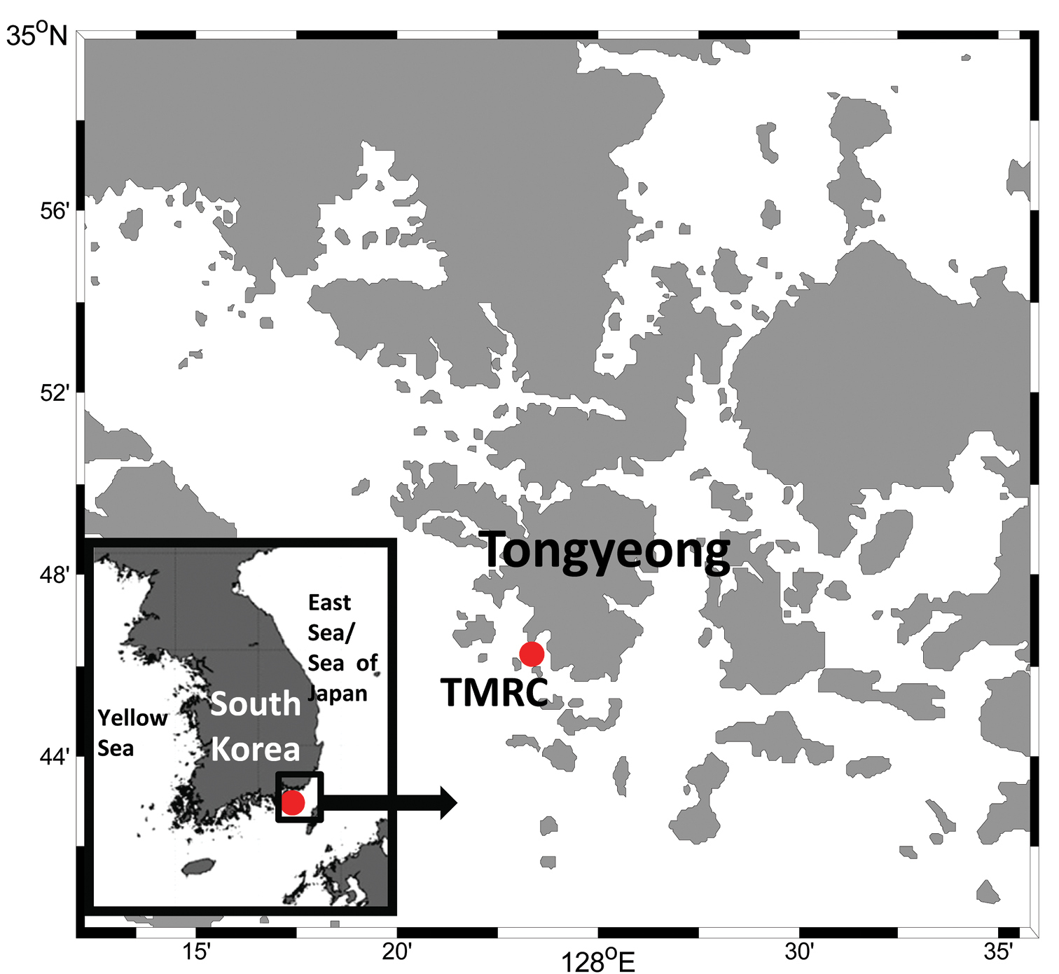
Map showing the marine ranched fish farming facility, Tongyeong marine living resources research & conservation center (TMRC), Tongyeong, Gyeongsangnam-do, Korea

Results Order Siphonostomatoida Burmeister, 1835 Family Pennellidae Burmeister, 1835 Genus Peniculus von Nordmann, 1832
Peniculus minuticaudae Shiino, 1956

http://species-id.net/wiki/Peniculus_minuticaudae

Figures 2, 3
Peniculus minuticaudae Shiino, 1956: 593; Nagasawa et al. 2011: 43; Yamaguti 1963: 1104
Peniculus sp. Fukuda 1999: 57
Material examined.

10 ♀♀ (NIBRIV0000245080) and 2 ♀♀ (MABIK CR00178439) from Thamnaconus modestus, Tongyeong, Gyeongsangnam-do, Korea, 20 September 2011.

Description.

Postmetamorphic adult female. Body (Figure 2A), 2.42 (2.12–2.73) mm long (n=10) comprising oval head, slender neck, large trunk and reduced abdomen. Head (cephalothorax) ovoid, longer than wide, with blunt pointed apex (Figure 2B, C). Short slender neck (Figure 2C) consisting of three somites bearing legs 1, 2 and 3. Fourth pedigerous somite incorporated into trunk. Trunk large, cylindrical, longer than wide, bearing leg 4 proximally (Figure 2C). Abdomen slightly triangular-shaped (Figure 2D, E) long with subterminal caudal rami on ventral surface and projecting posterior tip with anal indentation. Egg sacs long and uniseriate with 33–40 eggs (Figure 2F). Caudal rami (Figure 2G) bearing 2 long, 3 medium sized subequal, 1 small setae. Antennule not observed. Antenna (Figure 2H) 2-segmented, chelate; proximal segment consisting of 2 pointed projections overlapping each other; terminal segment claw-like, acutely pointed with minute seta at base.

Mandible (Figure 3A) broad with 10 teeth terminally. Maxillule (Figure 3B) with 2 lobes having one and two long setae. Maxilla (Figure 3C) 2-segmented; proximal segment broad with spiniform small process, 2 rows of setules distally; distal segment blunt and curved with transverse striations and rows of spinules. Maxilliped absent. Legs 1 to 4 (Figure 3D–G) all represented by broad plate-like structures derived from the protopodal segments, without rami or seta. Leg 5 absent.

Variability.

Some females showed variation on posterior end of trunk and abdomen (Figure 3H–J).

Attachment site.

All fins of host fish.

Remarks.

Careful comparison between our material and the original description of Peniculus minuticaudae provided by Shiino (1956) revealed some differences: (1) the abdomen was described as trapezoid and rhomboid; (2) the striation and fine setulose ornamentation of the maxilla was not shown. The mandible was not described. Our redescription revealed that the abdomen of Peniculus minuticaudae is triangular and protrudes, however, the two closely related congeners Peniculus ostraciontis and Peniculus truncatus both have a rudimentary abdomen. We also noted some variation in the posterior end of trunk and abdomen (Figure 3H–J). In the maxilla, fine striations and rows of setulose were found on the distal segment. In addition, the trunk is long and narrow in Peniculus minuticaudae and there is no major gap between cephalothorax and trunk so it has a short neck, where legs 1 to 3 are located (Figure 2C). Leg 4 (Figure 2C) is embedded on the anterior part of the trunk. In comparison, the closely related congener Peniculus ostraciontis has a stout trunk and short neck (Yamaguti 1939) while Peniculus truncatus has a long trunk and neck, and leg 1 has minute setal structure which are not present in Peniculus minuticaudae and Peniculus ostraciontis.

Figure 2.

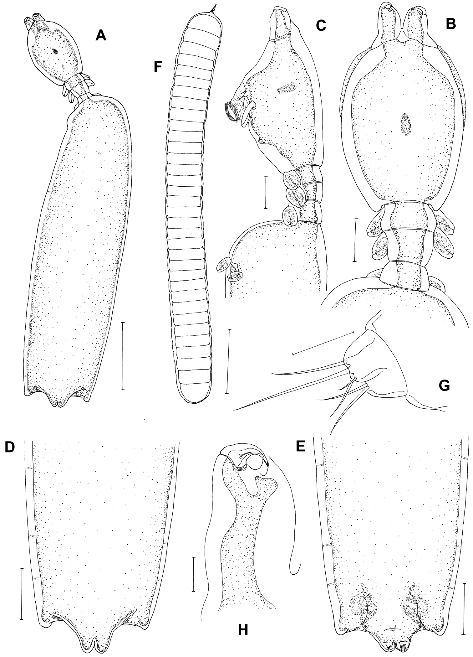
Peniculus minuticaudae Shiino, 1956. Postmetamorphic adult female. A Habitus, dorsal B Cephalothorax and free thoracic somites, dorsal C Cephalothorax and free thoracic somites, lateral D Posterior end of trunk with abdomen, dorsal E Posterior end of trunk with abdomen, ventral F Egg sac G Caudal ramus H Antenna, dorsal. Scale bars: A=500 μm; B–F=200 μm; G=25 μm; H=50 μm.

Figure 3.

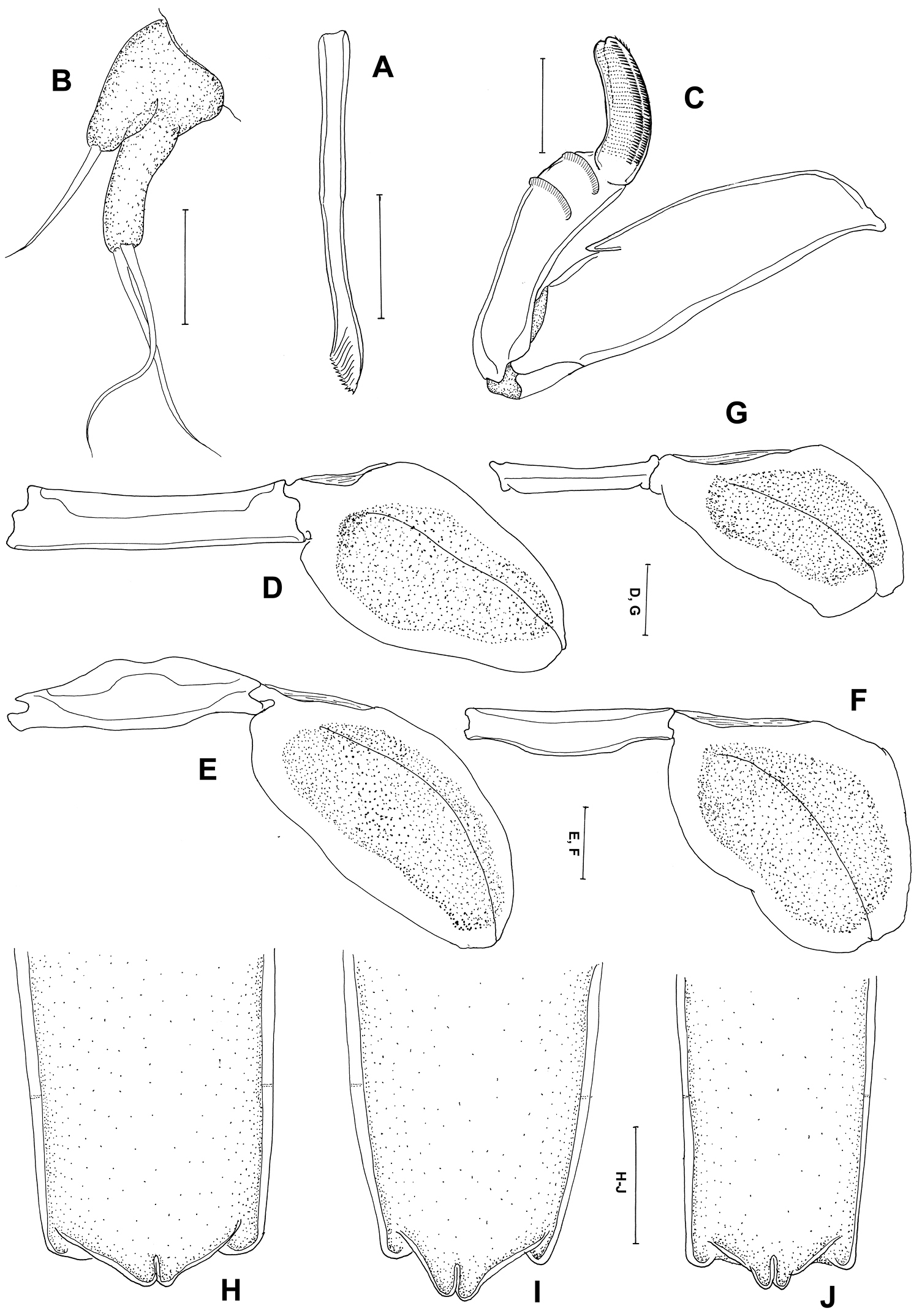
Peniculus minuticaudae Shiino, 1956. Postmetamorphic adult female. A Mandible, ventral B Maxillule, ventral C Maxilla, dorsal D Leg 1, ventral E Leg 2, ventral F Leg 3, ventral G Leg 4, ventral H–J variations of posterior end of trunk with abdomen, dorsal. Scale bars: A–C=25 μm; D–G=50 μm; H–J=200 μm.

Peniculus truncatus Shiino, 1956

http://species-id.net/wiki/Peniculus_truncatus

Figures 4, 5
Peniculus truncatus Shiino, 1956: 593; Yamaguti 1963: 1104.
Peniculus ostraciontis: Choi et al. 1996: 117
Material examined.

4 ♀♀ (NIBRIV0000252624) and 1 ♀ (MABIK CR00178440) from Sebastes schlegelii, Tongyeong, Gyeongsangnam-do, Korea, 15 February 2012.

Description.

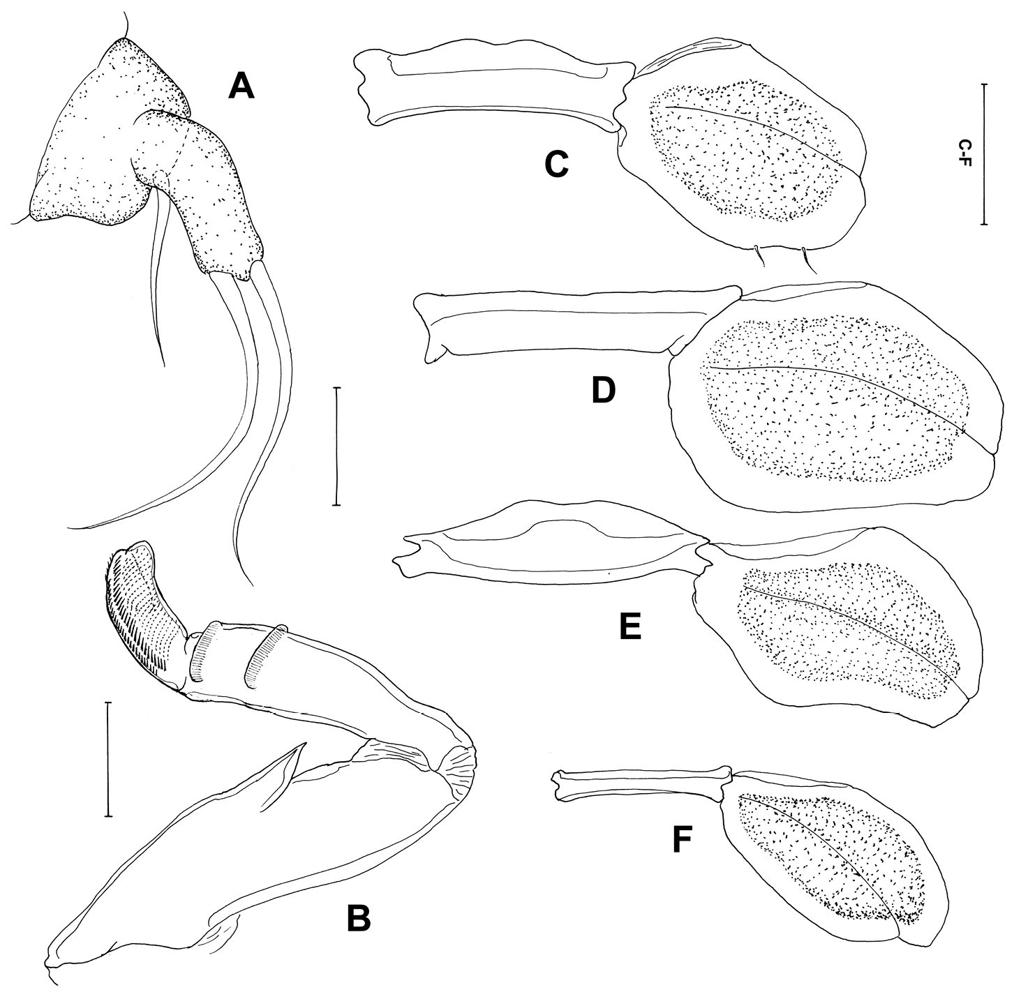
Postmetamorphic adult female. Body (Figure 4A), 4.59 (4.14–5.41) mm long (n=4) comprising oval head, long slender neck, large trunk and reduced abdomen. Head (cephalothorax) ovoid, flattened dorsally but convex ventrally with pair of rounded swellings anteriorly bearing antennae (Figure 4B, C). Mouth tube prominent, directed posteroventrally (Figure 4C). Neck long (0.47–0.55 mm) (Figure 4B, C), slender, comprising about one sixth of trunk length; consisting of three somites bearing legs 1, 2 and 3 (Figure 4B, C). Fourth pedigerous somite incorporated into trunk. Trunk slender, cylindrical, longer than wide, 6 times longer than neck, bearing leg 4 proximally. Abdomen (Figure 4D), reduced with subterminal caudal rami on ventral surface. Caudal rami (Figure 4E) bearing 6 setae. Egg sacs long and uniseriate with 30-37 eggs. Antennule not observed. Antenna (Figure 4F) 2-segmented, chelate; proximal segment bearing 2 pointed projections overlapping each other; terminal segment claw-like, acutely pointed with minute seta at base. Mandible (Figure 4G) moderate-sized, broad, provided with 10 teeth terminally.

Maxillule (Figure 5A) with 2 lobes having one short and two long setae. Maxilla (Figure 5B) 2-segmented; proximal segment broad with robust spiniform process, projecting laterally, 2 rows of setules distally; distal segment blunt and curved with transverse striations and rows of spinules. Maxilliped absent. Leg 1 (Figure 5C) forming blunt plate-like structure derived from protopodal segments, with 2 minute setae laterally. Legs 2–4 (Figure 5D–F) as for leg 1, but without seta. Leg 5 absent.

Attachment site.

Only on dorsal fin-rays.

Remarks.

Comparison between our material and the original description of Peniculus truncatus provided by Shiino (1956) revealed some omissions in that the antennae and mandibles were not shown, and possible differences, since the striation of setules on maxilla was not shown. The characteristic features of Peniculus truncatus are: (1) the rudimentary abdomen; (2) the long neck (more than half as long as cephalothorax); (3) the maxilla with transverse striations of setules and rows of spinules on the distal segment; (4) the leg 1 is tipped with 2 minute setae laterally. Peniculus truncatus differs from Peniculus minuticaudae in its rudimentary abdomen (vs. well developed abdomen); long neck (vs. short neck); and in the presence of setae on leg 1 (vs. absence of seta). It differs from Peniculus ostraciontis in its moderately slender trunk (vs. stout trunk); long neck, ie: neck more than half as long as cephalothorax (vs. short neck, ie: neck less than half as long as cephalothorax); and in the presence of setae on leg 1 (vs. absence of setae) (Yamaguti 1939; Shiino 1956).

Choi et al. (1996) reported the same pennellid collected from the fins of Stephanolepis schlegelii as Peniculus ostraciontis. We compared our material with their illustrations (specimens were not deposited in the museum). It showed the features of Peniculus truncatus: (1) long neck; (2) slender trunk [not as stout as like Peniculus ostraciontis illustrated by Yamaguti (1939)] and the host was Stephanolepis schlegelii (Choi et al. 1996), as in the present study.

Figure 4.

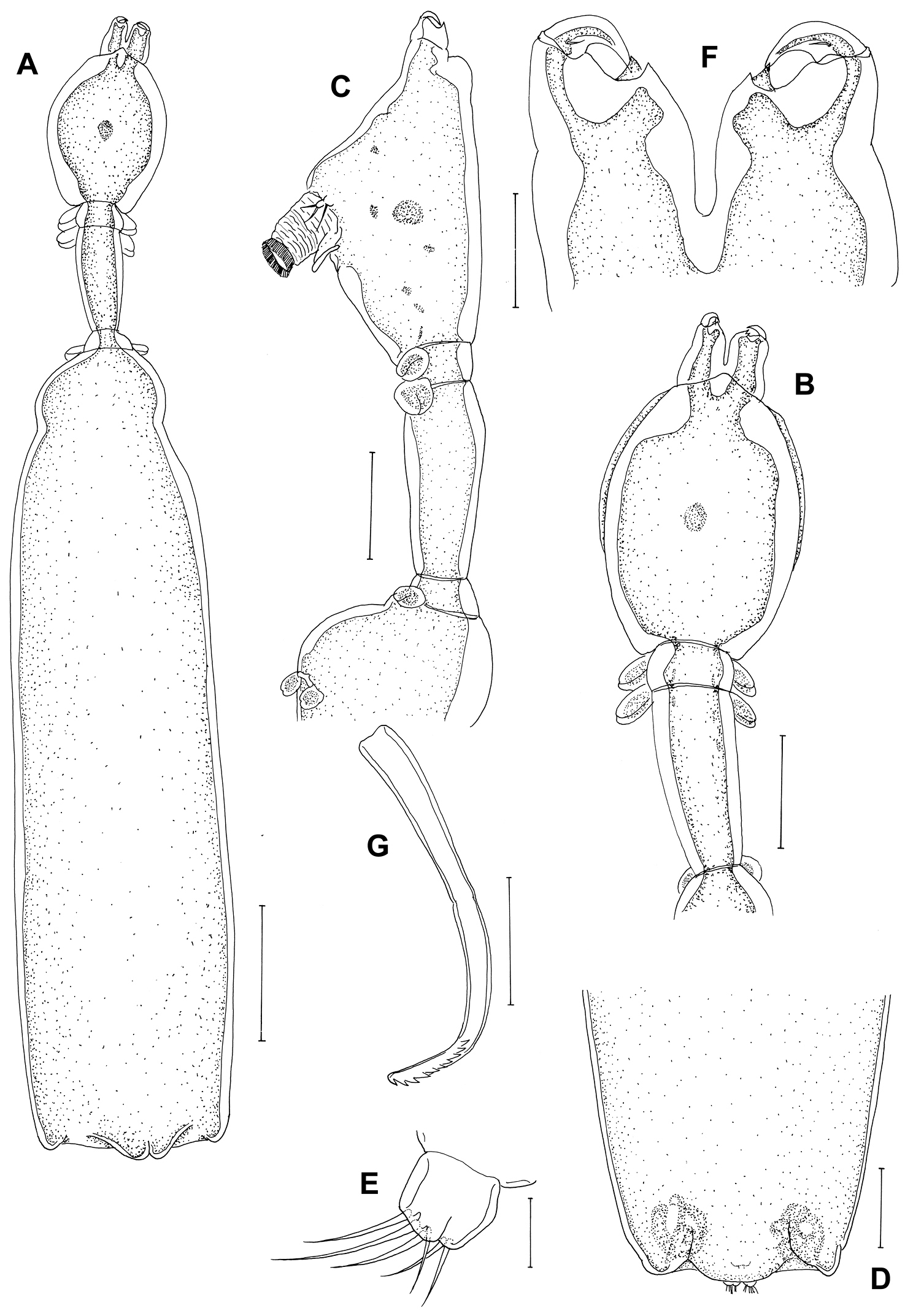
Peniculus truncatus Shiino, 1956. Postmetamorphic adult female. A. Habitus, dorsal B Cephalothorax and free thoracic somites, lateral C Cephalothorax and free thoracic somites, dorsal D Posterior end of trunk with abdomen, ventral E Caudal ramus, ventral F Antenna, dorsal G Mandible, ventral. Scale bars: A=500, μm; B–D=200 μm; E, G=25 μm; F=50 μm.

Figure 5.

Peniculus truncatus Shiino, 1956. Postmetamorphic adult female. A Maxillule, dorsal B Maxilla, dorsal C Leg 1, ventral D Leg 2, ventral E Leg 3, ventral F Leg 4, ventral. Scale bars: A, B=25 μm; C–F=50 μm.

Discussion

According to Boxshall and Halsey (2004), there are 14 species on the genus Peniculus: Peniculus asinus Kabata & Wilkes, 1977; Peniculus clavatus (Müller, 1779); Peniculus communis Leigh-Sharpe, 1934; Peniculus elegans Leigh-Sharpe, 1934; Peniculus elongatus Boxshall, 1986; Peniculus fistula Nordmann, 1832; Peniculus furcatus Krøyer, 1863; Peniculus minuticaudae Shiino, 1956; Peniculus ostraciontis Yamaguti, 1939; Peniculus scomberi Gnanamuthu, 1951; Peniculus stromatei Gnanamuthu, 1951; Peniculus theraponi Gnanamuthu, 1951; Peniculus trichuri Gnanamuthu, 1951; and Peniculus truncatus Shiino, 1956.Seven of these are reported from Asian countries.

Alexander (1983) reported Peniculus haemuloni from Brazil, however, it was subsequently treated as a separate genus, Metapeniculus Castro-Romero & Baeza-Kuroki, 1985 based on the presence of only 3 pairs of swimming legs (vs. 4 pairs for Peniculus) (Boxshall, 1986). According to Kabata (1979), two species of Peniculus, Peniculus calamus Nordmann, 1864 and Peniculus fissipes Wilson, 1917 should be regarded as species inquirendae, and in addition we treat Peniculus sciaenae Gnanamuthu, 1951 as species inquirenda since it is also reported with 3 pairs of swimming legs (Gnanamuthu 1951a; Alexander 1983). Thus there are 14 species considered valid and a key is provided for nominal species following Alexander (1983). Most Peniculus species were not described adequately by modern standards and most need to be redescribed. In Asia, all species are poorly described and detailed studies are necessary for the five species reported from India (Gnanamuthu 1951a; 1951b; Pillai 1985) and for the three from Japan (Yamaguti 1939; Shiino 1956).

The mean body length of Peniculus minuticaudae was 2.42 mm. It corresponds well to the body length (2.48 mm) of Peniculus minuticaudae reported from Oita Prefecture, Japan (Nagasawa et al. 2011). The morphological features (Figures 2, 3) agree with the original description of Peniculus minuticaudae (Shiino 1956). The present collection represents the first record of Peniculus minuticaudae from ranched Tetrosomus modestus in Korea. Thus, it is the third documented record of pennellid copepod from commercially cultured fishes.

Peniculus truncatus was originally reported from Stephanolepis oblongus in Japan (Shiino 1956). This parasite is shown here to utilize a second host species, Stephanolepis schlegelii, of the same host genus, although it was initially misidentified as Peniculus ostraciontis by Choi et al. (1996). The misidentification was revealed by comparison between Choi’s descriptions, our material and Yamaguti (1939) illustrations of Peniculus ostraciontis. We collected Peniculus truncatus from the same host species Stephanolepis schlegelii cultured in Korea. The host for Peniculus ostraciontis is Tetrosomus gibbosus (Table 1). In Choi et al. (1996) redescription, they overlooked the third seta on the maxillule and the setules on the maxilla, in addition to the minute setal structures on leg 1.

Peniculus truncatus has so far been reported from two species of the genus Sebastes, Stephanolepis schlegelii and S. oblongus and this pennellid appears to be host specific to rockfish (Table 1). Peniculus minuticaudae and Peniculus ostraciontis might be specific to file fish and puffer hosts, respectively (Yamaguti 1939; Shiino 1956; 1959; Nagasawa et al. 2011; present study). A key is provided for all 14 valid species below.

Key to the species of Peniculus

(Modified from Alexander 1983)

1 Cephalothorax with 4 large holdfast processes Peniculus asinus Kabata
Cephalothorax without such processes 2
2 Cephalothorax with rounded swelling on ventral surface posterior to mouth tube 3
Cephalothorax without posterior swelling on ventral surface 5
3 Swimming legs apparently absent Peniculus scomberi Gnanamuthu
Swimming legs with 4 pairs 4
4 Trunk about 11 times longer than wide Peniculus trichuri Gnanamuthu
Trunk about 8 times longer than wide Peniculus stromatei Gnanamuthu
5 Legs 3 and 4 closer together than legs 1 and 2 Peniculus communis Leigh-Sharpe
Legs 3 and 4 further apart than legs 1 and 2 6
6 Trunk conical-shaped Peniculus furcatus Krøyer
Trunk between 3 and 4.5 times longer than wide 7
7 Mouth tube forming a massive posteriorly-directed proboscis Peniculus clavatus Krøyer
Mouth tube not forming a massive posteriorly-directed proboscis 8
8 Cephalothorax ovoid 9
Cephalothorax cylindrical Peniculus theraponi Gnanamuthu
Cephalothorax widest near posterior margin and tapering anteriorly Peniculus elegans Leigh-Sharpe
9 Abdomen well developed; trunk longer than wide Peniculus minuticaudae Shiino
Abdomen well developed; trunk longer than wide; with swelling on the head 10
Abdomen reduced; posterior margin of trunk more or less straight 11
10 High degree of ventral swelling on the head; neck constricted Peniculus fistula Nordmann
Low degree of ventral swelling on the head; neck constricted Peniculus elongatus Boxshall
11 Trunk 4.3 times longer than wide; neck less than half as long as cephalothorax Peniculus ostraciontis Yamaguti
Trunk 3.3 times longer than wide; neck more than half as long as cephalothorax Peniculus truncatus Shiino
Conflict of interest statement

All authors declare that they do not have any conflict of interest.

Acknowledgments

We thank two anonymous reviewers for their constructive comments. Senior author is thankful to Korea Research Council of Fundamental Science and Technology (KRCF) and this work was formed part of Korea Institute of Ocean Science & Technology projects (PK08080, PE98746, PE98785). This work was partially supported by the National Institute of Biological Resources (NIBR), Korean project on the survey of Korean indigenous species and National Marine Life Collection program (project) sponsored by the Ministry of Land, Transport and Maritime Affairs, Korea (MABIK 2012-001-03) to SYM, HYS.

References
Alexander PD (1983) Peniculus haemuloni, a new species of copepod (Siphonostomatoida: Pennellidae) parasitic on Haemulon steindachneri from Ubatuba, Brazil. Bulletin of the British Museum (Natural History), (Zoology) 45: 381-385.
Boxshall GA (1986) A new genus and two new species of Pennellidae (Copepoda: Siphonostomatoida) and an analysis of evolution within the family. Systematic Parasitology 8: 215-225. doi: 10.1007/BF00009890
Boxshall GA, Halsey SH (2004) An Introduction to Copepod Diversity. The Ray Society, London, 940 pp.
Choi S-D, Suh H-L, Hong S-Y (1996) Two species of parasitic copepods (Neobrachiella incurva and Peniculus ostraciontis) from the marine fishes, Halichoeres poecilopterus and Sebastes schlegeli, of the south coast of Korea. Journal of Aquaculture 9: 117-123.
Froese R, Pauly D (2012) FishBase. World Wide Web electronic publication. www.fishbase.org [accessed 15.08.12]
Fukuda Y (1999) Diseases of marine fishes and shellfishes cultured in Oita Prefecture diagnosed from 1980 to 1997. Bulletin of the Oita Institute of Marine and Fisheries Science 2: 41-73. [in Japanese]
Gnanamuthu CP (1951a) Two new species of copepods of the genus Peniculus parasitic on Madras fishes. Records of the Indian Museum 49: 221-226.
Gnanamuthu CP (1951b) Lernaeid copepods parasitic on South Indian fish. Annals and Magazine of Natural History 12: 77-86. doi: 10.1080/00222935108654127
Humes AG, Gooding RU (1964) A method for studying the external anatomy of copepods. Crustaceana 6: 238-240. doi: 10.1163/156854064X00650
Kabata Z (1979) Parasitic Copepoda of British Fishes. The Ray Society, London, 468 pp.
Kabata Z (1981) Copepoda (Crustacea) parasitic on fishes: problems and perspectives. Advances in Parasitology 19: 1-71. doi: 10.1016/S0065-308X(08)60265-1
MOMAF (2007) Studies on the development of marine ranching program in Tongyeong, Korea. KORDI Report, BSPM40600–1884–3, Korea Ocean Research and Development Institute, 1082 pp.
Nagasawa K, Fukuda Y, Tanaka S (2011) Infection with Peniculus minuticaudae (Copepoda: Pennellidae) on threadsail filefish (Stephanolepis cirrhifer) and black scraper (Thamnaconus modestus) farmed in Japan. Biosphere Science 50: 43-47.
Pillai NK (1985) Fauna of India: Parasitic Copepods of Marine Fishes. Technical and General Press, Calcutta, 900 pp.
Shiino SM (1956) Copepods parasitic on Japanese fishes. 7. Peniculus and Peniculisa. Japanese Journal of Zoology 11: 593-608.
Shiino SM (1959) Sammlung der parasitischen Copepoden in der Prafekturuniversitat von Mie. Report of Faculty of Fisheries of the Prefectural University of Mie 3 (2): 334-374.
Venmathi Maran BA, Oh S-Y, Soh HY, Choi HJ, Myoung J-G (2012) Caligus sclerotinosus (Copepoda: Caligidae), a serious pest of cultured red seabream Pagrus major (Sparidae) in Korea. Veterinary Parasitology 188: 355-361. doi: 10.1016/j.vetpar.2012.03.023
Yamaguti S (1939) Parasitic copepods from fishes of Japan, Part 5. Caligoida, III. Volume Jubilare pro professore Sadao Yoshida 2: 443-487.
Yamaguti S (1963) Parasitic Copepoda and Branchiura of Fishes. Interscience Publishers, New York, 1104 pp.