


(C) 2012 Robert Wharton. This is an open access article distributed under the terms of the Creative Commons Attribution License 3.0 (CC-BY), which permits unrestricted use, distribution, and reproduction in any medium, provided the original author and source are credited.
For reference, use of the paginated PDF or printed version of this article is recommended.
Four new species of opiine Braconidae are described from Mexico. These are Diachasmimorpha martinalujai Wharton reared from Rhagoletis infesting fruits of Crataegus spp., Diachasmimorpha norrbomi Wharton reared from Euphranta mexicana infesting fruits of Ribes pringlei, Eurytenes (Stigmatopoea) norrbomi Wharton reared from Trypeta concolor mining leaves of Barkleyanthus salicifolia and Eurytenes (Stigmatopoea) maya Wharton reared from Rhagoletis pomonella infesting apples and fruits of Crataegus spp. Morphological features of the first metasomal segment and occipital carina, useful for placement of these species, are discussed relative to the genera Diachasmimorpha, Eurytenes, Lorenzopius, Tubiformopius, and Opius s.l. Descriptions and diagnoses are referenced to the Hymenoptera Anatomy Ontology. The following represent new combinations: Diachasmimorpha hildagensis, Lorenzopius euryteniformis, and Tubiformopius tubibasis. Revised diagnoses are provided for Diachasmimorpha hildagensis, Diachasmimorpha mexicana, Diachasmimorpha sanguinea, Eurytenes (Stigmatopoea), Lorenzopius, Lorenzopius euryteniformis, Tubiformopius, Tubiformopius tubigaster, Tubiformopius tubibasis, Opius incoligma, and Opius rugicoxis. Two species groups are delineated within Lorenzopius and a key to species of Diachasmimorpha occurring in the New World is provided.
Parasitoid, classification, Rhagoletis, HAO, Opius
The subfamily Opiinae is a diverse assemblage of relatively small braconids that develop as koinobiont endoparasitoids of various cyclorrhaphous Diptera, emerging from the puparium of their hosts. Opiines have long been recognized as a distinct taxon within the Braconidae (
Specimens. Reared material of several species, including the four newly described below, was kindly sent for study to the senior author by Martin Aluja and Juan Rull (Instituto de Ecologia, Xalapa, Mexico), Robert Jones (Universidad de Autónoma de Querétaro, Querétaro, Mexico), and Allen Norrbom (USDA Systematic Laboratory, Washington, D. C.). Other specimens used in this study, including type material of previously described species, were borrowed from or examined at the following institutions: American Entomological Institute, Gainesville, Florida, USA (AEIC), Canadian National Collection, Ottawa, Ontario, Canada (CNC), Hungarian Natural History Museum, Budapest, Hungary (HNHM), National Museum of Natural History, Leiden, The Netherlands (RMNH), Naturhistorisches Museum Wien, Vienna, Austria (NHNW), Texas A&M University Insect Collection, College Station, Texas, USA (TAMU), The Natural History Museum, London, England (BMNH), and U. S. National Museum of Natural History, Washington, D. C., USA (USNM).
In the material examined section under each species description, we record label data for the holotype exactly as they appear on the labels. We use a more standardized format for paratypes, additional specimens examined, and published data for other specimens.
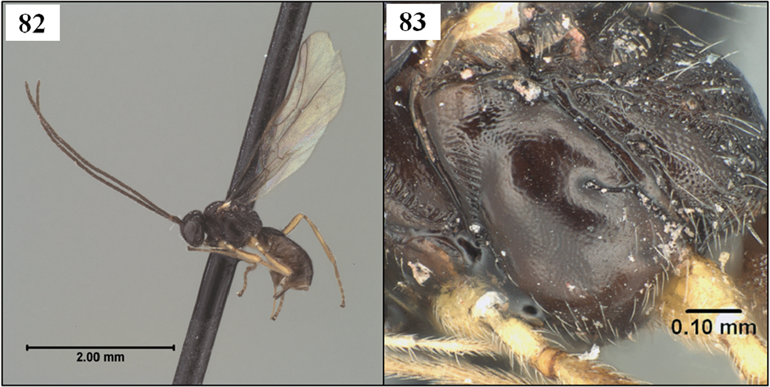
Figures. Images were acquired digitally using Syncroscopy’s AutoMontage® software, in combination with a ProgRes 3008 digital camera mounted on a Leica MZ APO dissecting microscope. All images were further processed using various minor adjustment levels in Adobe Photoshop® such as image cropping and rotation, adjustment of contrast and brightness levels, color saturation, and background enhancement. Automontage images are available in color and high resolution at http://peet.tamu.edu/projects/8/public/site/wharton_lab/home.
Database management, digital dissemination, and ontology reference. Illustrations and free-text diagnoses for morphospecies were assembled in mx, a web-based content management system that facilitates data management and dissemination for taxonomic and phylogenetic works (e.g.
Morphological terms used in this revision were matched to the Hymenoptera Anatomy Ontology (HAO,
Terminology and measurements. Terminology as linked through the HAO (Appendix) largely follows
Quantitative data in descriptions are based on 5 individuals of each sex, when available. Measurements largely follow
The new species described below are placed in the genera Diachasmimorpha Viereck and Eurytenes Foerster. The basis for these placements, with particular reference to the nature of the occipital carina, characteristics of the first metasomal segment, and tephritid parasitism, are discussed in this section. Diagnoses of relevant taxa and descriptions of the new species follow in the next section, alphabetically by genus.
The occipital carina varies from completely present to completely absent in the Opiinae with most species having the carina broadly absent mid-dorsally but well-developed laterally (Figs 1–4).
The first metasomal segment, often referred to as the petiole (
Neither
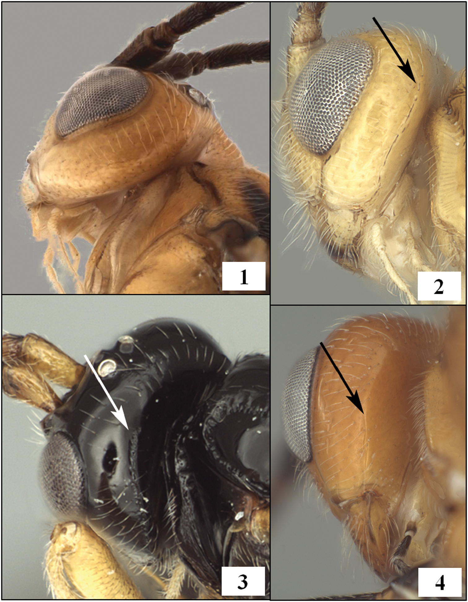
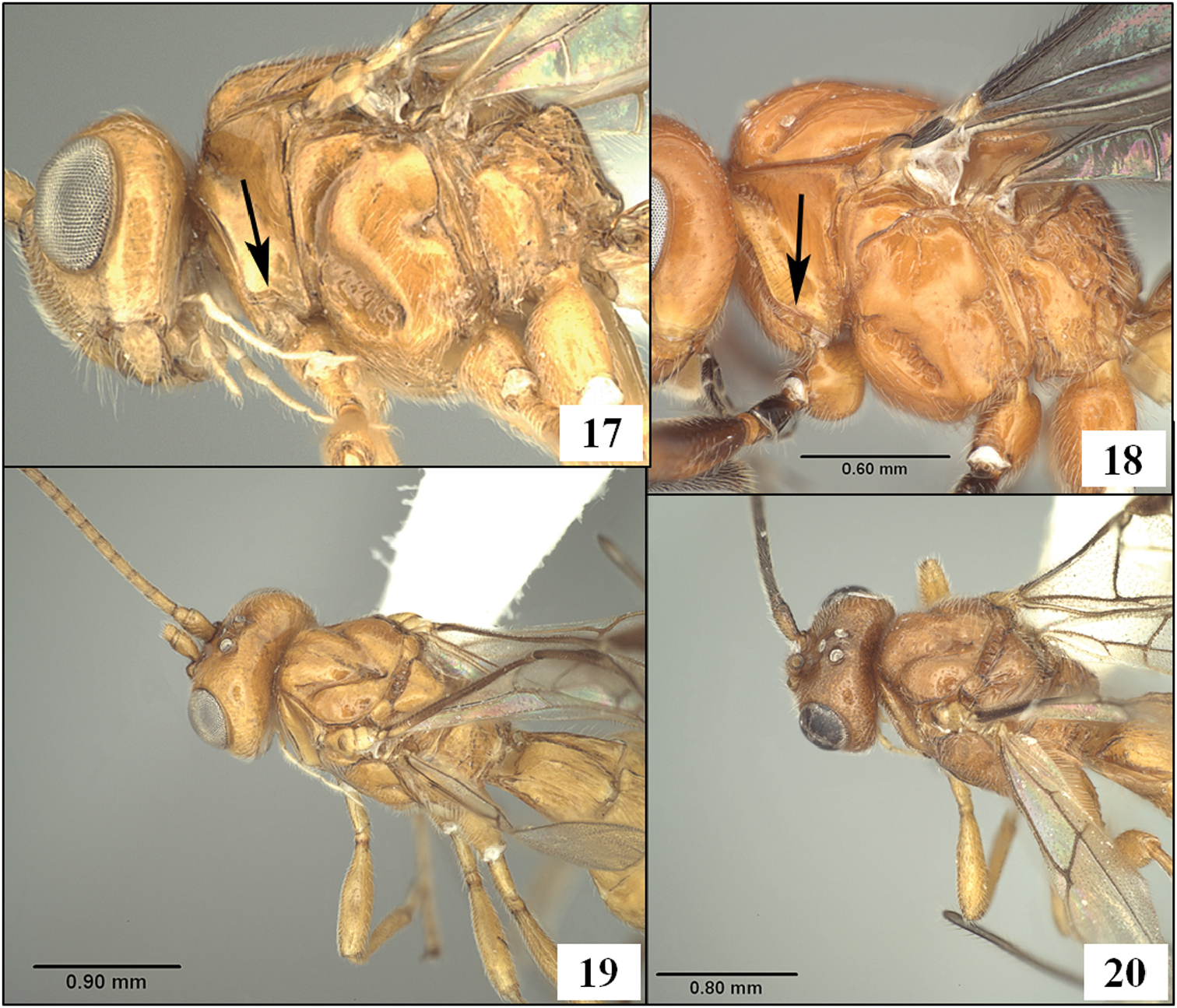
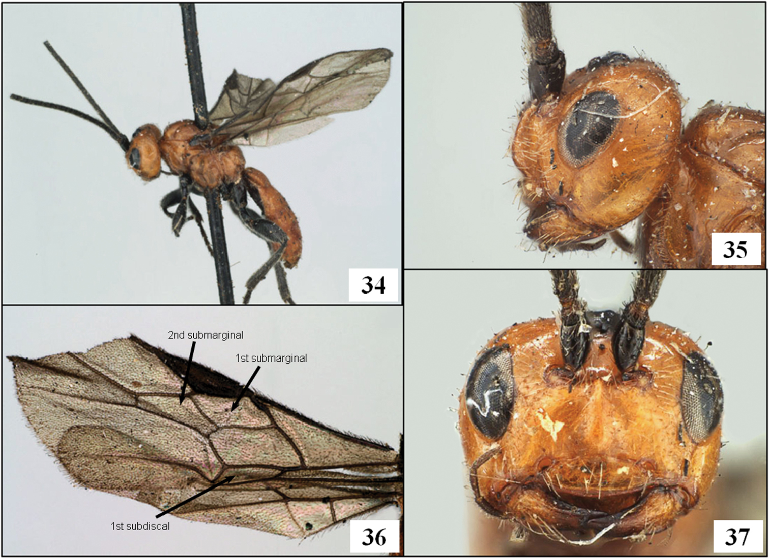
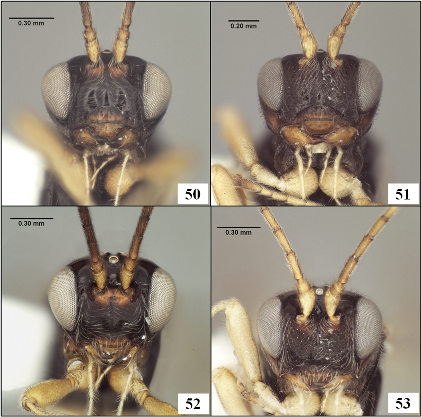
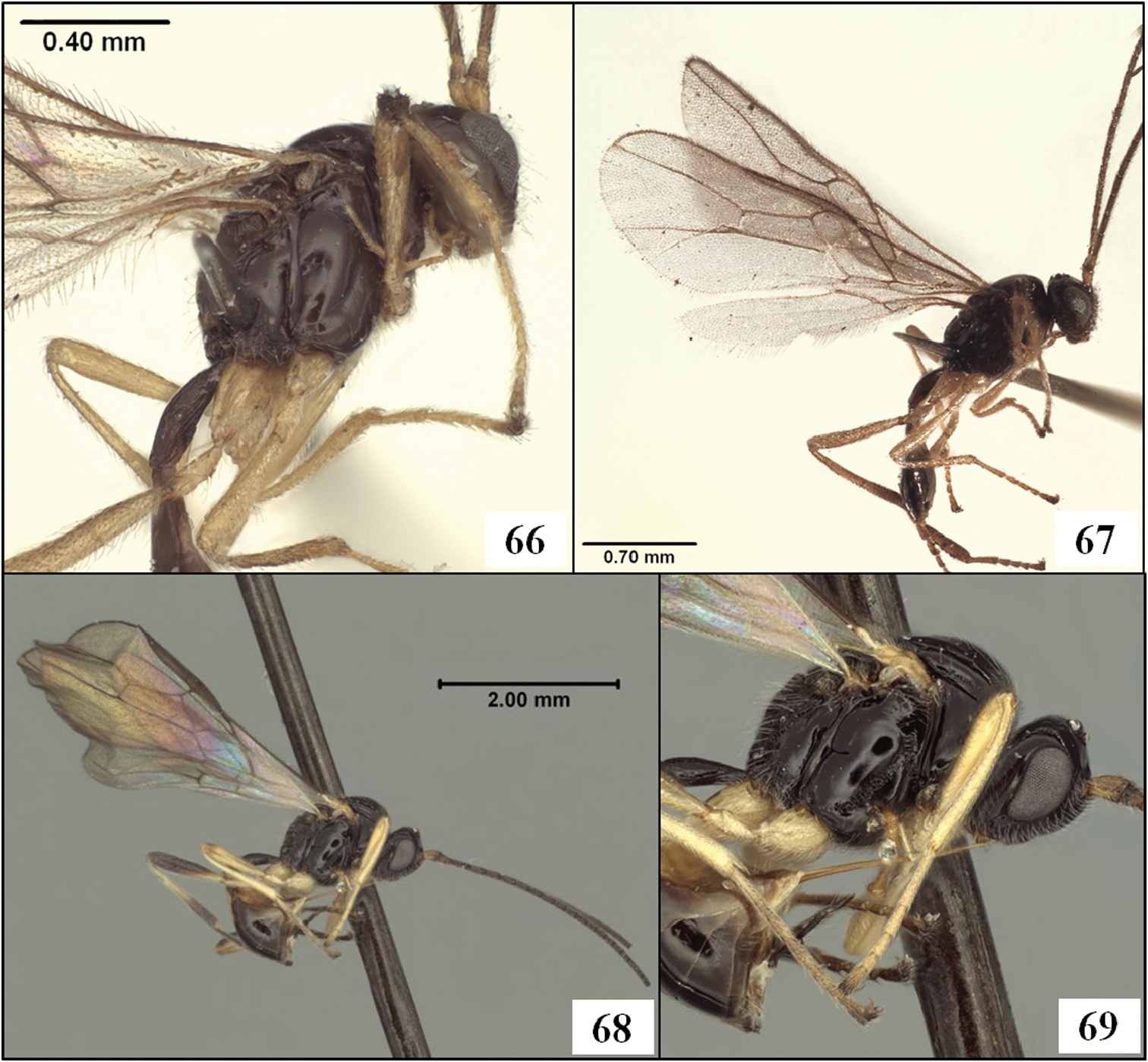
Occipital carina. 1 Opius (Bellopius) bellus Gahan, carina completely absent 2 Diachasmimorpha mellea (Gahan), arrow at dorsal end of carina 3 Lorenzopius tubulatus (Fischer), holotype female, arrow at dorsal end of carina 4 Diachasmimorpha sanguinea (Ashmead), arrow at dorsal end of weak carina.
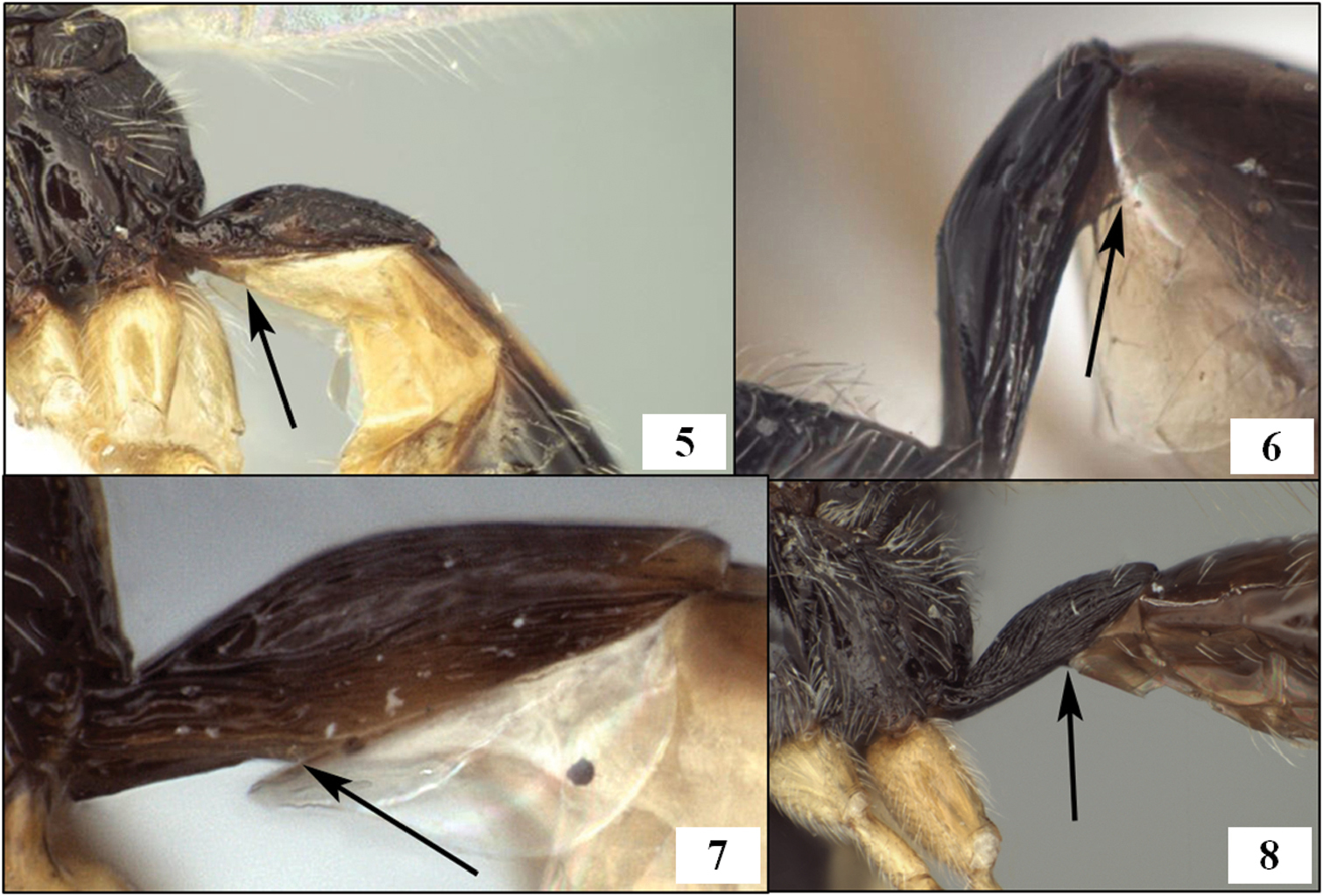
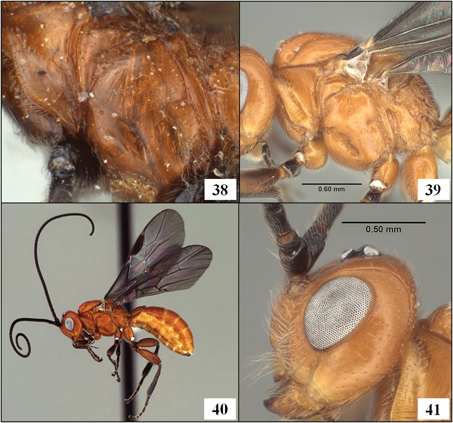
T1 and S1, arrows at posterior margin of S1. 5 Eurytenes (Stigmatopoea) macrocerus (Thomson) 6 Lorenzopius calicomyzae van Achterberg and Salvo, holotype female 7 Eurytenes (Stigmatopoea) maya Wharton sp. n., paratype female 8 Tubiformopius tubigaster (Fischer), holotype male.
http://species-id.net/wiki/Diachasmimorpha
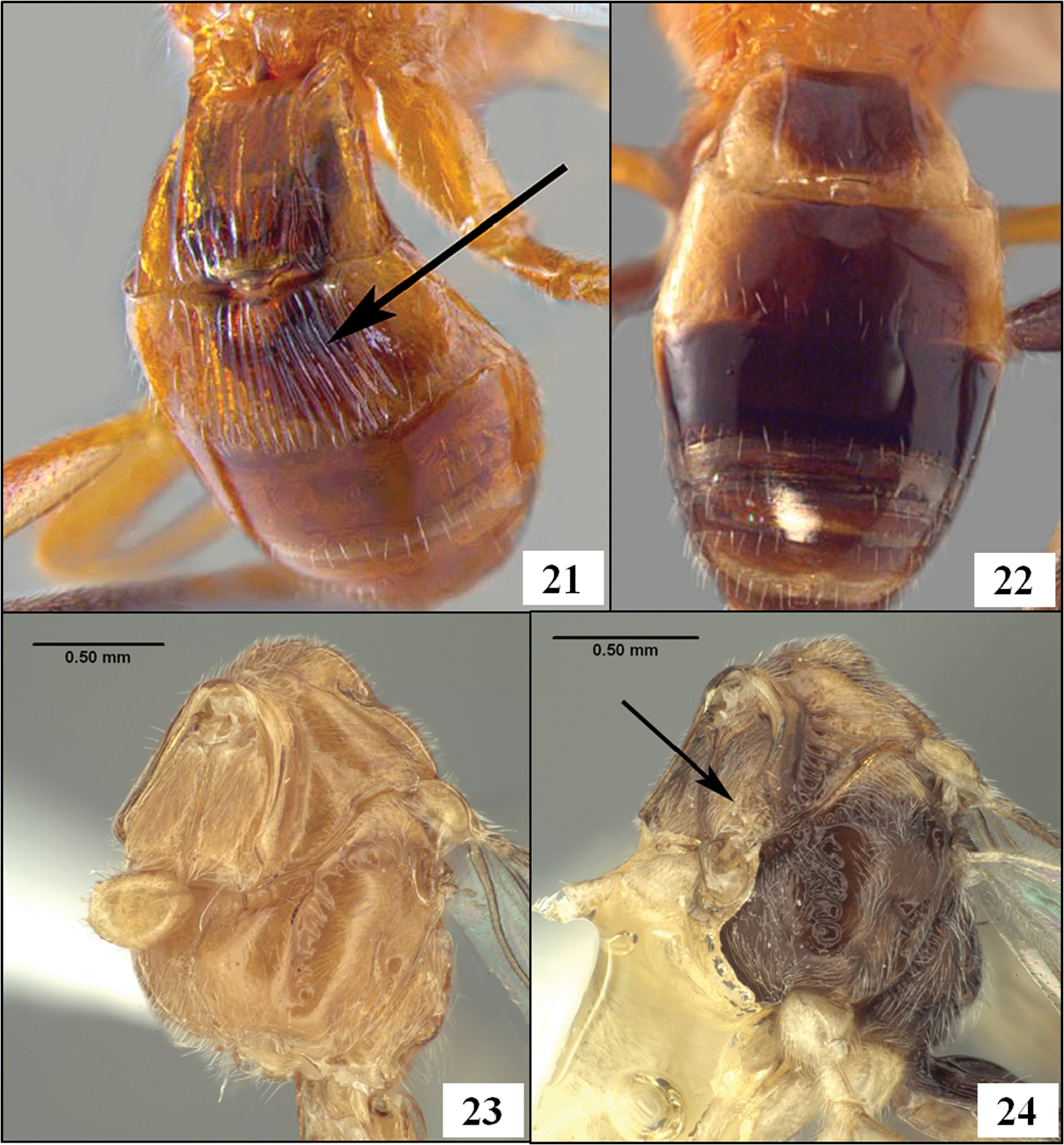
Mandible without basal lobe ventrally. Labrum concealed. Occipital carina broadly absent dorsally, present or absent laterally. Propleuron ventral-laterally without oblique carina. Notauli deep, unsculptured or nearly so, well developed anteriorly, varying posteriorly from absent to deep and complete to midpit; midpit always present. Fore wing stigma short, broad, discrete posteriorly, r1 arising at or distad its midpoint; second submarginal cell short; m-cu arising from second submarginal cell. Hind wing RS absent basally, sometimes present as a weakly pigmented crease distally; 2M distinctly pigmented nearly to wing margin; m-cu present, well-developed. Dorsope absent.
The species of Diachasmimorpha are most readily recognized by the pattern of fore and hind wing venation (Figs 9, 16) in combination with the concealed labrum (Fig. 12), unsculptured notauli (Figs 11, 14, 19, 20), and lack of oblique carina on the propleuron (Fig. 23). The species of Doryctobracon Enderlein, endemic to the New World, are similar but have the fore wing m-cu interstitial or arising from the first submarginal cell and the labrum is partially exposed. Fopius Wharton, an Old World genus with species that have been introduced to the New World, is also similar. The species of Fopius differ by the presence of completely sculptured notauli and the presence of an oblique carina on the propleuron (Fig. 24).
Both New and Old World species groups of Diachasmimorpha occur in Mexico. Diachasmimorpha longicaudata (Ashmead) and Diachasmimorpha tryoni, both representatives of the Old World longicaudata species group (
New World species have previously been referred to as the mexicana species group (
| 1 | Female (ovipositor clearly visible, extending well beyond apex of metasoma) | 2 |
| – | Male | 10 |
| 2 (1) | Ovipositor distinctly sinuate subapically (Fig. 28) | 3 |
| – | Ovipositor straight or nearly so subapically (Fig. 29) | 4 |
| 3 (2) | Metasomal tergum 2 distinctly striate medially (Fig. 21). Occipital carina well developed laterally, extending from base of mandible at least to mid eye height | Diachasmimorpha longicaudata (Ashmead) |
| – | Metasomal tergum 2 without striae or other sculpture (Fig. 22). Occipital carina poorly developed to absent, not extending dorsally to lower eye margin | Diachasmimorpha tryoni (Cameron) |
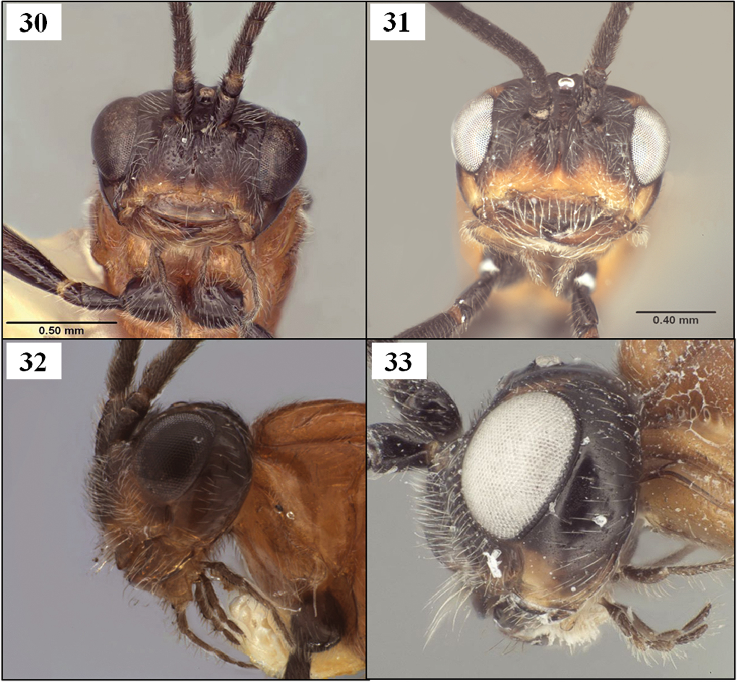
| 4 (2) | Head dark, at least on dorsal half (Fig. 9) | 5 |
| – | Head pale (Figs 2, 4), yellow or orange except sometimes ocellar field dark | 7 |
| 5 (4) | Ovipositor (total length) about 2.5 times longer than mesosoma. Notaulus extending anteriorly to margin of mesoscutum (Figs 10, 27) | 6 |
| – | Ovipositor (total length) less than 2.0 times longer than mesosoma. Notaulus rarely extending anteriorly to margin of mesoscutum, usually terminating just before reaching margin (Fig. 32) | Diachasmimorpha norrbomi, sp. n. |
| 6 (5) | Eye smaller than in Fig. 32, about 1.5–1.6 × longer than temple in lateral view | Diachasmimorpha hildagensis (Fischer) |
| – | Eye larger, 2.1–2.9 × longer than temple in lateral view (Fig. 33) | Diachasmimorpha martinalujai, sp. n. |
| 7 (4) | Wings darkly infumate (as in Figs 16, 36). Occipital carina represented at most as in Fig 4, usually present as a short spur near mandible, otherwise absent | Diachasmimorpha sanguinea (Ashmead) |
| Note: mexicana (Cameron) also keys here but is known only from the male, which has a much smaller eye than that of sanguinea. | ||
| – | Wings hyaline (Fig. 20). Occipital carina present laterally at least to lower margin of eye, usually as in Fig. 2 | 8 |
| 8 (7, 14) | Metasomal tergum 2 distinctly striate medially (as in Fig. 21) | 9 |
| – | Metasomal tergum 2 without striae or other sculpture (as in Fig. 22) | Diachasmimorpha juglandis (Muesebeck) |
| 9 (8) | Precoxal sulcus distinctly impressed, usually broad but very weakly sculptured, nearly smooth (as in Fig. 38). Hosts are walnut husk flies in species of Juglans D. sublaevis (Wharton) | |
| – | Precoxal sulcus distinctly impressed, broad, heavily sculptured: crenulate to foveolate (as in Fig. 17). Hosts are other species of Rhagoletis in other fruits | Diachasmimorpha mellea (Gahan) |
| 10 (1) | Head black at least over dorsal half | 11 |
| – | Head pale, yellow to orange except ocellar triangle sometimes black | 13 |
| 11 (10) | Eye in dorsal view as long as temple; eye in lateral view 1.3–1.4 × longer than temple | Diachasmimorpha hildagensis (Fischer) |
| – | Eye slightly larger, in dorsal view eye 1.4–1.9 × longer than temple, in lateral view 1.7–2.4 × longer than temple | 12 |
| 12 (11) | Notaulus extending anteriorly to margin of mesoscutum (Fig. 27) | Diachasmimorpha martinalujai, sp. n. |
| – | Notaulus rarely extending anteriorly to margin of mesoscutum, usually terminating just before reaching margin (Fig. 32) | Diachasmimorpha norrbomi, sp. n. |
| 13 (10) | Metasomal tergum 2 striate medially (Fig. 21) | 14 |
| – | Metasomal tergum 2 without striae or other sculpture (Fig. 22) | 15 |
| 14 (13) | Notauli deep posteriorly as it nears midpit (Fig. 19) | Diachasmimorpha longicaudata (Ashmead) |
| – | Notauli more shallow posteriorly as it nears midpit (Fig. 20) | 8 |
| 15 (13) | Metasomal terga mostly black (Fig. 22) | Diachasmimorpha tryoni (Cameron) |
| – | Metasoma with at least terga 3–5 pale: yellow to orange | 16 |
| 16 (15) | Wings hyaline | Diachasmimorpha juglandis (Muesebeck) |
| – | Wings darkly infumate | 17 |
| 17 (16) | Eye larger, about 1.3–1.5 × longer than temple in lateral view | Diachasmimorpha sanguinea (Ashmead) |
| – | Eye smaller, subequal to temple in lateral view (Fig. 35) | Diachasmimorpha mexicana (Cameron) |
http://species-id.net/wiki/Diachasmimorpha_hildagensis
Figs 9–12, 13–16Mexico, State of Mexico, Hidalgo National Park.
Holotype male (AEIC), first label, first line: Hidalgo Natl. Pk. second line: State of Mex., Mex. third line: x.12.62 3000 m. fourth line: H. & M. Townes Second label [purple]: Holotype Third label: Opius hildagensis [male symbol] sp. n. det. Fischer Fourth label: Type No. 336
Other specimens examined: 2 females, 1 male, Mexico, Mexico, Rt 890, km 9, 6 km W Lago Zempoala, 2.x.1991, A.L. Norrbom, reared from Oedicarina latifrons infesting fruits of Solanum brachycarpum (91M14B) (TAMU, USNM).
Holotype male. Eye in dorsal view as long as temple, temples neither receding nor expanded beyond eyes; eye in lateral view 1.3 × longer than temple. Frons irregularly rugulose along midline between antenna and median ocellus. Clypeus 2.8 × wider than high. Occipital carina distinct near base of mandible, short, not extending dorsally to ventral margin of eye. Antenna with 46 flagellomeres; first flagellomere 1.25 × longer than wide. Pronope deep, large, interrupting posterior crenulate groove middorsally. Notauli deep anteriorly, reaching anterior-lateral margin of mesoscutum and extending posteriorly about 0.5 × distance to deep, elongate midpit. Precoxal sulcus distinctly crenulate throughout, nearly extending to anterior margin of mesopleuron. Propodeum rugose, areola extending over posterior 0.6 but largely obscured by sculpture. Fore wing 2RS 0.95 × length of 3RSa; m-cu distinctly postfurcal. T1 with dorsal carinae weakly converging, widely separated at posterior margin, gradually weakening posteriorly. Meso- and metasoma orange, tegula black, head dark brown to black except narrow yellow-orange band along epistomal sulcus extending to and through malar sulcus and small orange spot on vertex adjacent eye; legs black except extreme base of hind coxa irregularly orange, joint between femora and trochantelli reddish orange, mid and hind tarsi dark brown. Body length about 4.3 mm, fore wing length 4.5 mm, mesosoma length 1.8 mm.
Specimens reared from Oedicarena latifrons (Wulp) vary as follows relative to the holotype: clypeus length/height ratio 2.6–2.8; eye/temple ratio, lateral view, 1.3–1.4 (males), 1.55 (female); antenna with 46–48 flagellomeres; 2RS/3RS ratio 0.95–1.0; ovipositor sheath 2.5 times longer than the mesosoma; mesosoma length 1.85–1.9 mm (male), 2.0 mm (female); one male with T1 dorsal carinae absent over posterior 0.5 and mandible, clypeus, face, and hind coxa more extensively orange; female with outer surface of hind coxa completely pale (dark medially), mandible, clypeus and lower part of face more extensively pale than in holotype.
This species is slightly larger and has a smaller eye than both of the similarly-colored species described below, Diachasmimorpha martinalujai, sp. n. and Diachasmimorpha norrbomi, sp. n. Based on the single female reared from Diachasmimorpha latifrons, Diachasmimorpha hildagensis alsohas a much longer ovipositor than Diachasmimorpha norrbomi. The ovipositors of Diachasmimorpha hildagensis and Diachasmimorpha martinalujai are similar in length. In Diachasmimorpha hildagensis and Diachasmimorpha martinalujai, the notaulus consistently extends anteriorly to the margin of the mesoscutum whereas in Diachasmimorpha norrbomi, the notaulus usually does not. Color variation in the specimens reared from Opius latifrons is similar to that in the paratype series of Diachasmimorpha martinalujai and Diachasmimorpha norrbomi. Both Diachasmimorpha hildagensis and the two newly described speciesare similar in having the head mostly dark in contrast to the orange heads of Diachasmimorpha mexicana and Diachasmimorpha sanguinea, the other two members of this species group. The holotype of Diachasmimorpha hildagensis exhibits subsurface discoloration on the metasoma, but the tergites are all entirely orange.
There is no biological information associated with the holotype. The non-type material listed above was reared from the tephritid Oedicarina latifrons infesting fruits of Solanum brachycarpum Correll. Collection data and host information can be found in
The name hildagensis is based on a misreading of the locality label on the holotype, which is correctly written as Hidalgo Nat. Park, not “Hildago Nat. Park” as given by
Both Diachasmimorpha hildagensis and Diachasmimorpha mexicana were described from single male specimens collected in the state of Mexico and the Distrito Federal, respectively, and unassociated with either hosts or host plants. Both have relatively small eyes, but are readily separated from one another on the basis of head coloration. Associating the name hildagensis with the many dark-headed specimens available for study, however, has been considerably more challenging. Reared material, representing over 50 specimens kindly made available to us by Allen Norrbom, Martin Aluja, and Juan Rull, provides clear evidence of sexual dimorphism in eye size as well as variation in ovipositor length associated with different hosts and host plants. This material has been especially critical for understanding color patterns and associating males with females. Based primarily on eye size and body size, the holotype of Diachasmimorpha hildagensis is closest to the series of three specimens listed above under “other specimens examined, ” that emerged from puparia of Opius latifrons infesting fruits of Solanum brachycarpum. From the remaining reared material, we describe two closely similar species below.
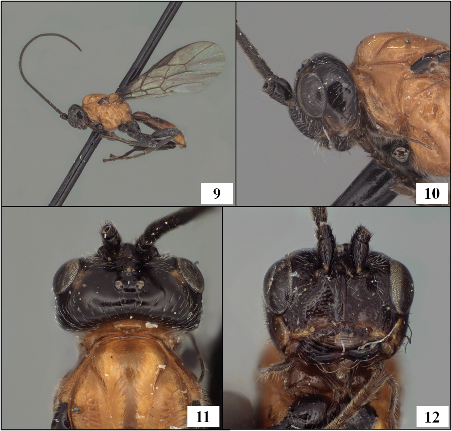
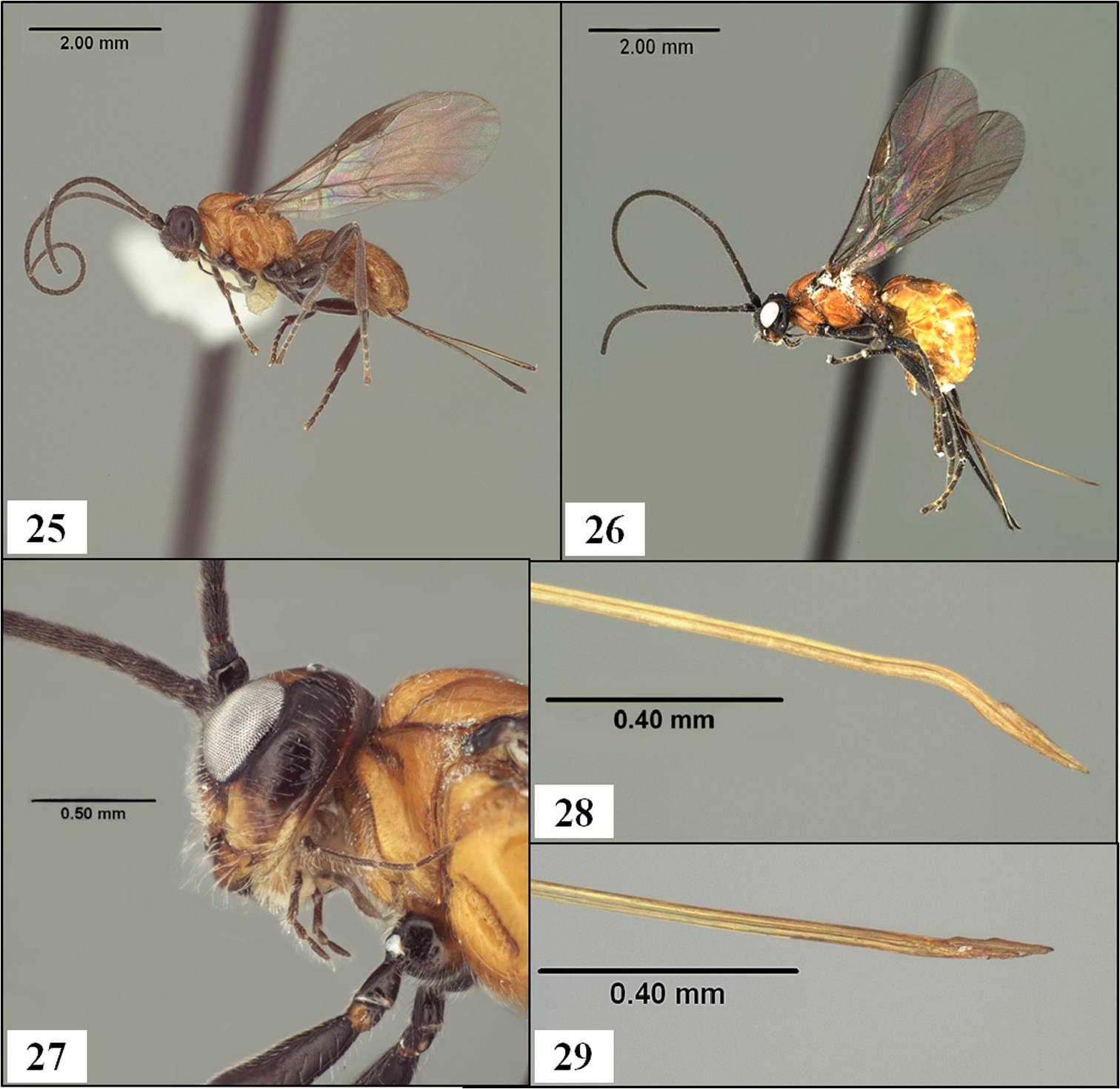
Diachasmimorpha hildagensis (Fischer), holotype male. 9 habitus 10 head and base of notaulus, lateral view 11 head, pronope, and base of notaulus, dorsal view 12 face.
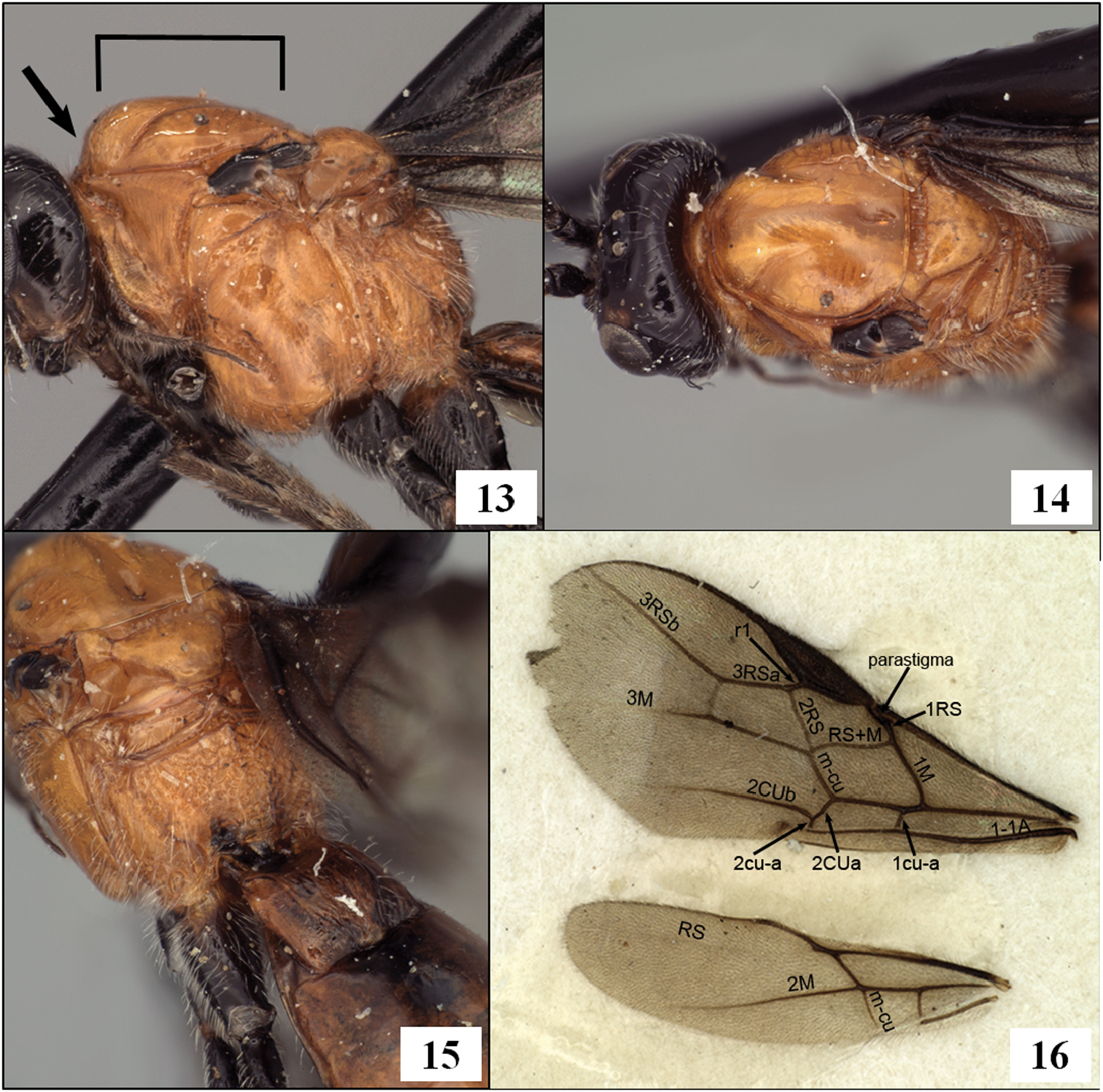
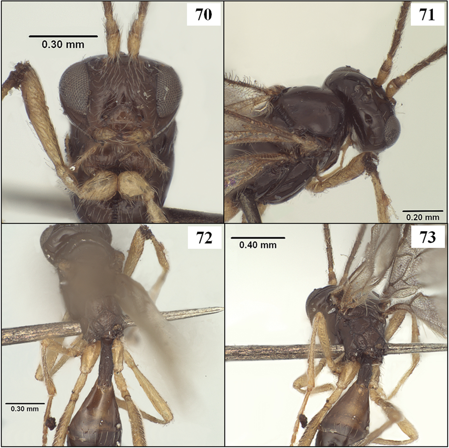
Diachasmimorpha hildagensis (Fischer), holotype male. 13 mesosoma, lateral view, arrow showing anterior declivity of mesoscutum, bracket showing mesoscutal disc 14 head and mesonotum, dorsal view 15 propodeal sculpture 16 left fore and hind wings illustrating wing vein terminology.
urn:lsid:zoobank.org:act:9E85B215-4032-4CEC-ADF4-6F068E83C029
http://species-id.net/wiki/Diachasmimorpha_martinalujai
Figs 26, 27, 31, 33Mexico, Distrito Federal.
Holotype. Female (UNAM), first and only data label, first line: Mexico, D. F. second line: Host = R. pomonella third line: Host plant=Crataegus sp. fourth line: Common name=Tejocote fifth line: 7.xi.2007 J. Rull
Paratypes: 1 male, same data as holotype (TAMU). 1 male, Mexico, Hidalgo, Atotonilco, 4.xi.2002, J. Rull, key 30, reared from Rhagoletis nr. pomonella infesting fruit of Crataegus spp. (TAMU). 1 male, Mexico, Puebla, San Martin, 24.xi.2003, M. Pale key 69, reared from Rhagoletis nr. pomonella infesting fruit of Crataegus mexicana (TAMU).
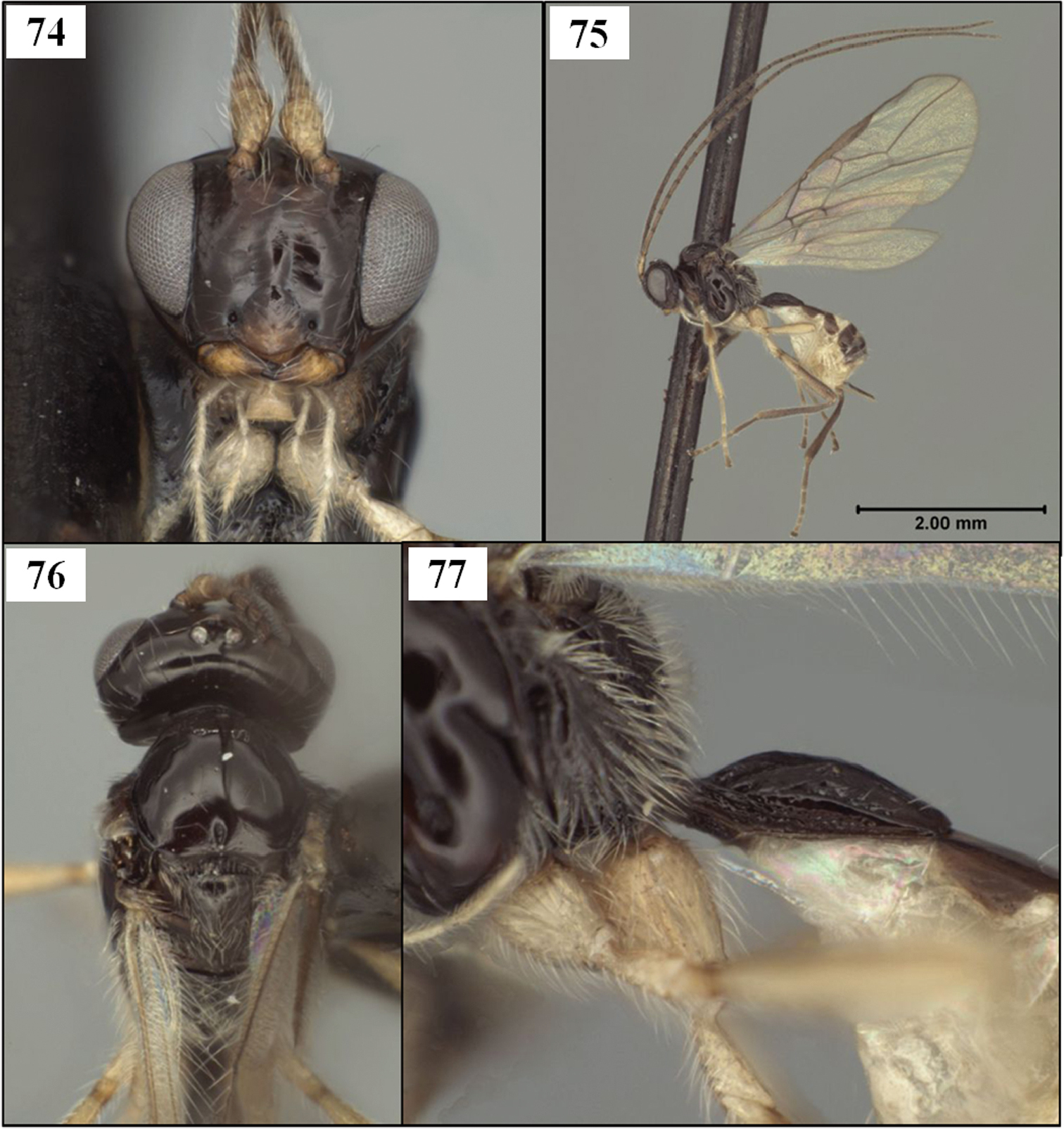
Female.Head in dorsal view 1.30 × broader than mesoscutum, 1.65 × broader than face; eye in dorsal view 2.0 × longer than temple, temples not receding, but width at eyes greater than width at temples; eye in lateral view 2.05 × longer than temple. Discrete facial midridge ending dorsally as a distinct elevation at base of antennae, continuing between antennae onto frons as low, sharp, bifurcating ridges. Frons irregularly rugulose along midline between bifurcating arms, otherwise polished, with moderately dense patch of decumbent, laterally-directed, white setae on either side of midline; bare on either side of ocellar field; width of ocellar field 0.95 × distance from ocellar field to eye. Face 2.2 × wider than high; uniformly setose (as in Figs 31, 33), distinctly punctate, punctures separated by about 1 × their diameter or slightly less. Malar sulcus deep, complete; malar space about 1.1 × basal width of mandible, 0.35 × eye height. Clypeus 2.65 × wider than high; very weakly convex, nearly flat. Occipital carina weak, difficult to discern near base of mandible, short, extending dorsally to ventral margin of eye. Hypostomal carina extending as short but distinct flange below mandible. Antenna with 45 flagellomeres; first flagellomere 1.3 × longer than second; 1.8 × longer than wide.
Mesosoma 1.4 × longer than high; 1.9 × longer than wide; 1.35 × higher than wide. Pronotum not visible dorsally; crenulae extending over dorsal 0.3–0.4 of pronotum laterally within narrow, shallow groove; groove not margined anteriorly by carina; anterior margin of pronotum laterally sinuate, not abruptly excavated. Notauli deep anteriorly, ending abruptly posteriorly, short, not quite extending posteriorly to level of anterior margin of tegula, not reaching long, narrow midpit, anterior end extending to anterior-lateral margin of scutum; mesoscutum without supra-marginal carina adjacent margin of mesoscutum between base of notaulus and tegula. Scuto-scutellar sulcus rectangular or nearly so; 4.75 × wider than midlength; crenulate-foveolate. Propodeum rugose, areola extending over posterior 0.8 but partially obscured by sculpture. Precoxal sulcus crenulate, distinctly separated from anterior margin of mesopleuron.
Wings. Fore wing stigma short, broad, discrete distally, 3.5 × longer than wide; r1 arising from midlength of stigma; 1RS (excluding parastigma) 0.30 × length of 1M; m-cu postfurcal by 0.25 × length of m-cu; second submarginal cell converging distally; 2RS 0.9 × length of 3RSa; 2CUa about 1.7 × longer than 2cu-a; 1cu-a distad 1M by about 1.0 × its length.
Metasoma not distinctly petiolate; head 1.8 × wider than apex of T1. T1 1.05 × as long as apical width; strongly diverging apically, with apex 2.1 × wider than base; surface smooth; dorsal carinae parallel-sided, widely separated posteriorly, distinctly elevated over anterior 0.6, weaker and becoming indistinct posteriorly; lateral carina weaker than dorsal carina basally, extending distinctly ventrad spiracle, rounded and barely distinguishable posteriorad spiracle; spiracle at midlength of T1; dorsope absent but lateral and dorsal carinae elevated at junction, giving appearance of a slight depression; laterope deep; S1 very short. T2 unsculptured, with sharp lateral margins. Ovipositor sheath 2.4 × longer than mesosoma, densely setose over apical half, with 4–5 irregular rows of setae, the setae longer than sheath width, more sparsely setose basally.
Color (Fig. 26). Very similar to Diachasmimorpha hildagensis. Meso- and metasoma orange, except tegula black; head dorsally black except for small orange spot on vertex adjacent eye; lower gena and most of occiput yellow-orange; narrow bands dorsad epistomal sulcus, along ventral margin of clypeus and vertically through middle of mandible orange; legs black to dark reddish brown except basal 0.5 of hind coxa orange, joint between femora and trochantelli reddish orange.
Male. Largely as in female with variation as follows: head in dorsal view 1.35–1.45 × broader than mesoscutum, 1.6–1.7 × broader than face; eye in dorsal view 1.6–1.85 × longer than temple, in lateral view 1.7–1.95 × longer than temple; face 1.95–2.1 × wider than high; malar space 0.3–0.45 × eye height; clypeus 2.6–2.8 × wider than high; antenna with 39–47 flagellomeres; first flagellomere 1.1–1.2 × longer than second, 2.0–2.1 × longer than wide; mesosoma 1.25–1.35 × longer than high; 1.85–1.95 × longer than wide; 1.4–1.5 × higher than wide; pronope deep, moderately large but not interrupting posterior crenulate groove middorsally; crenulae extending over dorsal 0.2–0.4 of pronotum laterally; scuto-scutellar sulcus 4.0–5.0 × wider than midlength; areola of propodeum variably obscured, short and triangular rather than pentagonal in topotypic paratype; precoxal sulcus occasionally extending to anterior margin of mesopleuron; fore wing stigma 3.3–3.8 × longer than wide; 1RS 0.2–0.25 × length of 1M; m-cu postfurcal by 0.15–2.0 × length of m-cu; 2RS 0.8–1.05 × length of 3RSa; head 1.85–2.2 × wider than apex of T1; T1 0.95–1.05 × as long as apical width, apex 2.1–2.25 × wider than base; surface of T1 between dorsal carinae weakly rugulose; dorsal carinae weakly sinuate, weakly converging at posterior margin of T1; S1 extending posteriorly only to level of dorsal tendon attachment; head varying from darker as in female to more extensively pale (as in Fig. 31) with ventral 0.5 of face orange, outer surface of mandible entirely dark orange and clypeus reddish brown; hind coxa varying from almost entirely orange to almost entirely black; hind femur and tibia varying from black to reddish brown.
Body length 4.9 mm (female), 3.1–4.7 mm (male), fore wing length 4.0 mm (female), 2.7–4.1 mm (male), mesosomal length 1.55 mm (female), 1.0–1.7 mm (male).
This species is nearly identical to Diachasmimorpha hildagensis based on the similarly long ovipositor and the notaulus that consistently extends all the way to the anterior margin of the mesoscutum. The eye is distinctly larger in Diachasmimorpha martinalujai than in Diachasmimorpha hildagensis. Diachasmimorpha norrbomi is also similar, but has a shorter ovipositor and the notaulus only rarely extends anteriorly to the margin of the mesoscutum.
This is the species that has been referred to as Diachasmimorpha mexicana (vide Wharton) in previous publications on parasitoids of Rhagoletis Loew in Mexico (e.g.
This species is named after Martin Aluja in recognition of his many contributions to tephritid biology, particularly in Mexico.
The male paratypes, though only three in number, are remarkably variable in size, with larger individuals closely approaching the size of Diachasmimorpha hildagensis. Quantitative measures are also highly variable, which is not surprising given the variation in size.
Detailed assessment of the available reared material suggests the presence of a diverse assemblage of Diachasmimorpha species in Mexico, associated with different hosts and host plants. The relatively small morphological differences between Diachasmimorpha hildagensis and Diachasmimorpha martinalujai are consistent among the available material and the differences in host and host plant associations lend support to the recognition of these as separate species.
Diachasmimorpha spp. 17 Diachasmimorpha longicaudata (Ashmead), arrow showing sharply indented margin of pronotum laterally 18 Diachasmimorpha sanguinea (Ashmead), arrow showing less sharply indented margin of pronotum laterally 19 Diachasmimorpha longicaudata, dorsal view 20 Diachasmimorpha mellea (Gahan), dorsal view.
Propleuron and T2. 21 Diachasmimorpha longicaudata (Ashmead), T2 with striae (arrow) 22 Diachasmimorpha tryoni (Cameron), T2 without sulpture 23 Diachasmimorpha longicaudata, propleuron without oblique carina 24 Fopius arisanus (Sonan), propleuron with oblique carina (arrow).
urn:lsid:zoobank.org:act:4900256F-3E99-41FC-8CCF-3A42E06D2033
http://species-id.net/wiki/Diachasmimorpha_norrbomi
Figs 25, 29, 30, 32Mexico, State of Mexico, Parque Lago de Zempoala.
Holotype. Female (UNAM), first label, first line: Mexico, Parque second line: Lag. de Zempoala, path third line: along L. Zempoala, 10–11. fourth line: VIII.1989, A.L.Norrbom Second label, first line: reared ex. Euphranta second line: mexicana (Tephritidae) third line: ex. fruit of Ribes fourth line: pringlei Rose (89M13)
Paratypes: 27 females, 20 males, same data as holotype, one of these with an additional ALN 31 label and a Biosteres sp. 1 det P. Marsh label (TAMU, UNAM, USNM).
Other specimens examined (not paratypes): 1 female, 1 male, Mexico, D.F., Delegacion Tlapan, Fracc. Tlapuente, 19.ix.2003, M. Aluja #50, reared from fruit of Granadilla (TAMU).
Female.Head in dorsal view 1.25–1.30 × broader than mesoscutum, 1.80–1.85 × broader than face; eye in dorsal view 1.7–2.0 × longer than temple, temples not receding, but width at eyes greater than width at temples; eye in lateral view 2.1–2.9 × longer than temple. Facial midridge ending dorsally in short, very weak bifurcation between antennae. Frons irregularly rugulose along midline near bifurcation, otherwise polished, with moderately dense patch of decumbent, laterally-directed, white setae on either side of midline; bare on either side of ocellar field; width of ocellar field 1.0–1.2 × distance from ocellar field to eye. Face 1.80–1.95 × wider than high; uniformly setose (as in Figs 30, 32), distinctly punctate, punctures separated by at least 1 × their diameter. Malar sulcus deep, complete; malar space about 0.9–1.0 × basal width of mandible, 0.30–0.35 × eye height. Clypeus 2.8–3.2 × wider than high; very weakly convex, nearly flat. Occipital carina weak but distinct near base of mandible, short, extending dorsally to ventral margin of eye and often slightly beyond, not reaching mid eye height. Hypostomal carina extending as short but distinct flange below mandible. Antenna with 41–47 flagellomeres; first flagellomere 1.05–1.2 × longer than second; 1.8–2.0 × longer than wide.
Mesosoma 1.35–1.45 × longer than high; 1.85–1.95 × longer than wide; 1.35–1.40 × higher than wide. Pronope deep, large, interrupting posterior crenulate groove middorsally; crenulae extending along dorsal 0.2 of pronotum laterally within narrow, shallow groove; groove not margined anteriorly by carina; anterior margin of pronotum laterally sinuate, not abruptly excavated. Notauli deep anteriorly, gradually weakening posteriorly, extending posteriorly to level of tegula, not reaching long, narrow midpit, anterior end usually just short of and only rarely reaching anterior-lateral margin of scutum; mesoscutum usually without supra-marginal carina between base of notaulus and tegula, rarely with short, weak trace of a carina. Scuto-scutellar sulcus nearly rectangular, a little narrower medially; 4.2–4.8 × wider than midlength; crenulate-foveolate. Propodeum rugose, areola extending over posterior 0.8 but largely obscured by sculpture. Precoxal sulcus crenulate, widely separated from anterior margin of mesopleuron.
Wings. Fore wing stigma short, broad, discrete distally, 3.15–3.30 × longer than wide; r1 arising from midlength of stigma; 1RS (excluding parastigma) 0.30–0.35 × length of 1M; m-cu postfurcal by 0.2–0.3 × length of m-cu; second submarginal cell distinctly converging distally; 2RS 1.0–1.2 × longer than 3RSa; 2CUa 1.6–1.8 × longer than 2cu-a; 1cu-a distad 1M by about 1.0 × its length.
Metasoma not distinctly petiolate; head 1.6–1.9 × wider than apex of T1. T1 0.95–1.05 × as long as apical width; strongly diverging apically, with apex 2.0–2.5 × wider than base; surface smooth to weakly strigose posterior-medially, almost completed smooth laterally; dorsal carinae weakly converging, widely separated at posterior margin, strongly elevated over anterior 0.5, gradually weakening posteriorly; lateral carina weaker, extending distinctly ventrad spiracle, rounded and barely distinguishable posteriorad spiracle; spiracle at midlength of T1; dorsope absent but lateral and dorsal carinae elevated at junction, giving appearance of a slight depression; laterope deep; S1 very short, extending posteriorad to level of dorsal tendon attachment. T2 unsculptured, with sharp lateral margins. Ovipositor sheath 1.7–1.8 × longer than mesosoma, setal pattern about as in Diachasmimorpha martinalujai, with slightly greater density basally.
Color (Fig. 25). Very similar to Diachasmimorpha hildagensis. Meso- and metasoma orange, except tegula black; head dorsally dark brown to black except for small orange spot on vertex adjacent eye, lower occiput mostly yellow-orange, similar in color to broad band extending through epistomal sulcus, clypeus, lower gena (often), and mandibles; clypeus usually with narrow, transverse brown band, mandible with apical teeth dark, rarely with entire mandible brownish; legs black except extreme base and most or all of dorsal side of hind coxa orange, joint between femora and trochantelli reddish orange.
Male as in female except head in dorsal view 1.3–1.4 × broader than mesoscutum, 1.70–1.75 × broader than face; eye slightly smaller, in dorsal view eye 1.45–1.60 × longer than temple, in lateral view 1.9–2.4 × longer than temple; antenna with 41–43 flagellomeres, first flagellomere 0.95–1.2 × longer than second. Mesosoma slightly narrower, 1.95–2.05 × longer than wide; 1.4–1.5 × higher than wide; scuto-scutellar sulcus somewhat more variable in size, 4.0–5.5 × wider than midlength. Fore wing stigma 3.1–3.4 × longer than wide. T1 slightly smaller, head 1.9–2.2 × wider than apex of T1, T1 1.75–1.90 × wider at apex than at base.
Body length 3.3–4.3 mm, fore wing length 3.5–4.1 mm, mesosoma length 1.15–1.65 mm.
This species is similar in coloration to Diachasmimorpha hildagensis and Diachasmimorpha martinalujai but the ovipositor (with sheath 1.7–1.8 × longer than mesosoma) is slightly but distinctly shorter and the notaulus only rarely extends all the way to the anterior margin. The notaulus always reaches the anterior margin in the other two species. Diachasmimorpha norrbomi is smaller and has a larger eye than Diachasmimorpha hildagensis, and 2RS tends to be longer (relative to 3Ra) in Diachasmimorpha norrbomi than in Diachasmimorpha hildagensis and Diachasmimorpha martinalujai.
The type series of Diachasmimorpha norrbomi was reared from Euphranta mexicana Norrbom infesting fruits of Ribes pringlei Rose (
This species is named for Allen Norrbom, who reared many Opiinae from various fruit, stem, and flower-infesting tephritids in Mexico and Central America.
Size variation in this species is similar to that exhibited by Diachasmimorpha martinalujai, with males dominating the small end of the range.
Diachasmimorpha spp. 25 Diachasmimorpha norrbomi Wharton sp. n., paratype female, habitus showing relatively shorter ovipositor 26 Diachasmimorpha martinalujai Wharton sp. n., holotype, habitus showing relatively longer ovipositor 27 Diachasmimorpha martinalujai paratype male, base of notaulus 28 Diachasmimorpha tryoni (Cameron) apex of ovipositor showing subapical sinuation 29 Diachasmimorpha norrbomi, paratype female, apex of ovipositor.
Diachasmimorpha spp., heads. 30 Diachasmimorpha norrbomi Wharton, sp. n., paratype female, face 31 Diachasmimorpha martinalujai Wharton, sp. n., paratype male, face 32 Diachasmimorpha norrbomi paratype female, lateral view 33 Diachasmimorpha martinalujai, holotype female, face.
http://species-id.net/wiki/Diachasmimorpha_mexicana
Figs 34–38Mexico, D. F., Chapultepec.
Holotype male (BMNH), first label [round, white with red margin], first line: Type second line: H. T. Second label, first line: B. M. TYPE second line: HYM third line: 3.c.705 Third label, first line: B.C.A. Hymen. I. second line: Opius third line: mexicanus fourth line: Cam. Fourth label, first line: Opius second line: mexicanus third line: Cam. Type fourth line: BCA ii 409 Fifth label, first line: Bilimek second line: Mexico third line: 1871. fourth line: Chapul fifth line: tepek.
Holotype male. Eye in dorsal view shorter than temple, temples weakly expanded beyond eyes; eye in lateral view 0.95 × length of temple. Frons unsculptured along midline between antenna and median ocellus. Clypeus 3.4 × wider than high. Occipital carina distinct near base of mandible, short, not extending dorsally to ventral margin of eye. Antenna broken. Pronope deep, large, interrupting posterior crenulate groove middorsally. Notauli deep anteriorly, reaching margin of mesoscutum anteriorly, apparently extending about half distance from anterior-lateral margin to elongate midpit but pin obliterates midpit and surrounding area of mesonotum. Precoxal sulcus very weakly crenulate, nearly smooth, short, not extending close to anterior margin of mesopleuron. Propodeum largely smooth, with rugulose sculpture largely confined to midline, especially around apex, and along border of metapleuron. Fore wing 2RS 0.8 × 3RSa; m-cu distinctly postfurcal. T1 with dorsal carinae widely separated, short, barely extending to level of spiracle, T1 otherwise unsculptured. Head, meso- and metasoma orange, tegula black; legs black as in holotype of Diachasmimorpha hildagensis. Body length about 4.0 mm. This species has a much smaller eye (Figs 35, 37) than the similarly-colored Diachasmimorpha sanguinea (Fig. 41) and is also less heavily sculptured. Females are unknown.
Unknown.
The body of the Diachasmimorpha mexicana holotype is remarkably smooth relative to that of other species in the mexicana species group. The precoxal sulcus, for example, is very weakly crenulate, the propodeum is very weakly sculptured in general but completely smooth and polished anterior-laterally, and T1 is unsculptured except for the very short dorsal carinae. Sculpture is variable to some extent in other species of this species group, and thus it would be useful to obtain additional specimens of the true Diachasmimorpha mexicana to determine the extent of sculptural variation in this species and ascertain whether reduction in sculpture is a useful diagnostic feature.
See additional remarks under Diachasmimorpha hildagensis above.
Diachasmimorpha mexicana (Cameron), holotype male. 34 habitus 35 head, lateral view 36 wings, showing names of cells used in descriptions 37 face.
Diachasmimorpha spp. 38 Diachasmimorpha mexicana (Cameron) holotype male, mesopleuron 39 Diachasmimorpha sanguinea (Ashmead), male mesosoma, lateral view 40 Diachasmimorpha sanguinea habitus 41 Diachasmimorpha sanguinea, male head, lateral view.
http://species-id.net/wiki/Diachasmimorpha_sanguinea
Figs 4, 18, 39–41USA, Washington, D. C.
Syntype female (USNM), first label, first line: 3737x second line: Oct. 3. 85 Second label (red with black print), first line: Type second line: No2989 third line: U.S.N.M. Third label, first line: Phaedrotoma second line: sanguinea third line: Ashm ms. Syntype male, with same label data as syntype female except Third label = first line: Opius second line: sanguineus third line: Gahan Ashm Syntype male with first label, first line: 3737x second line: Aug. 5. 86 Second label: same as other two syntypes, no third label.
Other specimens examined. USA, Texas, 1 female, 1 male, Brazos Co., Yancey, xi.2010, emerged 9.iv & 3.v.2011, L. Ward, reared from Zonosemata vittigera infesting fruits of Solanum eleagnifolium (TAMU); 1 female, Hidalgo Co., Bentsen Rio Grande Valley State Park, 10.?.1978, C. Porter (TAMU); 5 females, 1 male, Hidalgo Co., Donna, J. W. Monk, reared from Zonosemata vittigera; 5 females, 1 male, Jeff Davis Co., 14 mi. S. Ft. Davis, 16–19.viii.1985, L. E. Carroll, reared from Zonosemata infesting fruits of Solanum; 5 males, Jeff Davis Co., Davis Mts. State Park, 12.vii.1995, R. Wharton; 1 female, Swisher Co., Happy, 17.viii.1977, W. F. Chamberlin.
Male. Eye in dorsal view 1.1–1.3 × longer than temple, temples not expanded beyond eyes; eye in lateral view 1.3–1.5 × longer than temple. Frons between short, low, bifurcating ridges varying from unsculptured to irregularly strigose, frons otherwise smooth, polished. Clypeus 2.5–2.8 × wider than high. Occipital carina distinct near base of mandible, short, not extending dorsally to ventral margin of eye. Antenna with 38–48 flagellomeres. Pronope deep, large, interrupting posterior crenulate groove middorsally. Notauli deep anteriorly, reaching margin of mesoscutum anteriorly, extending about half distance from anterior-lateral margin to elongate midpit. Precoxal sulcus heavily sculptured, crenulate to foveolate, usually extending to or nearly to anterior margin of mesopleuron. Propodeum rugose, areola, when partially visible, extending over posterior 0.6–0.7 but frequently completely obscured by sculpture. Fore wing 2RS 0.9–1.05 × length of 3RSa; m-cu distinctly postfurcal. T1 with dorsal carinae weakly converging, widely separated at posterior margin, gradually weakening posteriorly, T1 smooth to strigose between carinae. Head, meso- and metasoma orange; tegula orange to brown, legs varying from black except hind coxa mottled black and orange to more extensively orange. Female about as in male except eye in lateral view 1.2–1.6 × longer than temple. Ovipositor sheath 1.6–1.75 × longer than mesosoma. Body length 3.6–5.3 mm, fore wing length 3.3–4.6 mm, mesosoma length 1.2–1.9 mm. This species has a larger eye than the similarly-colored Diachasmimorpha mexicana and is generally more heavily sculptured.
This species was originally described from several specimens reared from a tephritid infesting fruits of Solanum carolinense L. (
The diagnosis is based on the material from Texas listed in the other material examined section.
The sculpture is somewhat variable in this species, with smaller individuals having a tendency towards rugulose rather than rugose sculpture on the propodeum. The precoxal sulcus is always heavily sculptured, however, never approaching the reduction in sculpture seen in the holotype of Diachasmimorpha mexicana (Fig. 39 vs. Fig. 38). The syntypes from Washington, D. C. are as variable in sculpture of the propodeum and T1 as are the specimens from Texas. Specimens from Texas, even within the same reared series, are exceptionally variable in leg coloration. The syntypes from Washington, D. C. have black legs with mostly orange hind coxa. Some specimens from Jeff Davis Co., Texas also have this pattern while in others only the tarsi are dark with the remaining parts orange. Similarly, the tegula is usually orange, but varies from orange to brown even within the same reared series.
Diachasmimorpha sanguinea is nearly identical to Diachasmimorpha mexicana and additional material from the type locality of the latter is needed for a better understanding of the relationship between these two nominal species.
http://species-id.net/wiki/Eurytenes
Mandible without basal lobe ventrally. Labrum broadly exposed. Occipital carina broadly absent dorsally, present laterally. Propleuron ventral-laterally without oblique carina. Notauli deep, well developed anteriorly, varying posteriorly from largely absent to deep and extending to scuto-scutellar sulcus or nearly so; midpit present. Fore wing stigma long, narrow, parallel-sided, discrete posteriorly, r1 arising distinctly basad its midpoint; second submarginal cell with 2RS shorter than 3RSb; 2CUb arising above middle of hind margin of first subdiscal cell. Dorsope present; S1 0.2–0.3 × length of T1, never fused to T1.
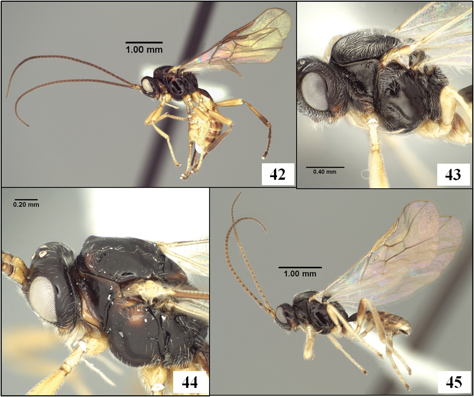
The new species described below have been placed in Eurytenes (Stigmatopoea) based on the relative length of S1 (Figs. 5, 7) and the specific characteristics of T1 (Figs 5, 7, 54, 56, 57), wing venation (Fig. 64), mesoscutal sculpture (Figs 44, 48, 49), clypeus (Figs 50–53), and mandibles (Figs 50, 51) listed in the diagnosis. The wing venation is similar to that in Lorenzopius but in Lorenzopius, the dorsope is absent and S1 is longer and apparently fused to T1 (Fig. 6). We follow
Aulonotus Ashmead has usually been characterized on the basis of well-developed notauli (
urn:lsid:zoobank.org:act:A5E2449E-78E5-48A3-B4CD-B4FC77A410A4
http://species-id.net/wiki/Eurytenes_maya
Figs 7, 42, 44, 46, 48, 50, 52, 56, 59, 64Mexico, Chiapas, San Cristobal de las Casas.
Holotype. Female (TAMU), first label, first line: MEXICO: Chiapas second line: San Cristobal de las third line: Casas, xi.2001, #37A fourth line: J. Marquez, M. Aluja Second label, first line: host: Rhagoletis second line: pomonella third line: ex fruit of: fourth line: Crataegus mexicana
Paratypes: 2 females, same data as holotype but collected 26.xi.2001, #35A (TAMU); 1 female, same locality, 14.xi.2001, M. Aluja, Key 30A, host: Rhagoletis sp. on tejocote, manzanita (TAMU); 1 female, same locality, 14.xi.2001, J. Marquez, ex: Rhagoletis pomonella on Crataegus sp., #27 (TAMU); 1 female, Chiapas, Rancho Nuevo, 5 km to San Cristobal de las Casas-freeway 190, 15.xi.2002, J. L. Marquez, M. Aluja, # 42, host: Rhagoletis pomonella ex fruit of Crataegus mexicana (TAMU); 2 males, Chiapas, 3 km E. San Cristobal, 15.xi.1994, R. Jones, ex pupa of Rhagoletis pomonella (TAMU); 3 females, Chiapas, Huixtan, 15.ix.2002, J. Marquez, Key 34, host: Rhagoletis pomonella ex fruit of Crataegus spp. (TAMU); 1 male, 1 female, Chiapas, Cruz Quemada, 15.xi.2002, host: Rhagoletis pomonella ex fruit of Malus sp., J. Marquez, Key 35, and J. L. Marquez, M. Aluja, #45 (TAMU); 1 male, 1? (abdomen missing), Chiapas, Teopisca, 26.xi.2001, J. L. Marquez, ex: Rhagoletis pomonella on Crataegus sp. #26 (TAMU).
(not paratype): 1 male, Mexico: San Luis Potosi, Rio Verde, 7.x.2003, M. Pale, Key 71, Rhagoletis nr. pomonella on Crataegus parrayana (TAMU) [sequenced].
Female. Head in dorsal view 1.25–1.30 × broader than mesoscutum, 1.80–1.95 × broader than face; eye in dorsal view 2.5–3.2 × longer than temple, temples distinctly receding behind eyes. Frons and vertex highly polished, unsculptured except for shallow, median depression between toruli; frons bare, vertex and occiput with a few, short, scattered setae; width of ocellar field 1.05–1.3 × distance from ocellar field to eye. Face 1.55–1.70 × wider than high; slightly less polished than frons; uniformly setose (as in Figs 50, 52), with very fine punctures, these separated by at least 2 × their diameter. Frons and face delimited by slight change in sculpture resulting in weak, shallow sulcus between torulus and eye; distance between antennal toruli equal to distance from torulus to eye, eye not distinctly emarginate in region of antenna. Malar sulcus deep, complete; malar space about 0.5 × basal width of mandible, 0.2 × eye height. Face weakly convex, bulging slightly medially along the low midridge. Epistomal sulcus weak mid-dorsally, more distinct laterally. Clypeus 2.2–2.5 × wider than high; weakly convex, slightly protruding in profile; ventral margin sharp, truncate to very weakly concave in frontal view. Labrum broadly exposed, gap between ventral margin of clypeus and dorsal margin of mandible varying from 0.5–1.0 × height of clypeus, depending on how tightly closed the mandibles are. Occipital carina distinctly curved medially at dorsal end, broadly absent mid-dorsally, the space where the carina is absent distinctly wider than width of ocellar field; occipital and hypostomal carinae widely separated at base of mandible, the latter extending as a flange beneath about basal 0.2 of mandible. Mandible without basal lobe ventrally; bidentate apically, lower tooth much smaller than dorsal tooth and slightly twisted beneath dorsal tooth; ventral margin carinate throughout. Antenna 1.35–1.45 ×longer than fore wing, with 39–43 flagellomeres; first flagellomere 1.1–1.3 × longer than second, 1.2–1.3 × longer than third; flagellomeres 2.3–2.7 × longer than wide basally, twice longer than wide apically. Maxillary palps a little longer than head height; fifth and sixth segments equal in length or nearly so, fourth segment 1.1–1.15 × longer than both fifth and sixth.
Mesosoma 1.4 × longer than high; 1.9 × longer than wide; 1.35–1.40 × higher than wide. Pronotum dorsally a narrow, polished, smooth band with crenulate groove along posterior margin; rarely with discernible, slightly enlarged pit in middle of crenulate groove; crenulae extending in narrow, shallow groove onto pronotum laterally, but only covering dorsal 0.2–0.4; groove margined anteriorly by sharp carina that continues ventrally along full length of pronotum. Anterior declivity of mesoscutum completely vertical, bare or nearly so; anterior-lateral corners of mesoscutum at upper edge of declivity elevated, rounded, sparsely setose; notauli extending 0.4 × distance from anterior declivity to scuto-scutellar sulcus, extending posteriorly from lateral side of elevated anterior-lateral corners, not extending to mesoscutal margin anteriorly, very weakly converging posteriorly; narrow, crenulate throughout; mesoscutum with distinct supra-marginal carina extending from elevated anterior-lateral corner to tegula. Lateral and median mesoscutal lobes bare except scattered setae along notauli; midpit deep, round to somewhat elongate, never extending to notauli. Scuto-scutellar sulcus nearly rectangular, a little narrower medially; 3.75–4.25 × wider than midlength; crenulate-foveolate, with 7 ridges; all sides vertical, clearly delineated. Scutellum very weakly convex, nearly flat, not strongly elevated; bare except for scattered setae posteriorly; unsculptured, even along posterior margin. Propodeum with median carina over anterior 0.3, bifurcating at this point to form an inverted v-shaped transverse carina extending to pleural carina just posteriad spiracle; pleural carina complete from base to apex though sometimes partly obscured by sculpture posteriad spiracle; lateral longitudinal carina parallel to and narrowly separated from pleural carina anteriad spiracle, more medially displaced when visible posteriad transverse carinae, forming part of broad areola; area between pleural and lateral longitudinal carinae rugose and sparsely setose anteriorly; lateral propodeal areas anteriorly on either side of median carina smooth, bare, unsculptured; areola broad, varying from distinct (with surface irregularly, weakly rugulose) to indistinct (surface rugose, disrupting carinate margin of areola); lateral propodeal areas posteriorly varying from nearly unsculptured and distinct to rugose and indistinct; propodeum largely bare medially, with a few scattered setae. Mesopleuron largely bare, with sparse setae in unsculptured subalar region and a small patch of setae dorsad mid coxa; posterior margin unsculptured. Precoxal sulcus weakly impressed but distinct; unsculptured. Metapleuron bare on dorsal half except for small patch below wing, with a few long setae medially, and patches of setae among rugulose sculpture along ventral margin and in groove on ventral half of anterior margin; otherwise unsculptured.
Wings. Fore wing stigma parallel-sided, discrete posteriorly, 7.50–7.75 × longer than wide; r1 arising from basal 0.35; 1RS (excluding parastigma) 0.20–0.25 × length of 1M; RS+M straight or nearly so; m-cu postfurcal, extending into basal corner of second submarginal cell; second submarginal cell weakly converging distally; 3RSa 1.10–1.25 × longer than 2RS; 2RS 2.5–3.4 × longer than r, the two not forming a continuous line; 2RS with distinct median bend; 3RSb very weakly bowed, nearly straight; 3M variable, but often pigmented and sclerotized for most of its length; 2CUa 0.5–0.7 × length of 2cu-a, 2CUb arising well above middle of first subdiscal cell; 1cu-a distad 1M by about 1.0 × its length; 1–1A bowed toward wing margin, and separated therefrom by its width. Hind wing RS a weak but distinct, unpigmented crease, extending nearly to wing margin in most specimens; 2M extending to wing margin as a more deeply impressed line, very weakly pigmented for much of its length; m-cu usually a deeply impressed, curved line extending about half distance to wing margin.
Metasoma distinctly petiolate; head 3.5–3.8 × wider than apex of T1. T1 2.15–2.35 × longer than apical width; nearly parallel-sided, with apex 1.20–1.35 × wider than base; surface striate throughout, above and below lateral carina; one or two very shallow, subapical depressions usually present dorsally; dorsope distinct, deep; laterope completely absent; dorsal carina present only at base, lateral carina usually distinct throughout; spiracle positioned 0.6 × length of T1 from the base; S1 extending about 0.25–0.30 × length of T1; dorsal surface of petiole in profile evenly convex from base to apex. T2 and following without sharp lateral margins; spiracle of second metasomal tergum laterally displaced, not visible in dorsal view. Ovipositor as long as mesosoma; ovipositor sheath 0.6–0.7 × length of mesosoma, with 2–3 irregular rows of long setae along its length.
Color: head, including antenna, mesosoma, petiole and ovipositor sheath dark brown except scape yellow; mandible, lower gena, ventral portion of clypeus, pedicel (occasionally), face adjacent antennal base, propleuron, anterior margin of pronotum, spot on mesopleuron below wing and a smaller spot above mid coxa, two streaks on either side of midpit on mesoscutum, posterior margins of scutellum and metapleuron, and petiole laterally (occasionally) dark yellow to orange; palps pale yellow, nearly white. Legs and metasoma beyond T1 yellow except hind tibia, hind tarsi, lateral margin of metasomal terga 2 + 3 and often anterior half of terga 4–6 brown, the hind tibia often paler medially.
Male. As in female except antenna with 41–45 flagellomeres, head 4.0–4.6 × wider than apex of T1 and T1 2.5–2.9 × longer than apical width. Body somewhat darker in color, with metasomal terga 6, 7, and most or all of 5 dark brown.
Body length 3.2–4.3 mm; wing length 3.5–4.2 mm.
This species runs to Opius (Nosopoea) in
All specimens were reared from Mexican populations of Rhagoletis pomonella (Walsh) infesting either hawthorns (species of Crataegus L.) or apples (Malus domestica Borkh.).
The species name is in reference the Mayan Indians of this region.
This species is similar in general appearance to members of the genus Lorenzopius, but T1 is not distinctly tubular as it is in the latter genus (see discussion below under Lorenzopius). The overall resemblance to Lorenzopius is enhanced by the presence of weak depressions on T1 that are similar in position in Eurytenes maya and Lorenzopius calycomyzae van Achterberg and Salvo (Figs 55, 56). The depressions are variable within members of the same reared series of Eurytenes maya: being absent, for example, in the holotype, but well developed in some of the paratypes.
The limited information on hosts suggests that species with a more tubular petiole, such as those in Lorenzopius, are parasitoids of leaf-mining Agromyzidae while the species of Stigmatopoea attack both leaf-mining and fruit-infesting tephritids.
Eurytenes (Stigmatopoea) spp. 42 ES.) maya Wharton sp. n., paratype female, habitus 43 ES.) norrbomi Wharton sp. n., holotype female, mesosoma 44 ES.) maya, paratype female, head and mesosoma, dorsal-lateral view 45 ES.) norrbomi, holotype female, habitus.
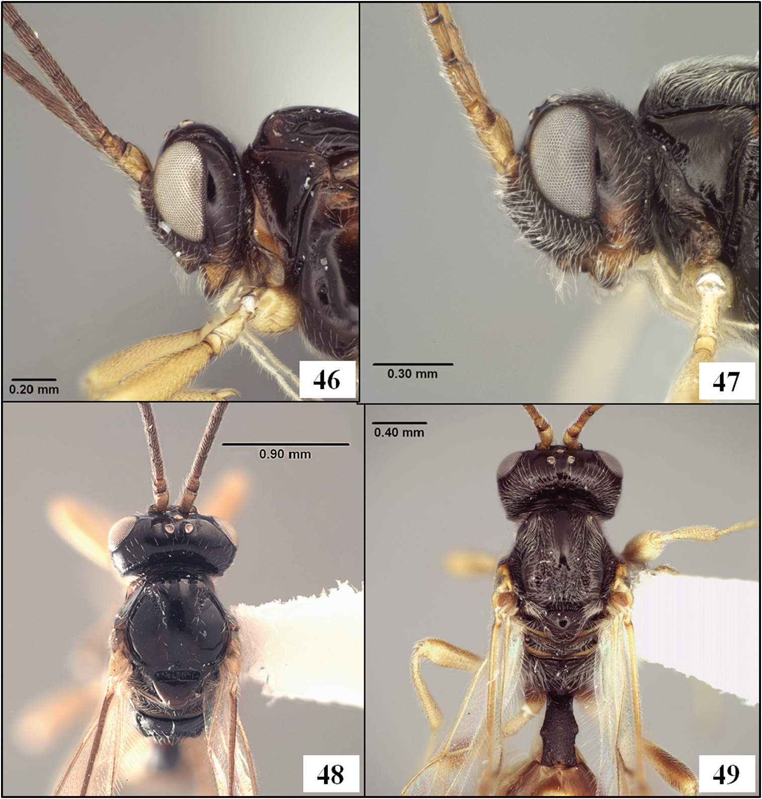
Eurytenes (Stigmatopoea) spp. 46 ES.) maya Wharton sp. n., paratype female, head, lateral view 47 ES.) norrbomi Wharton sp. n., holotype female, head, lateral view 48 ES.) maya, paratype female, head and mesosoma, dorsal view 49 ES.) norrbomi, holotype female, dorsal view.
urn:lsid:zoobank.org:act:FC57AAB3-6290-4076-BB44-6289F8ABF2EC
http://species-id.net/wiki/Eurytenes_norrbomi
Figs 43, 45, 47, 49, 51, 53, 57Mexico, Morelos, Km. 9–10 between Huitzilac and Lago Zempoala.
Holotype. Female (UNAM), first label, first line: MEXICO: Morelos second line: Km 9–10, btw. Huitzilac third line: & Lago Zempoala fourth line: roadside, 22–24.ix.1991 fifth line: A. L. Norrbom #42
Paratypes: Mexico, 4 females, same data as holotype (TAMU, USNM); 1 female, Mexico, Rt. 890, Km 9 area, 6 km W Lago Zempoala 2.x.1991, Norrbom, #43, reared ex. Trypeta concolor ex. leafmines on Barkleyanthus salicifolius (91M1D) (TAMU). 3 males, Distrito Federal, Rt. 95 (libre), Km 42–43, 1 km N. La Cima, 20–26.ix.1991 A. L. Norrbom, #41, reared ex. Trypeta concolor ex. leafmines on Barkleyanthus salicifolius (91M1) (TAMU, USNM).
Female. Head in dorsal view 1.2–1.3 × broader than mesoscutum, 1.75–1.85 × broader than face; eye in dorsal view 1.2–1.5 × longer than temple, temples weakly receding behind eyes. Frons and vertex as in Eurytenes maya except vertex and outer part of occiput densely covered with long, decumbent setae; width of ocellar field 1.20–1.35 × distance from ocellar field to eye. Face 1.75–1.85 × wider than high; slightly less polished than frons; uniformly setose (as in Figs 51, 53), distinctly punctate, the punctures separated by about 1 × their diameter. Frons and face delimited by a slightly more distinct change in sculpture in area between torulus and eye. Malar space about 0.6 × basal width of mandible, 0.25 × eye height. Clypeus 3.0–3.4 × wider than high; protruding in profile. Occipital carina distinctly curved medially at dorsal end, absent mid-dorsally, the space where the carina is absent approximating width of ocellar field. Antenna 1.15–1.30 ×longer than fore wing, with 31–33 flagellomeres; first flagellomere 1.05–1.10 × longer than second, 1.05–1.20 × longer than third; flagellomeres 3.1–4.1 × longer than wide basally, 2.3–2.7 longer than wide apically. Head otherwise as described for Eurytenes maya.
Mesosoma 1.35–1.45 × longer than high; 1.8–1.9 × longer than wide; 1.3–1.4 × higher than wide. Pronotum dorsally as in Eurytenes maya but with slightly enlarged pit in middle of crenulate groove consistently present; crenulae extending in shallow groove onto pronotum laterally, covering dorsal 0.2–0.6; groove margined anteriorly as in Eurytenes maya. Anterior declivity of mesoscutum completely vertical, densely covered with white, decumbent setae except for bare median band extending posteriorly to midpit; anterior-lateral corners of mesoscutum at upper edge of declivity elevated, rounded, densely setose, the setal pattern extending in broad bands all along notauli and laterally from anterior declivity to tegula; notauli complete, extending from anterior margin to scuto-scutellar sulcus, weakly converging posteriorly alongside but not into tear-drop shaped midpit; crenulate throughout, with sculpture extending laterally around margin to tegula, sculpture largely obscured by dense setae; lateral lobes of mesoscutum bare posterior-medially. Scuto-scutellar sulcus 4–5 × wider than midlength, lateral margins difficult to discern due to setal density; with low midridge and indistinct crenulae on either side; otherwise as in Eurytenes maya. Scutellum as in Eurytenes maya except with long marginal setae extending medially to cover most of posterior 0.5. Propodeum extensively rugulose, obscuring nearly all traces of carinae; pleural carina weak, often indistinct, very short median carina often present basally; transverse carina rarely weakly indicated across middle; propodeum uniformly setose anteriorly, with a few scattered setae posteriorly. Mesopleuron as in Eurytenes maya except subalar region densely setose and groove below subalar ridge varying from nearly smooth to weakly rugulose. Precoxal sulcus distinctly impressed, unsculptured. Metapleuron a little more extensively setose but otherwise as in Eurytenes maya.
Wings. Fore wing stigma parallel-sided, discrete posteriorly, 6.3–6.6 × longer than wide; r1 arising from basal 0.35; 1RS (excluding parastigma) 0.25–0.35 × length of 1M; RS+M weakly sinuate; 3RSa 1.05–1.30 × longer than 2RS; 2RS 2.6–3.1 × longer than r; 2RS and 3RSb straight; 3M variable, but often pigmented and sclerotized for most of its length; 2CUa 0.8–0.9 × length of 2cu-a, 2CUb arising slightly above middle of first subdiscal cell; position of m-cu, 1cu-a, and 1–1A, shape of second submarginal cell, and angle between r1 and 2RS as in Eurytenes maya. Hind wing as in Eurytenes maya.
Metasoma distinctly petiolate; head 3.75–4.10 × wider than apex of T1. T1 2.2–2.5 × longer than apical width; nearly parallel-sided, with apex 1.20–1.35 × wider than base; surface granular coriaceous throughout; completely without subapical depressions dorsally; dorsope, laterope, dorsal carinae, dorsal surface of T1 in profile, as in Eurytenes maya; lateral carina at least partially present but difficult to distinguish from surrounding sculpture. S1 extending about 0.25–0.30 × length of T1; T2 and following without sharp lateral margins; spiracle of second metasomal terga laterally displaced, only partially visible in dorsal view. Ovipositor shorter than mesosoma, base not visible in type series, but total length approximately 0.6–0.7 × length of mesosoma; ovipositor sheath 0.30–0.35 × length of mesosoma, with setal pattern as in Eurytenes maya.
Color: Mesosoma, T1, S1, ovipositor sheath, and most of head dark brown to black; antenna yellow basally, apical 0.3 brown; mandibles yellow; palps white; lower gena adjacent malar sulcus brown to brownish red; ventral 0.3–0.4 of clypeus yellow to brownish red. Tegula reddish brown with yellow margin. Legs yellow to pale yellow except most of hind coxa, apical 0.6–0.7 of hind femur, and fifth tarsomere of all legs brown; hind tibia varying from weakly infumate to light brown, basal 0.2 nearly always pale yellow. T2 mostly brownish red with median yellow blotch posteriorly; T3 yellow with anterior and lateral margins brownish red; T4-T6 yellow with anterior and lateral margins dark brown; visible parts of remaining terga yellow.
Male. As in female except antenna with 37 flagellomeres; eye in dorsal view 1.55–1.75 × longer than temple; width of ocellar field 1.05–1.10 × distance from ocellar field to eye. Color same except visible parts of apical terga dark brown.
Body length 2.8–3.5 mm; wing length 3.2–3.6 mm.
This species shares with Eurytenes maya and Eurytenes macrocerus the diagnostic features noted above for Stigmatopoea. Eurytenes norrbomi is most readily differentiated from Eurytenes maya on the basis of the more densely setose head and body (Figs 43, 47, 49), particularly the vertex, occiput, and mesoscutum, and the more extensively rugose propodeum. It also has a shorter ovipositor than Eurytenes maya (Fig. 45 vs. Fig. 42). The setal pattern on the mesoscutum also differentiates Eurytenes norrbomi from Eurytenes macrocerus. The latter has shorter setae that are more sparsely distributed laterally (Fig. 54).
Four of the specimens from the type series were reared from puparia of Trypeta concolor (Wulp) (Tephritidae) mining leaves of Barkleyanthus salicifolius (H.B.K.) H. Robins & Brett (Asteraceae). The remaining specimens were collected from flowers of this same plant together with Trypeta concolor and Trypeta reducta Han and Norrbom. See
This species is named after the collector, Allen Norrbom, who has provided many valuable host records for tephritid parasitoids.
This species attacks leaf-mining tephritids, as does Eurytenes macrocerus, while Eurytenes maya attacks fruit-infesting tephritids. Despite the difference in host habitat, all three species share many morphological features, and readily fit the characterization of Eurytenes (Stigmatopoea) as given above.
Eurytenes (Stigmatopoea) spp. 50 ES.) maya Wharton sp. n., paratype female, face, frontal view 51 ES.) norrbomi Wharton sp. n., holotype female, face, frontal view 52 ES.) maya, paratype female, face, slightly deflected 53 ES.) norrbomi, holotype female, face, slightly deflected.
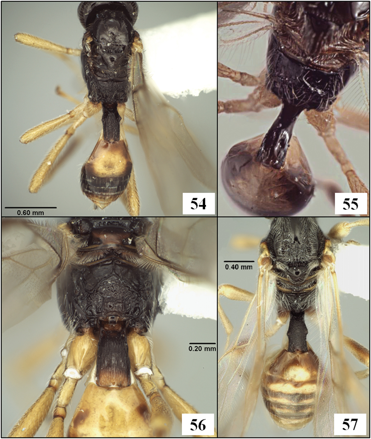
Eurytenes (Stigmatopoea) and Lorenzopius. 54 ES.) macrocerus (Thomson), mesosoma and metasoma, dorsal view 55 Lorenzopius calycomyzae van Achterberg and Salvo, holotype female, T1, dorsal view 56 ES.) maya Wharton sp. n., paratype female, propodeum and T1, dorsal view 57 ES.) norrbomi Wharton sp. n., holotype female, mesosoma and metasoma, dorsal view.
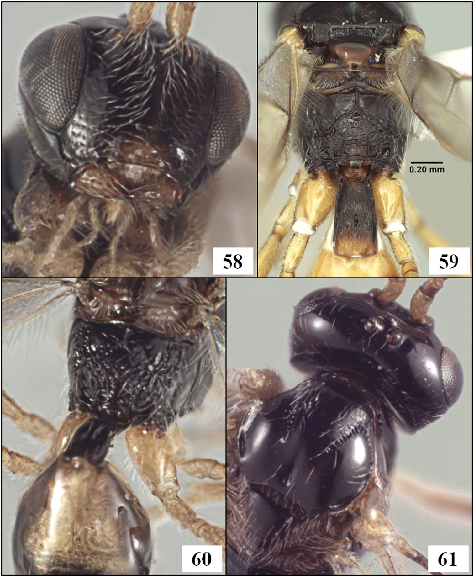
Lorenzopius and Eurytenes (Stigmatopoea). 58 Lorenzopius calycomyzae van Achterberg and Salvo, holotype female, face 59 ES.) maya Wharton sp. n., propodeum and T1 60 Lorenzopius calycomyzae, propodeum 61 Lorenzopius calycomyzae, head and mesoscutum, dorsal view.
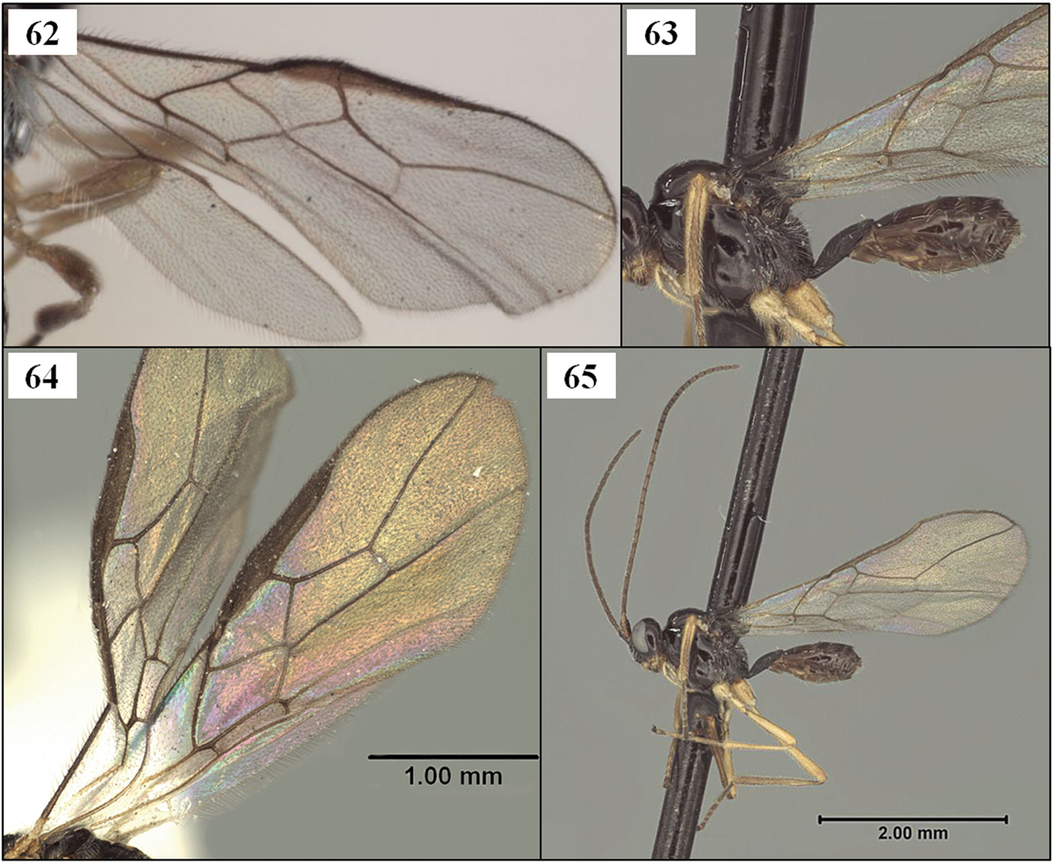
Opiinae. 62 Lorenzopius calycomyzae van Achterberg and Salvo, holotype female, wings 63 Tubiformopius tubigaster (Fischer), holotype male, lateral view showing T1, S1, and wing base 64 Eurytenes (Stigmatopoea) maya Wharton, sp. n., paratype male, fore wing 65 Tubiformopius tubigaster, holotype male, habitus.
http://species-id.net/wiki/Lorenzopius
Mandible distinctly narrowed from base to apex, without basal lobe ventrally. Labrum exposed. Clypeus relatively flat, not distinctly protruding in profile; ventral margin sharp, truncate to weakly concave. Malar sulcus a sharp, weakly curved groove. Occipital carina broadly absent dorsally, present laterally; widely separated from hypostomal carina ventrally. First flagellomere longer than second. Propleuron ventral-laterally without oblique carina; pronotum dorsally without pronope or otherwise enlarged pit, posterior margin transversely rugulose. Notauli deep, narrow, well developed anteriorly, usually extending onto disc posteriorly; midpit present. Precoxal sulcus distinctly impressed. Propodeum with large areola, posterior portion often obscured by rugose sculpture. Fore wing stigma long, narrow, parallel-sided, discrete posteriorly, r1 arising distinctly basad its midpoint but not from extreme base; m-cu entering base of second submarginal cell; second submarginal cell with 2RS shorter than 3RSb; 2CUb arising above middle of hind margin of first subdiscal cell. Dorsope and laterope of T1 absent; S1 at least 0.7 × length of T1 in females, slightly shorter in males, apparently fused to T1; T1 long and narrow throughout; T2 and following terga unsculptured. Ovipositor tapering evenly to a fine point, without dorsal nodes or ridges.
Lorenzopius and Tubiformopius are both characterized by having a tubular petiole with a long S1 which appears fused to T1 (Figs 6, 8). In the material available, S1 is longer in Lorenzopius than in Tubiformopius but there are more significant differences in the shape of the mandible, wing venation, and mesoscutal sculpture, as noted above in the section discussing genus group characters. Lorenzopius also shares many features with Eurytenes (Stigmatopoea), but the petiole is less tubular in the latter, with a distinctly shorter S1 that is clearly separated by membrane from T1 (Fig. 5).
The shape of the stigma has been proposed as a useful feature for assessing relationships among opiines (
We recognize two distinct species groups within Lorenzopius: the calycomyzae species group containing the orginially included species Lorenzopius calycomyzae, Lorenzopius tubulatus, and Lorenzopius sanlorenzensis and a second group typified by Lorenzopius euryteniformis (Fischer), new combination. All have same basic wing venation and petiole. The precoxal sulcus is distinctly sculptured in the calycomyzae species group (Fig. 69) but the distinctly impressed sulcus is unsculptured or nearly so in the euryteniformis species group (Fig. 66). The smallest specimens of the calycomyzae species group examined during this study are slightly larger than the largest available specimens of the euryteniformis species group and perhaps as a consequence they tend to have slightly longer notauli and more sculpture bordering the supra-marginal carina extending from the base of the notaulus to the tegula. Most of the species we have examined from the euryteniformis species group have reduced propodeal sculpture with the areola clearly visible (Figs 72, 73). In addition to holotypes of Lorenzopius tubulatus and Lorenzopius sanlorenzensis and the holotype and paratypes of Lorenzopius calycomyzae, we have seen two additional specimens from Argentina (TAMU), and one specimen each from Peru and Costa Rica (both CNC) representing the calycomyzae species group. RAW has examined 17 specimens representing the euryteniformis species group in addition to the holotype of Lorenzopius euryteniformis. The material examined includes specimens housed in TAMU and CNC collected in Bolivia, Colombia, Costa Rica, Dominican Republic, Guatemala, and Mexico (as far north as Monterrey in Nuevo Leon).
Lengthy descriptions (
The type species of Lorenzopius was described from specimens reared from Calycomyza mikaniae Spencer, a leafminer in the family Agromyzidae. RAW has also seen specimens from Colombia of a species nearly identical to Lorenzopius euryteniformis that was also reared from an agromyzid leafminer. No other host records are known for this genus but given the general similarity of the habitus and the length and shape of the ovipositor, we predict that other species will also prove to be agromyzid leafminer parasitoids.
http://species-id.net/wiki/Lorenzopius_euryteniformis
Figs 66, 67, 70–73Costa Rica, Mount Irazu, 2200–2300 m.
Holotype. Male (NHMW), first label, first line: Costa Rica, Irazu, second line: 2200–2300 m, 21–28. third line: V.’30. Reimoser Second label, first line: Opius second line: euryteniformis third line: sp. n. fourth line: det. Fischer Third label: Holotype [purple], Fourth label: NHMW
Holotype male. Head in dorsal view with temples neither receding nor expanded beyond eyes; in lateral view, eye about 1.6 × longer than temple. Labrum partly exposed between clypeus and mandibles (Fig. 70); clypeus about twice as wide as tall, flat or nearly so, not distinctly protruding in profile, ventral margin truncate to very weakly concave. Mandible without basal lobe. Malar space well developed, longer than basal width of mandible; malar sulcus deeply impressed. Antenna with 27 flagellomeres. Pronotum dorsally not visible in holotype. Disc of mesoscutum nearly bare, with scattered setae along margin of anterior declivity and a single pair of setae arising about midlength of notauli; notaulus extending posteriorly along anterior 0.3 of disc, less than half distance to small, deep, round midpit; supra-marginal carina distinct anteriorly, not extending to level of tegula. Scuto-scutellar sulcus relatively narrow (Fig. 71), densely crenulate throughout. Precoxal sulcus distinctly impressed, long, narrow, completely unsculptured. Propodeum largely smooth with broad, pentagonal areola on posterior 0.65, anterior 0.35 with median carina. Fore wing stigma long, narrow, with some postmortem curling, but at least 4.5 × longer than width at r1; r1 arising from basal 0.3; second submarginal cell long, weakly converging distally, 3RSa 1.7 × longer than 2RS; 1RS 0.2 × length of 1M; m-cu postfurcal; 2CUb arising a little above middle of hind margin of first subdiscal cell, 2cu-a present, tubular. T1 long, narrow, apparently fused ventrally with S1 for most of its length, 4x longer than apical width, apex as wide as base; surface completely striate. T2 and following smooth, polished.
Unknown.
Placement of this species in Lorenzopius is based on the wing venation and long S1, which is 0.65 × length of T1 in the male holotype; S1 appears fused to T1. See additional comments on species groups under the remarks section for the genus.
The holotype bears a single data label containing the information given above. However, the label data listed in the original description are as follows: “Costa Rica, La Caja bei San José, H. Schmidt”. As this species was described from a single male specimen, and the specimen from Irazu labeled as the holotype matches the original description, it is likely that the locality data in the original publication is an inadvertent error. The new species described immediately before euryteniformis in the same publication is from the La Caja locality. The type locality should therefore be Irazu (a mountain in Costa Rica), somewhere in the 2200–2300 m range in elevation.
Lorenzopius spp. 66 Lorenzopius euryteniformis (Fischer), holotype male, dorsal-lateral view showing unsculptured precoxal sulcus 67 Lorenzopius euryteniformis, holotype male, habitus 68 Lorenzopius tubulatus (Fischer), holotype female, habitus 69 Lorenzopius tubulatus, holotype female, mesosoma, lateral view showing sculptured precoxal sulcus.
Lorenzopius euryteniformis, holotype male. 70 face 71 mesoscutum and head, dorsal-lateral view 72 propodeum and T1–3 73 propodeum and metasoma.
http://species-id.net/wiki/Opius
Diagnoses are presented below for two species that represent a fairly diverse group of neotropical Opiinae that differ from both Lorenzopius and Tubiformopius in several features. These species all have a narrow, parallel-sided T1 and distinctly visible S1, though S1 is never as long as in Lorenzopius, and seldom as long as in Tubiformopius. Ultimately, the relationships of genus group taxa such as Eurytenes s.l., Lorenzopius, and Tubiformopius will have to be carefully considered in order to place the many neotropical species with a distinct S1.
http://species-id.net/wiki/Opius_incoligma
Figs 74–77, 81Colombia, Magdalena, 41 km south of Sta. Marta, 7000 ft.
Holotype. Female (AEIC), first label, first line: 41Km S.St. Marta second line: Magd., Colombia third line: V.6.1973 7000 ft. fourth line: Howden&Campbell second label [red]: Holotype third label, first line: [female symbol] Opius second line: incoligma third line: Holotype fourth line: det Fischer sp. n.
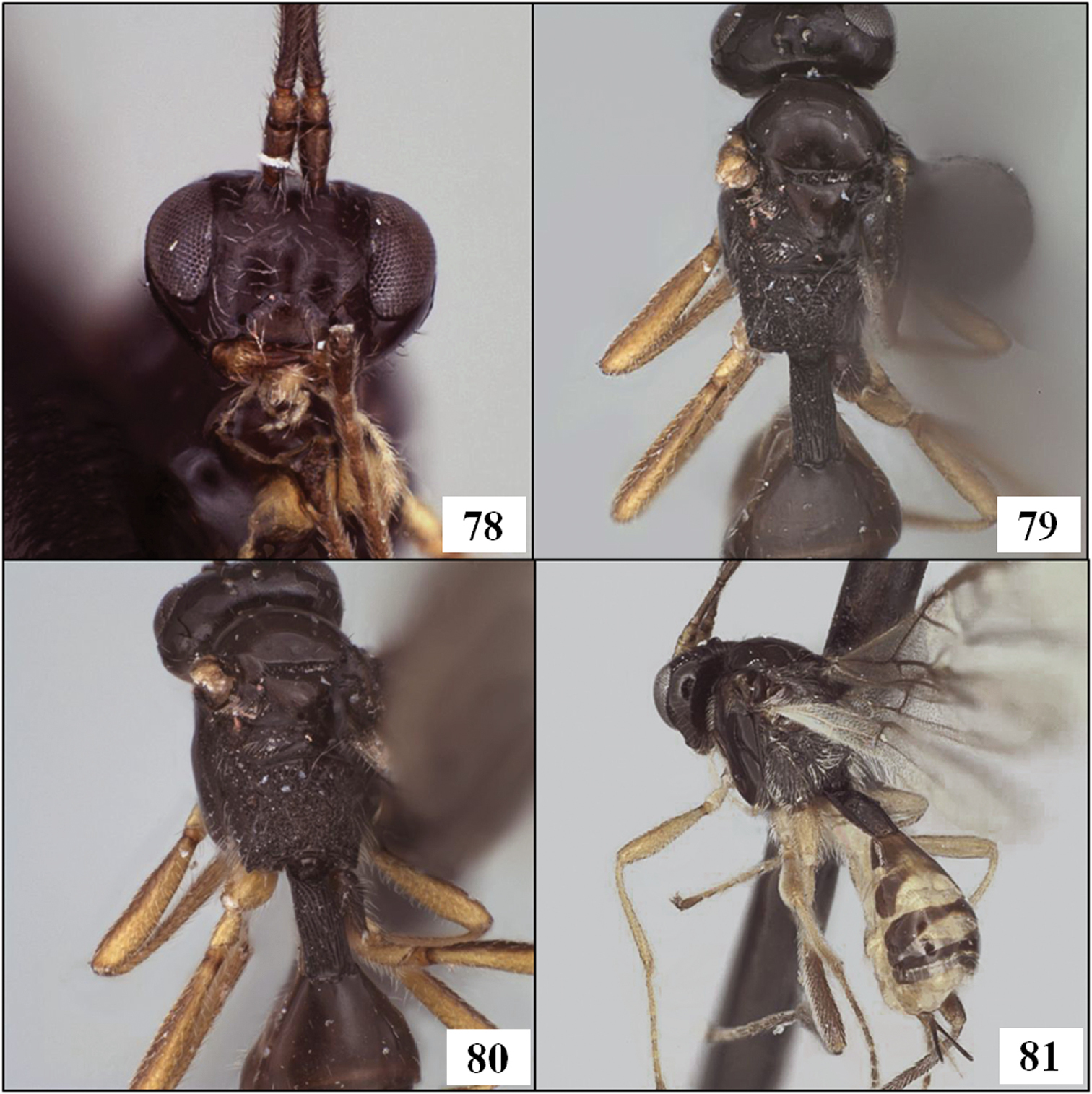
Holotype female. Labrum completely concealed by mandibles; clypeus nearly as tall as wide, flat, not protruding, ventral margin convex. Mandible without basal lobe, distinctly narrowing apically to narrow, bifid tooth. Malar space distinct, malar sulcus deep, distinct. Antenna with 33 flagellomeres. Pronotum dorsally without pronope or distinct pit, mostly unsculptured, crenulate posterior margin broadly interrupted medially. Disc of mesoscutum nearly bare, with a few setae along traces of notauli; midpit small, distinct, narrowly elongate; notauli weak, present as very short, weakly sculptured grooves directed posterior-medially from and along edge of anterior declivity, not extending posteriorly onto disc of mesoscutum; distinct supra-marginal carina extending laterally from base of notaulus to tegula. Scuto-scutellar sulcus narrow (about 6–7 × wider than long but difficult to measure), crenulate throughout. Precoxal sulcus distinct, moderately deep, long, completely unsculptured, somewhat vertically oriented as in Lorenzopius. Propodeum granular rugose, with very short median carina anteriorly, densely setose throughout. Fore wing stigma parallel-sided to weakly expanded apically; r1 longer than stigma width; second submarginal cell long, weakly narrowing distally; m-cu weakly postfurcal; 2CUb arising distinctly above middle of first subdiscal cell, 2CUa nearly absent. Hind coxa smooth; hind femur slender, weakly bilobed. T1 weakly strigose, irregularly sculptured with smooth patches; dorsal carina short but distinct; lateral carina very well developed, extending from junction with dorsal carina to apex, passing ventrad spiracle; dorsope shallow, indistinct, laterope shallow, weakly indicated by a long, narrow groove; T1 spiracle situated slightly posteriad midlength of T1; T1 narrow, parallel-sided, 2.6 × longer than apical width; no visible membrane between S1 and T1, though lateral margin between the two clearly visible; S1 0.35 × length of T1.
The venation (Fig. 75) and features of the first metasomal segment (Figs 77, 81) suggest a relationship to Eurytenes (Stigmatopoea), but this species differs most remarkably by the completely concealed labrum (Fig. 74). Also, unlike the other species of Eurytenes, Lorenzopius, and Tubiformopius treated here, the individual flagellomeres are long throughout in Opius incoligma but notably decreasing in length in the other species.
Opius incoligma Fischer, holotype female. 74 face 75 habitus 76 head and mesonotum, dorsal view 77 T1, lateral view.
Opius spp. 78 Opius rugicoxis Fischer, holotype female, face 79 Opius rugicoxis, holotype female, dorsal view 80 Opius rugicoxis, holotype female, propodeum and T1 81 Opius incoligma Fischer, holotype female, dorsal-lateral view.
http://species-id.net/wiki/Opius_rugicoxis
Figs 78–80, 82, 83Ecuador, Troya, 2900 m.
Holotype. Female (AEIC), first label, first line: Troya, Ecuador second line: VI. 10–13. 65 2900m. third line: Luis Pena second label [purple]: Holotype third label, first line: Opius [female symbol] second line: rugicoxis third line: det Fischer sp. n. fourth label, first line: Type no. second line: 659
Holotype female. Labrum completely concealed by mandibles (Fig. 78); clypeus tall, flat, not protruding, ventral margin truncate. Mandible with broad, discrete basal lobe, apical half narrow, nearly parallel-sided. Malar space distinct; malar sulcus weak but present. Antenna with 25 flagellomeres. Pronotum not visible dorsally. Disc of mesoscutum (Figs 79, 80) bare, midpit small, round; notauli weak, present as very short, weakly sculptured grooves directed posterior-medially from and along edge of anterior declivity, not extending posteriorly onto disc of mesoscutum; weak supra-marginal carina extending laterally from base of notaulus nearly to tegula. Scuto-scutellar sulcus narrow (5–6 × wider than long), crenulate throughout. Precoxal sulcus absent, thus unsculptured (Fig. 83). Propodeum (Fig. 80) completely granular rugose, without carinae, very sparsely setose. Fore wing (Fig. 82) with stigma folded, shape not readily discernible; r1 shorter than stigma width; second submarginal cell long, distinctly narrowing distally; m-cu distinctly postfurcal; 2CUb arising below middle of first subdiscal cell. Hind coxa granular-rugose, hence the species name; hind femur slender, distinctly bilobed. T1 (Figs 79, 80) completely striate, the striae curving medially from basal-lateral area adjacent dorsal tendon attachment, obscuring dorsal and lateral carinae; dorsope absent, laterope not apparent; T1 spiracle indistinct, situated posteriad midlength of T1; T1 nearly parallel-sided, 2.25 × longer than apical width; S1 appears fused to T1; S1 0.3 × length of T1.
Opius rugicoxis Fischer, holotype female. 82 habitus 83 mesopleuron.
The hind coxa is smooth to weakly punctate in other species treated here.
http://species-id.net/wiki/Tubiformopius
Mandible very weakly narrowing, nearly parallel-sided over distal 0.5, more abruptly widening basally, with weak to distinct basal lobe. Labrum narrowly exposed to concealed. Clypeus relatively weakly but distinctly protruding in profile; ventral margin truncate. Malar sulcus absent or represented only by a short, weak indentation adjacent eye; malar space distinct, at least as long as basal width of mandible. Occipital carina broadly absent dorsally, present laterally, distinctly separate from hypostomal carina ventrally. First flagellomere much longer than second. Propleuron ventral-laterally without oblique carina. Notauli short, shallow, narrow, confined to anterior declivity, not extending onto disc posteriorly; distinct midpit absent. Precoxal sulcus broad, very weakly impressed, unsculptured. Propodeum granular rugose, without areola. Fore wing stigma long, narrow, curled in holotypes of both species treated below, but not as discrete distally as in Lorenzopius and Stigmatopoea; r1 arising distinctly basad midpoint of stigma but not from extreme base; m-cu entering first submarginal cell, widely separated from second submarginal cell; second submarginal cell with 2RS much shorter than 3RSb; 2CUb arising near middle of hind margin of first subdiscal cell, the posterior-distal corner of the latter broadly open. Dorsope and laterope of T1 absent; S1 about 0.5–0.6 × length of T1, apparently fused to T1; T1 long and narrow throughout; T2 and following terga unsculptured. Ovipositor not tapering evenly to a fine point.
Remarks. The diagnosis above is based on the holotypes of Tubiformopius tubigaster (Fischer) and Tubiformopius tubibasis (Fischer), new combination.
There is as yet no host data for either of the species currently included in Tubiformopius.
http://species-id.net/wiki/Tubiformopius_tubigaster
Figs 8, 63, 65, 85–87Ecuador, Cerro Tinajillas, 3200 m.
Holotype. Male (AEIC), first label, first line: Cerro Tinajillas second line: 3200m Ecuador third line: III. 18–21. 65 fourth line: Luis Peña second label [purple]: Holotype third label, first line: Opius [male symbol] second line: tubigaster third line: det Fischer sp. n. fourth label: first line: Type no. second line: 589
Holotype male. Labrum partly concealed by mandibles (Fig. 85); clypeus nearly twice as wide as tall, protruding in profile, ventral margin truncate to very weakly concave. Mandible with basal lobe, apically nearly parallel-sided. Malar space distinct, malar sulcus not evident except as a small impression adjacent eye. Antenna with 26 flagellomeres. Pronotum dorsally not readily visible in holotype. Disc of mesoscutum nearly bare, with a sparse row of setae between notauli and transscutal articulation; midpit completely absent; notauli weak, present as very short, unsculptured grooves on anterior declivity, not extending posteriorly onto disc of mesoscutum; supra-marginal carina between base of notaulus and tegula absent. Scuto-scutellar sulcus relatively narrow (Figs 86, 87), crenulate throughout. Precoxal sulcus indistinct, short, broad, very shallow, completely unsculptured. Propodeum granular rugose, without median carina anteriorly, moderately setose. Fore wing stigma long, curled in holotype, but appears to be very gradually tapered distally; r1 equal to or slightly longer than stigma width; second submarginal cell long, distinctly narrowing distally; m-cu widely antefurcal (Fig. 63, 65); 2CUb arising about middle of hind margin of first subdiscal cell, 2cu-a absent, first subdiscal cell broadly open at posterior-distal corner. Hind coxa smooth; hind femur very long, slender, weakly bilobed. T1 (Figs 63, 65, 86, 87) completely striate, the striae curving medially from basal-lateral area adjacent dorsal tendon attachment, completely obscuring dorsal and lateral carinae; dorsope and laterope absent; T1 spiracle indistinct, situated posteriad midlength of T1; T1 nearly parallel-sided, 2.1 × longer than apical width; S1 appears fused to T1; S1 0.5 × length of T1.
Remarks. This species is very similar to Tubiformopius tubibasis, but differs in having a little more of the labrum exposed between the apex of the clypeus and the tightly closed mandibles. The hind coxae are yellow in Tubiformopius tubigaster and distinctly infumate in Tubiformopius tubibasis. Both species were described from Ecuador.
http://species-id.net/wiki/Tubiformopius_tubibasis
Fig. 84Ecuador, Cañar, Naupán, 3200 m.
Holotype. Female (AEIC), first label, first line: W. Naupán(Cañar) second line: 3200m. Ecuador third line: XII. 10. 70 fourth line: Luis Peña second label [red]: Holotype third label, first line: [female symbol] Opius second line: tubibasis third line: Holotype sp. n. fourth line: det. Fischer fourth label [yellow] Type 1195
Holotype female. Labrum completely concealed by mandibles; clypeus tall, narrow, weakly protruding in profile, ventral margin truncate. Mandible with weak basal lobe, apically nearly parallel-sided. Malar space distinct, malar sulcus not evident except as a small impression adjacent eye. Antenna with 24 flagellomeres. Pronotum dorsally not readily visible in holotype. Disc of mesoscutum nearly bare, with a very sparse row of setae between notauli and transscutal articulation; midpit absent or nearly so, with faint indication of a depression when viewed in certain angles; notauli weak, present as short, weakly sculptured grooves on anterior declivity, not extending posteriorly onto disc of mesoscutum; supra-marginal carina between base of notaulus and tegula absent. Scuto-scutellar sulcus relatively narrow as in Opius tubigaster, crenulate throughout. Precoxal sulcus barely visible as a short, broad, very shallow, completely unsculptured indentation. Propodeum granular rugose, without median carina anteriorly, moderately setose. Fore wing with stigma long, curled in holotype, but very gradually tapered distally; r1 equal to or slightly longer than stigma width; second submarginal cell long, distinctly narrowing distally; m-cu widely antefurcal; 2CUb arising slightly below middle of hind margin of first subdiscal cell, 2cu-a absent, first subdiscal cell broadly open at posterior-distal corner. Hind coxa smooth; hind femur very long, slender, weakly bilobed. T1 completely striate, the striae curving medially from basal-lateral area adjacent dorsal tendon attachment, completely obscuring dorsal and lateral carinae; dorsope and laterope absent; T1 spiracle indistinct, situated at 0.65 length of T1; T1 parallel-sided, 2.5 × longer than apical width; S1 appears fused to T1; S1 0.6 × length of T1.
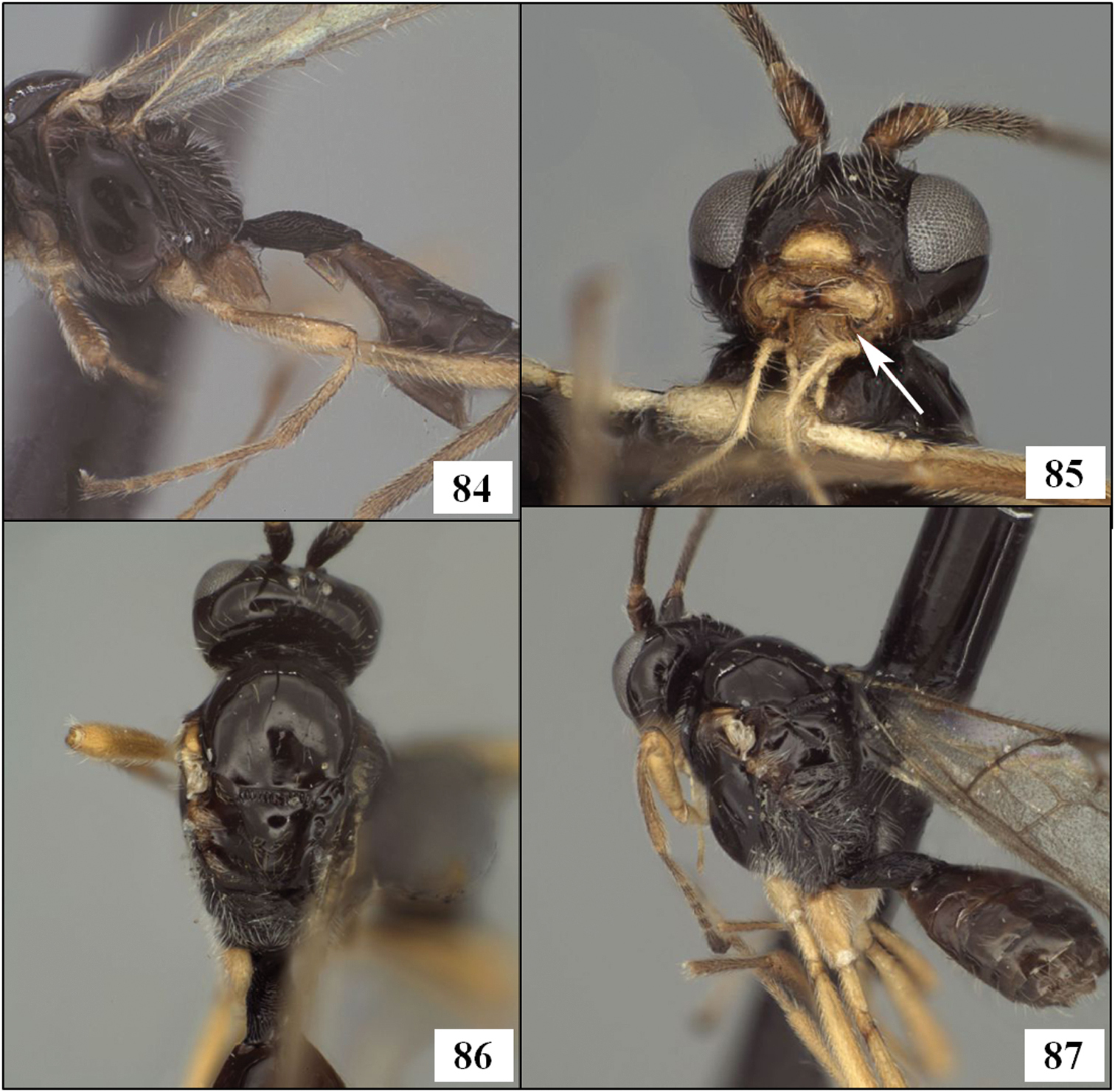
Tubiformopius spp. 84 T. tubibasis (Fischer), holotype female, T1, lateral view 85 T. tubigaster (Fischer), holotype male, face, arrow showing basal lobe of mandible 86 T. tubigaster, holotype male, head and mesoscutum, dorsal view 87 T. tubigaster, holotype male, dorsal-posterior view.
We thank David Wahl (AEIC) and Kees van Achterberg (RMNH) for extended loans of holotypes, as well as Dominique Zimmermann and Manuela Vizek (NHMW), Gavin Broad (BMNH), Robert Kula and Paul Marsh (USNM), Jenö Papp (HNHM) and Henri Goulet (CNC) for assistance with loans of types and/or other material in their care. Ana DalMolin provided notes on the holotype of Lorenzopius sanlorenzensis during her visit to HNHM. This work could not have been accomplished without the collecting efforts of several people, most notably Martin Aluja and his lab, Juan Rull, Al Norrbom, and Robert Jones. We are also sincerely grateful to Dr. Max Fischer for assistance with information on the type locality of Lorenzopius euryteniformis. Imaging and plate assembly was considerably facilitated by Trent Hawkins, Karl Roeder, Cheryl Hyde, Patricia Mullins, and Sophia Daniels. Patricia Mullins and Matt Yoder provided assistance with databasing and the HAO. This work was supported in part by NSF DEB 0949027 and NSF/PEET DEB 0328922, with REU supplements 1213790 and 0616851 respectively (all to RAW). The HAO is funded by NSF DBI 0850223 to Andy Deans, formerly at North Carolina State University. RW prepared the descriptions of new species, LW assisted RW with opiine taxonomy and general manuscript preparation, and IM contributed the HAO linkages and a critical review of the morphological terms used throughout the manuscript.
Hymenoptera Anatomy Ontology Table. (doi: 10.3897/zookeys.243.3990.app) File format: Microsoft Exsel file (xls).
Explanation note: Morphological terms used in text, referenced to the Hymenoptera Anatomy Ontology (HAO).