


(C) 2014 Natdanai Likhitrakarn. This is an open access article distributed under the terms of the Creative Commons Attribution License (CC BY 4.0), which permits unrestricted use, distribution, and reproduction in any medium, provided the original author and source are credited.
For reference, use of the paginated PDF or printed version of this article is recommended.
Citation: Likhitrakarn N, Golovatch SI, Panha S (2014) The millipede genus Orthomorpha Bollman, 1893 in Laos (Diplopoda, Polydesmida, Paradoxosomatidae), with descriptions of new species. ZooKeys 374: 1–22. doi: 10.3897/zookeys.374.6711
The genus Orthomorpha is currently represented in Laos by nine species, including three, O. paviei Brölemann, 1896, O. communis Likhitrakarn, Golovatch & Panha, 2011 and O. cambodjana (Attems, 1953), which are new to the fauna of the country, and further three new to science: O. suberectoides sp. n., O. gladiata sp. n. and O. sutchariti sp. n.
Millipede, Orthomorpha, taxonomy, new species, Laos
The large Southeast Asian millipede genus Orthomorpha Bollman, 1893 has recently become the subject of a thorough review (
The present paper puts on record a few more Orthomorpha from Laos, including three species heretofore known from the adjacent parts of Indochina (one also from as far south as Indonesia), and further three new to science.
The material was collected during a field trip to southern Laos undertaken in 2013 by SP and members of the Animal Systematics Research Unit, Chulalongkorn University. Live animals were photographed on the spot. Specimens were preserved in 75% ethanol, and morphological investigations were carried out in the laboratory using an Olympus stereomicroscope. Scanning electron micrographs (SEM) of gonopods coated with gold were taken using a JEOL, JSM–5410 LV microscope, returned to alcohol upon examination. Digital images of the specimens were taken in the laboratory and assembled using the “CellD” automontage software of the Olympus Soft Imaging Solution GmbH package. In addition, line drawings of gonopods were also prepared. All material is housed in the Museum of Zoology, Chulalongkorn University (CUMZ), Bangkok, Thailand.
Collecting sites were located by GPS using the WGS84 datum.
In the catalogue sections, D stands for the original description, subsequent descriptive notes or appearance in a key while M for a mere mention.
http://species-id.net/wiki/Orthomorpha_paviei
Figs 1–31 ♂ (CUMZ), Laos, Champasak Province, Khong District, Khone Phapheng Waterfall, 82 m a.s.l., 13°57'47"N, 105°59'17"E, 23.07.2013, leg. W. Siriwut.
According to the key in
Length 25 mm (♂), width of midbody pro- and metazona 2.06 and 3.23 mm, respectively.
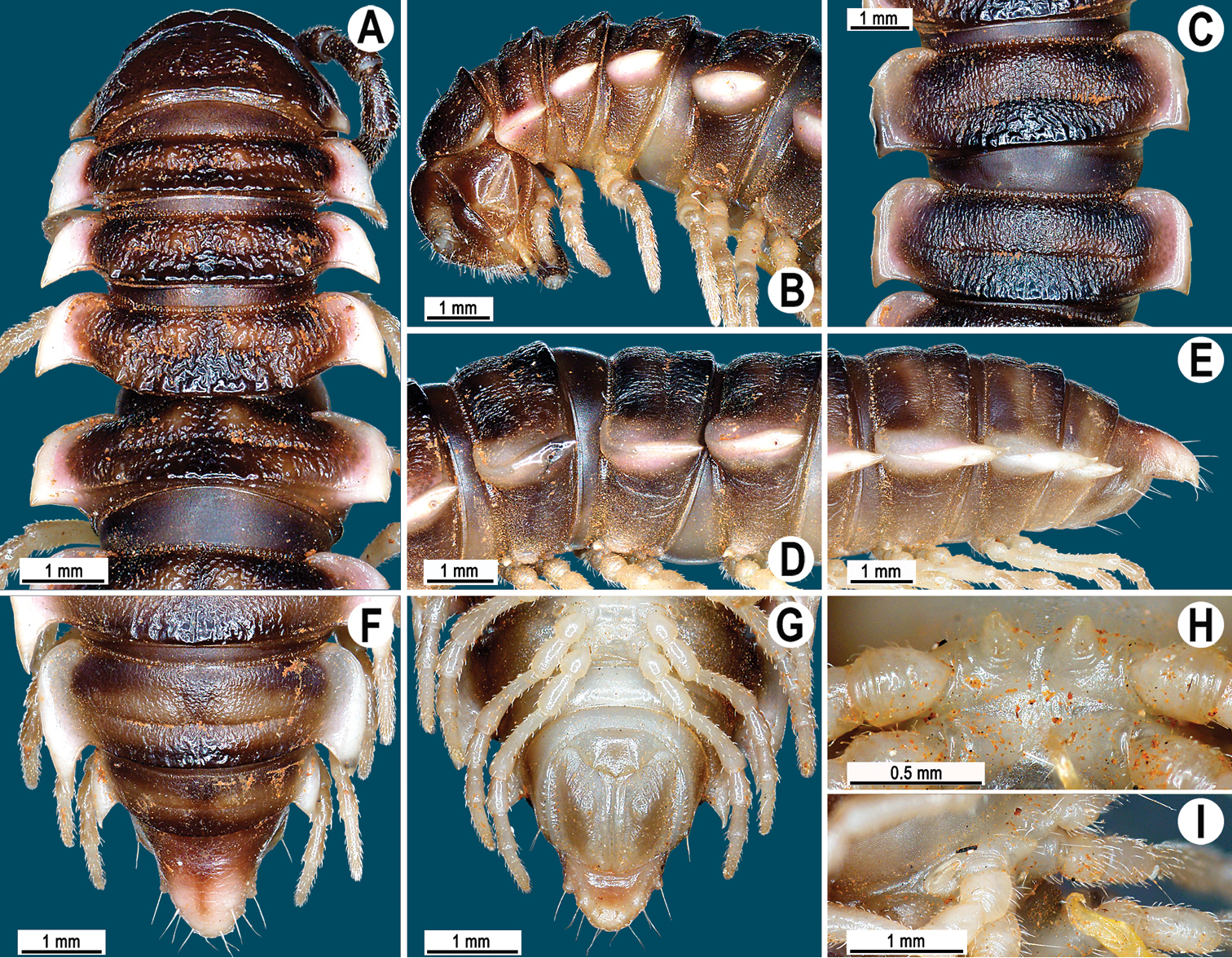
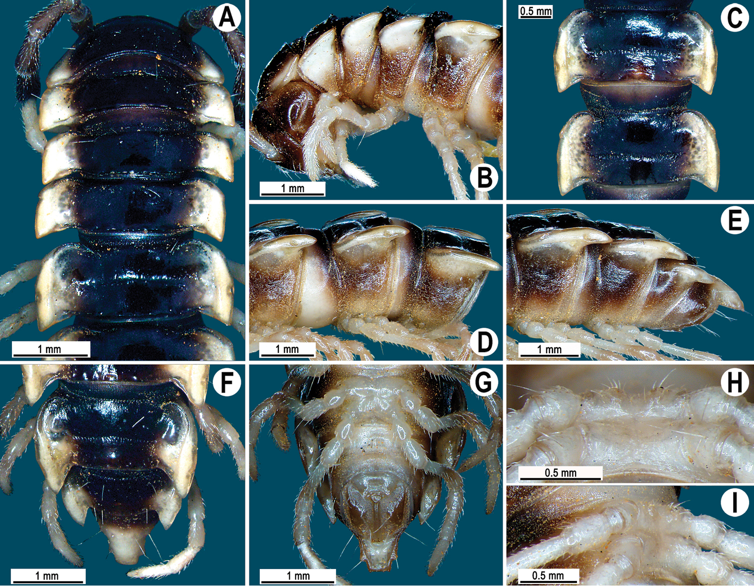
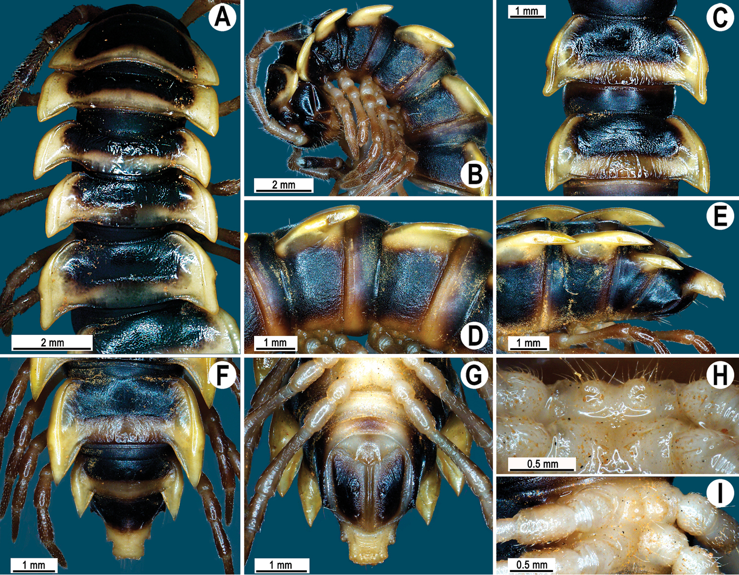
Coloration of live animals blackish (Fig. 1A), paraterga and epiproct contrasting dark yellow, head and antennae brownish, legs pale brownish; coloration in alcohol, after three months of preservation, faded to black-brown (Fig. 1B–H), paraterga and epiproct pale whitish yellow or pale brown, legs whitish to pale brown distally.
Clypeolabral region densely setose, vertigial region with a few setae only; epicranial suture distinct. Antennae long (Fig. 1A), reaching posterior end of body segment 4 when stretched dorsally. In width, head < segment 3 = 4 < collum < segment 2 < 5–17, gently and gradually tapering thereafter. Collum with three transverse rows of setae: 4+4 in anterior, 3+3 in intermediate, and 1+1 in posterior row; a very faint incision laterally near midway; caudal corner of paraterga pointed, dentiform, slightly upturned, but not drawn behind rear margin (Fig. 1B, C).
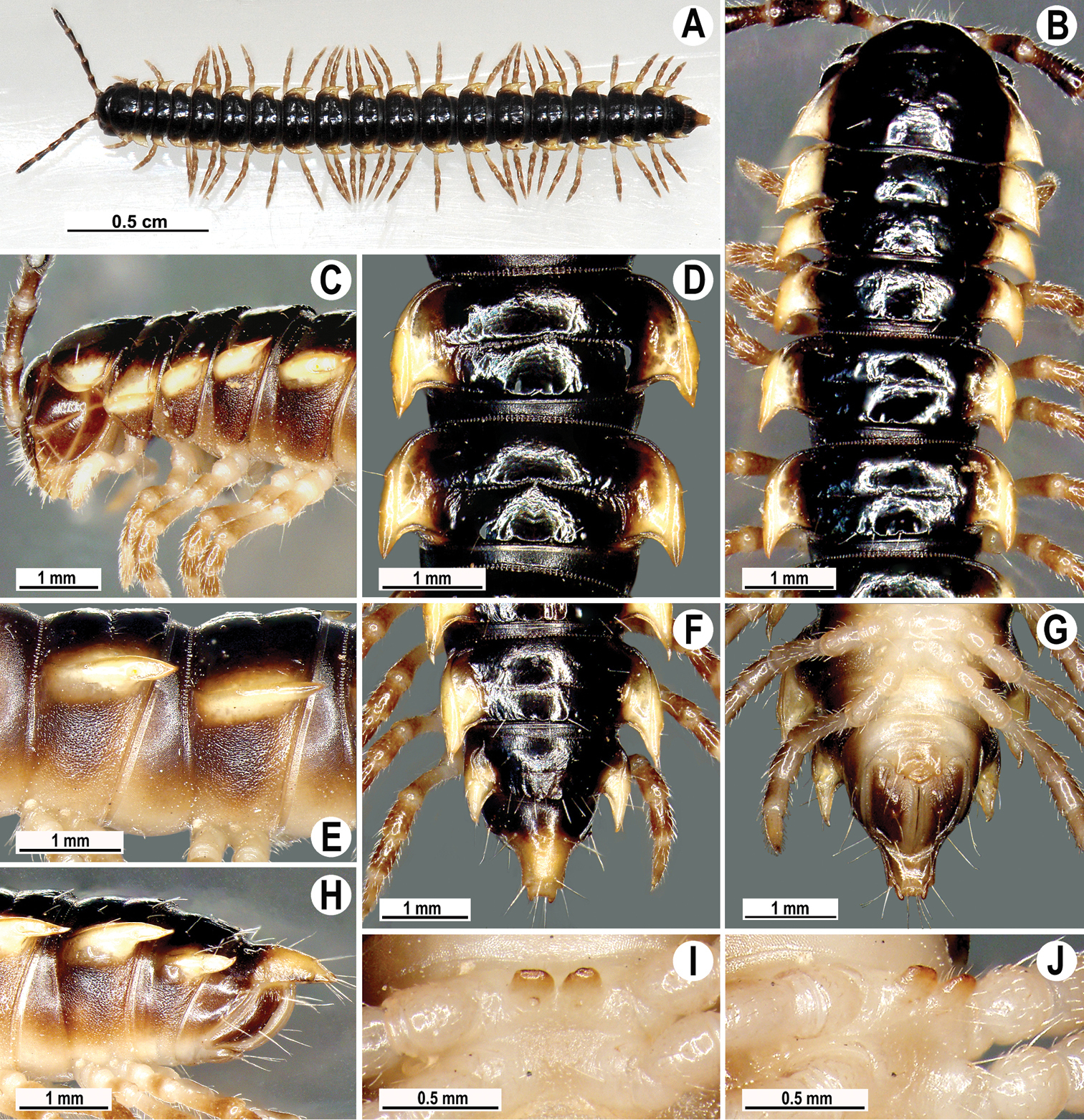
Orthomorpha paviei Brölemann, 1896, ♂ from Laos. A habitus, live coloration; B, C anterior part of body, dorsal and lateral views, respectivelyD, E segments 10 and 11, dorsal and lateral views, respectively F–H posterior part of body, dorsal, ventral and lateral views, respectivelyI, J sternal cones between coxae 4, subcaudal and sublateral views, respectively.
Tegument smooth and shining, prozona finely shagreened, metaterga smooth and delicately rugulose, leathery; surface below paraterga finely microgranulate. Postcollum metaterga with two transverse rows of setae traceable at least as insertion points when setae broken off: 2+2 in anterior (pre-sulcus), 3–4+3–4 in posterior (post-sulcus) row. Tergal setae long, strong, slender, about 1/3 of metatergal length. Axial line visible only on metaterga, slightly better developed on their posterior halves. Paraterga very strongly developed (Fig. 1B–H), mostly slightly upturned, all lying faintly below dorsum, set at about upper 1/3 of midbody height, subhorizontal, caudal corner almost or fully pointed, increasingly well spiniform and produced behind rear tergal margin until segment 18; paraterga very thin in lateral view, blunt blades, modestly enlarged in pore-bearing segments, thinner in poreless ones. Calluses on paraterga 2–4 delimited by a sulcus only dorsally, on following paraterga both dorsally and ventrally. Paraterga 2 broad, anterior edge angular, lateral edge with one larger and two smaller, but evident incisions in anterior 1/3; posterior edge well concave (Fig. 1B, D, F). Anterior edges of paraterga 3–9 clearly convex, of paraterga 10–18 nearly straight and slightly bordered. Lateral edge of paraterga with two slight, but evident incisions, one in anterior 1/3, the other in posterior 1/3. Posterior edge of paraterga clearly concave, especially well so in segments 16–19 (Fig. 1F–H). Ozopores evident, lateral, lying in an ovoid groove at about 1/4 of metatergite’s length in front of caudal corner. Transverse sulcus usually distinct (Fig. 1C, E, H), complete on metaterga 5–19, incomplete and nearly wanting on 19th, shallow, nearly reaching bases of paraterga, rather faintly beaded at bottom. Stricture between pro- and metazona wide, clearly beaded at bottom down to base of paraterga (Fig. 1B, D, F). Pleurosternal carinae complete crests with a sharp caudal tooth in segment 2, a very sharp, caudal tooth in segments 3 and 4, a small, mostly sharp tooth in segments 5–9 (Fig. 1C, E). Epiproct (Fig. 1F, G) conical, flattened dorsoventrally, with two evident apical papillae; tip subtruncate; pre-apical papillae small, but visible, lying rather close to tip. Hypoproct roundly subtriangular, setiferous knobs at caudal edge evident and well-separated.
Sterna sparsely setose, without modifications; two rather large, fully separated, sternal cones between ♂ coxae 4 (Fig. 1I, J). A paramedian pair of evident tubercles in front of gonopod aperture. Legs moderately long and slender, midbody ones ca 1.1–1.2 times as long as body height, prefemora without modifications, ♂ tarsal brushes present until legs of segment 8.
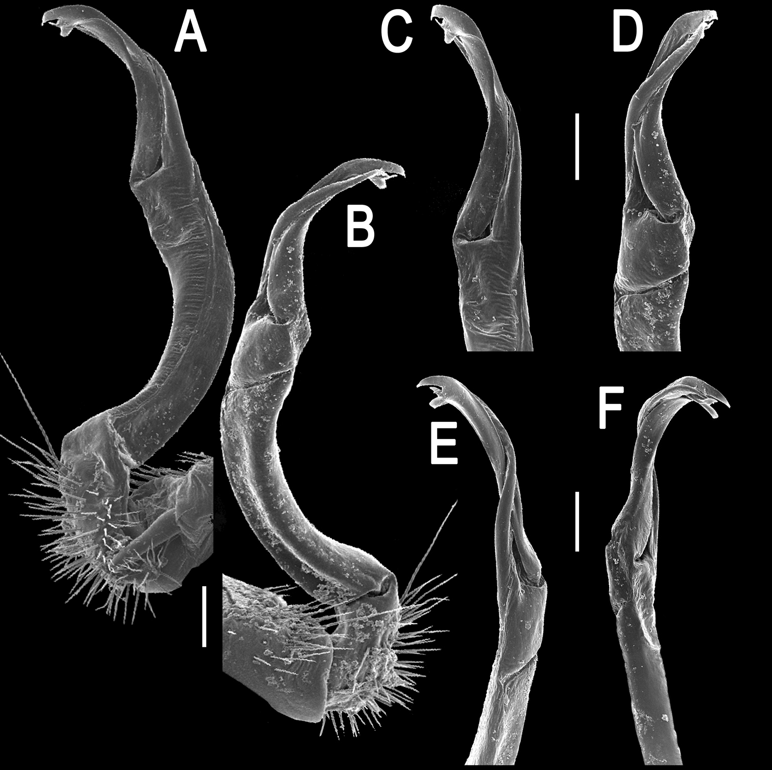
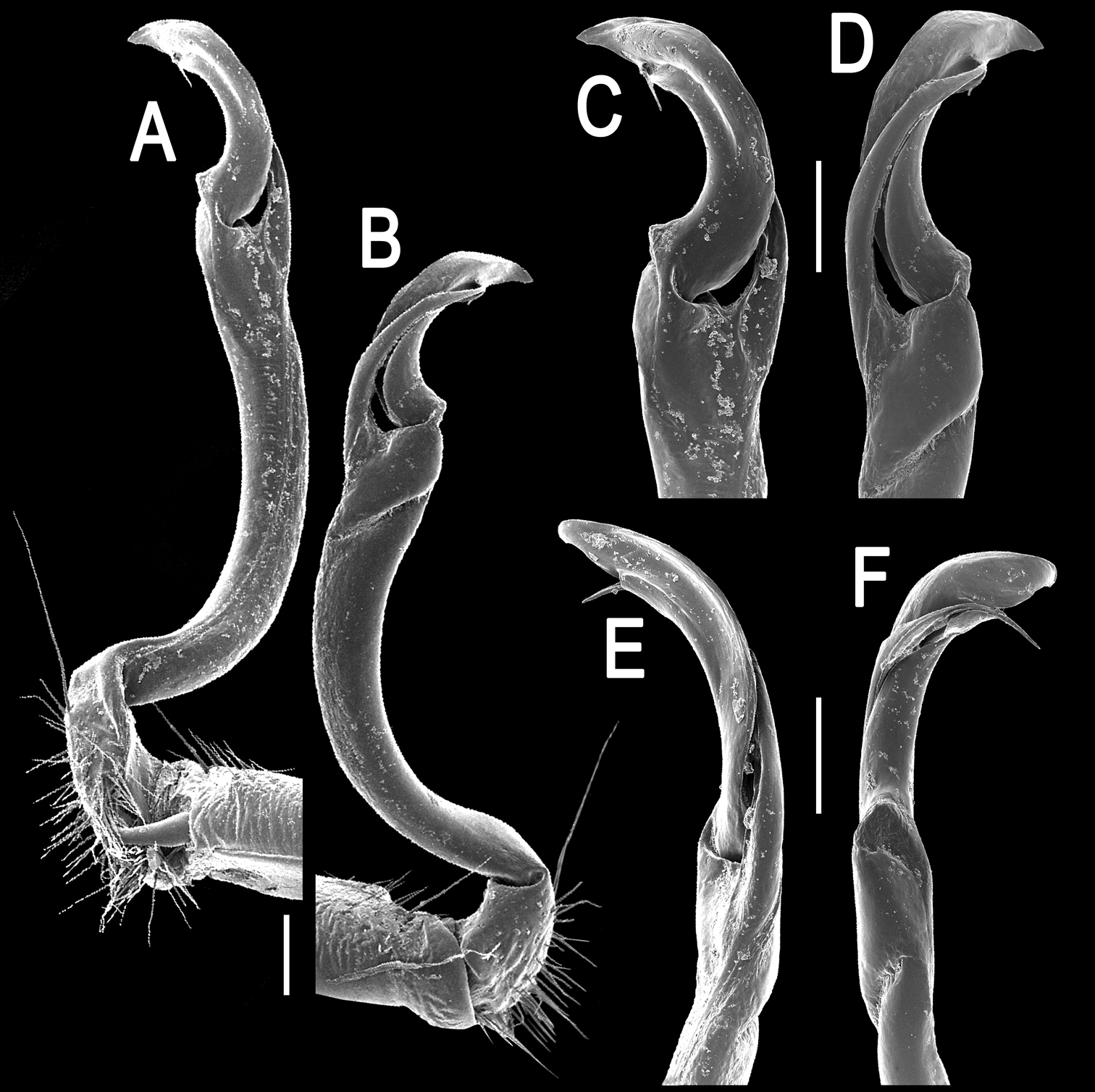
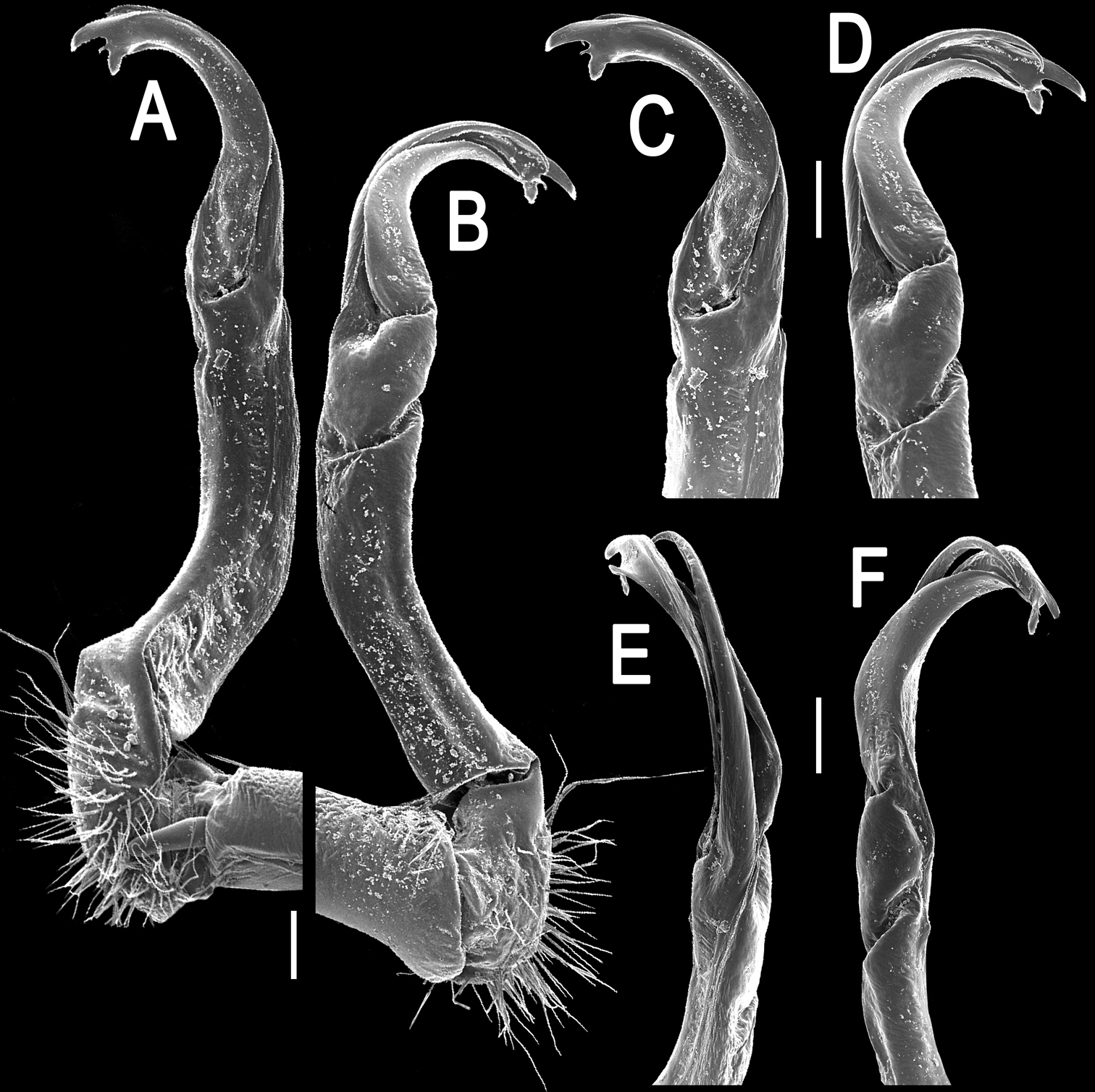
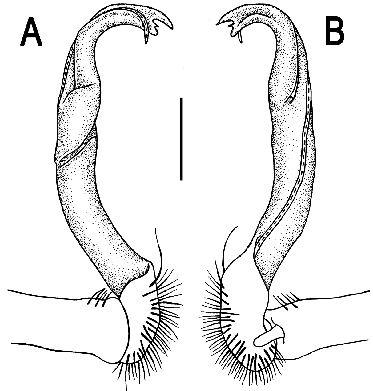
Gonopods (Figs 2, 3) with slender and long coxae, the latter with several setae distoventrally. Femorite about 3 times as long as prefemoral (= strongly setose) part. Femorite slender, slightly curved, postfemoral portion demarcated by an oblique lateral sulcus; tip of solenophore indistinctly trifid, both dorsal (d) and middle (m) prongs being sharp and deeply split, middle prong slightly stronger, supplied with a minute, but evident subterminal knob/lobule ventrally at about midway; solenomere long and flagelliform.
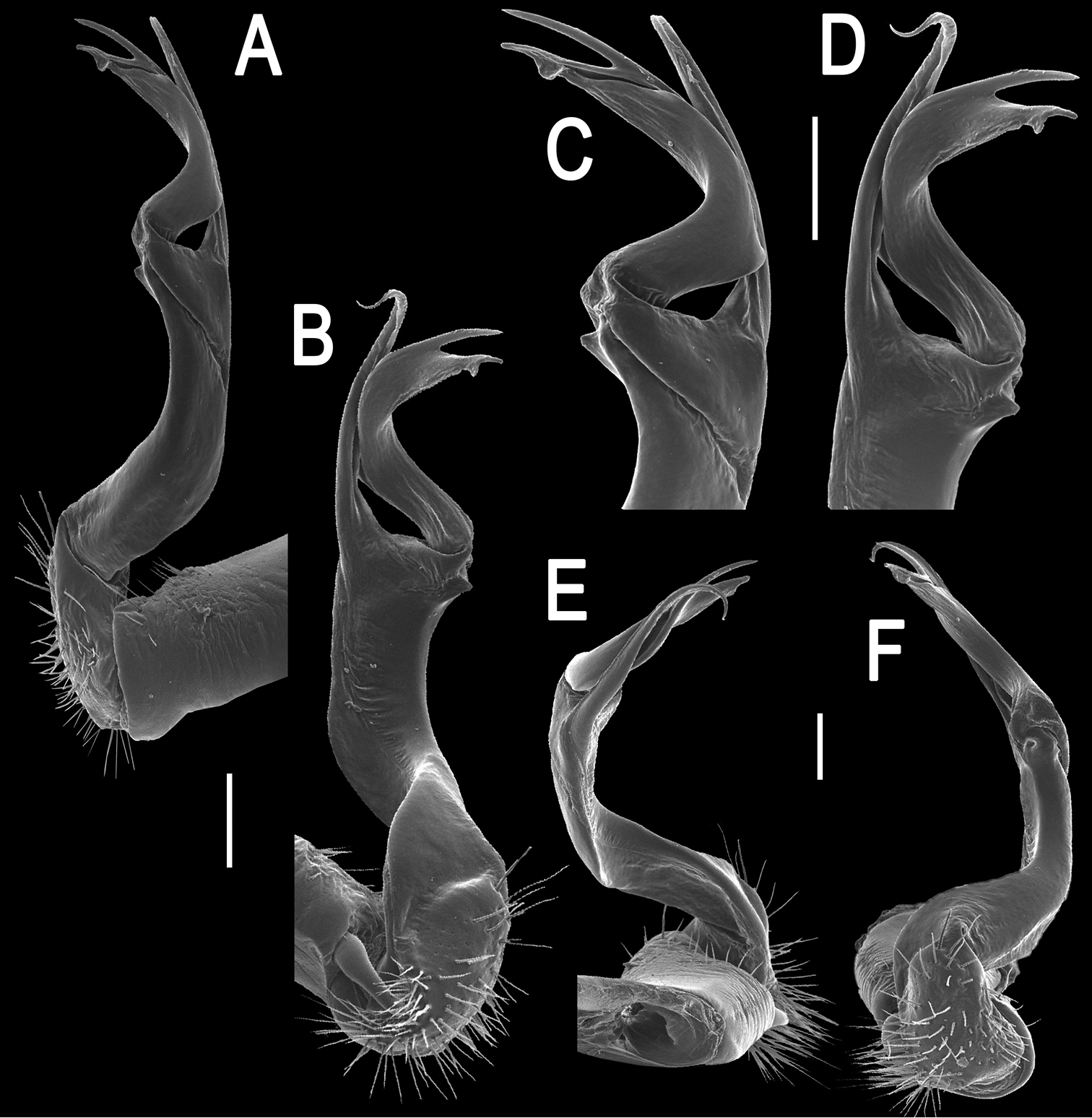
Orthomorpha paviei Brölemann, 1896, ♂ from Laos, left gonopod. A, B lateral and mesal views, respectively C, D telopodite, lateral and mesal views, respectively E, F distal part, subcaudal and suboral views, respectively. Scale bar: 0.2 mm.
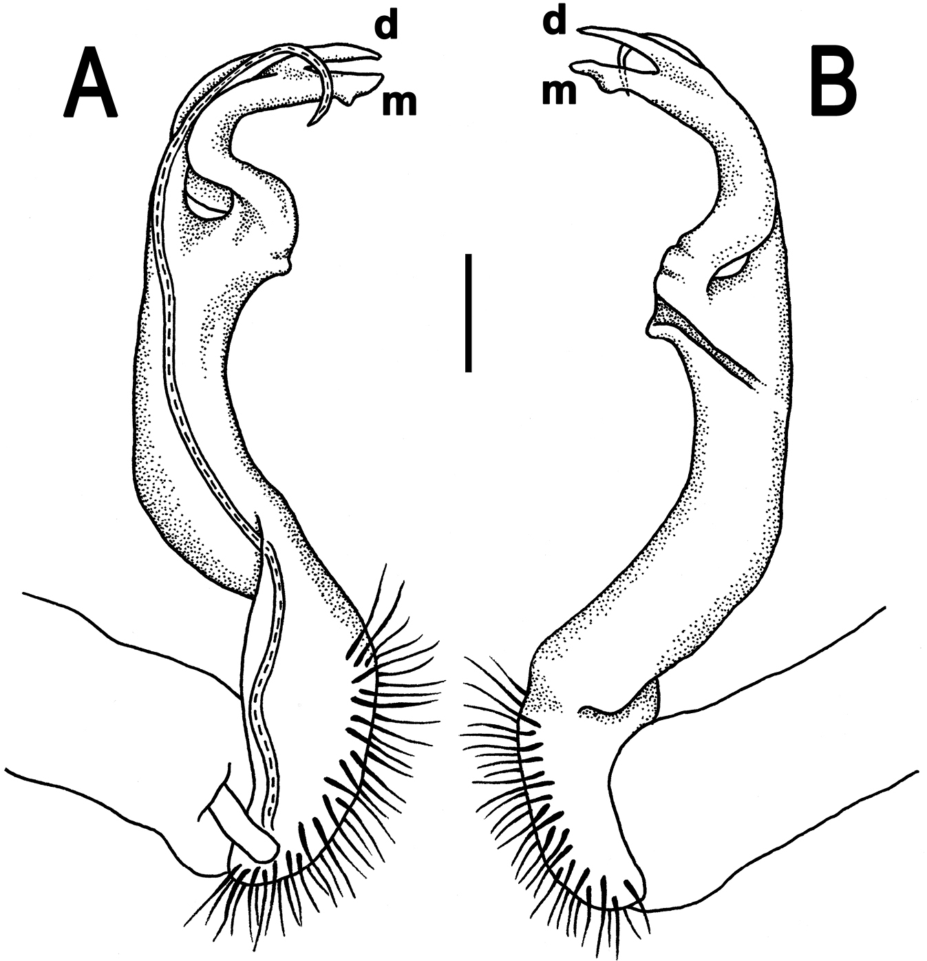
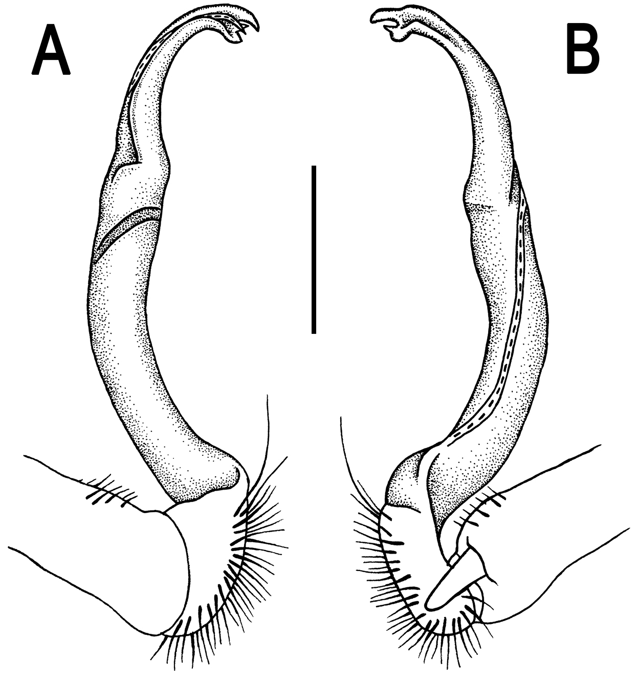
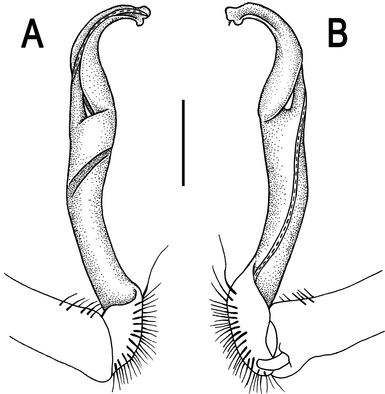
Orthomorpha paviei Brölemann, 1896, ♂. A, B left gonopod, mesal and lateral views, respectively. Scale bar: 0.5 mm.
The new specimen agrees nearly fully with the most detailed and beautifully illustrated descriptions of the species given by
In having both apical prongs of the gonopods rather deeply split, this species is somewhat intermediate between the genera Antheromorpha Jeekel, 1968 and Orthomorpha Bollman, 1893 (
The above detailed redescription is given not only to better outline the quite modest variations found in Orthomorpha paviei, but also to serve as a pattern for further, more condensed descriptions or descriptive notes below.
http://species-id.net/wiki/Orthomorpha_cambodjana
Figs 4–62 ♂ (CUMZ), Laos, Attapu Province, Xaysetha District, Ban Kasom, 116 m a.s.l., 15°06'27"N, 106°51'14"E, 16.10.2013, leg. C. Sutcharit and W. Siriwut.
Length 32–35 mm (♂), width of midbody pro- and metazona 2.6–2.7 and 3.8–3.9 mm (♂), respectively.
Coloration in alcohol, after one month of preservation, blackish (Fig. 4A–F), paraterga and epiproct light yellow to whitish yellow; venter, legs and antennae whitish to pale brown distally (Fig. 4A–I).
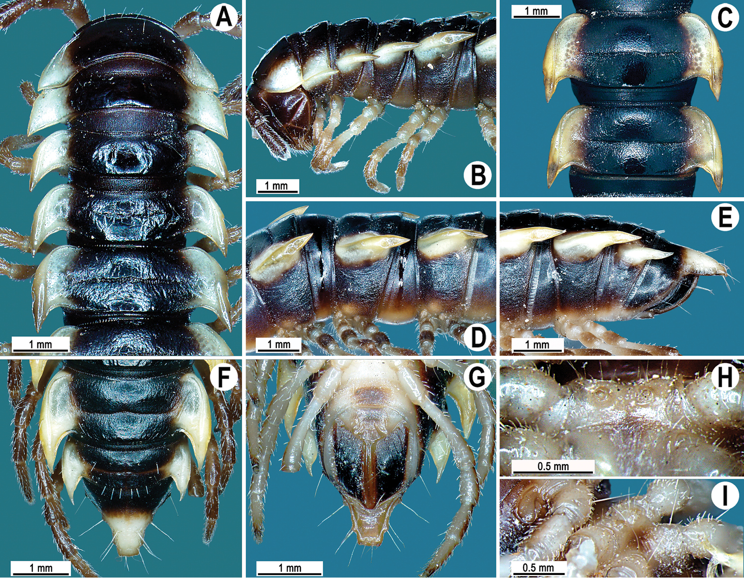
Orthomorpha cambodjana (Attems, 1953), ♂. A, B anterior part of body, dorsal and lateral views, respectively; C segments 10–11, dorsal view D segments 9–11, lateral view E–G posterior part of body, lateral, dorsal and ventral views, respectively; H, I sternal cones between coxae 4, subcaudal and sublateral views, respectively.
Antennae reaching segment 3 when stretched dorsally. Paraterga very strongly developed (Fig. 4A–G), anterolateral edge clearly convex. Calluses on paraterga 2 and 3 delimited by a sulcus only dorsally, following paraterga by a sulcus both dorsally and ventrally, strongly bordered. Pleurosternal carinae complete crests only in segment 2 (Fig. 4B, D, E), a small, sharp, caudal tooth in segments 3–7, a very faint tubercle until segment 15. Legs long and slender, midbody ones ca 1.1–1.3 times as long as body height, prefemora without modifications, tarsal brushes present until ♂ leg 7.
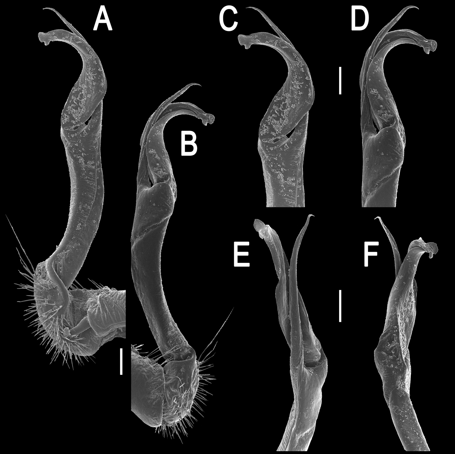
Gonopod solenophore with a tridentate tip (Figs 5, 6), dorsalmost prong being subacuminate, longer than both others, whereas middle denticle shortest and pointed.
Orthomorpha cambodjana (Attems, 1953), ♂, right gonopod. A, B mesal and lateral views, respectively C–F distal part of left gonopod, sublateral, submesal, subcaudal and suboral views, respectively. Scale bar: 0.2 mm.
Orthomorpha cambodjana (Attems, 1953), ♂. A, B right gonopod, lateral and mesal views, respectively. Scale bar: 0.5 mm.
This species has hitherto been known only from a ♂ lectotype and a ♀ paralectotype, currently both rather strongly faded, taken at Sre-Umbell, Kampot Province, southern Cambodia (
http://species-id.net/wiki/Orthomorpha_communis
1 ♂, 2 ♀ (CUMZ), Laos, Savannakhet Province, Atsaphangthong District, Ban Than Bang, 170 m, 16°42'12"N, 105°12'06"E, 19.10.2013, leg. C. Sutcharit and W. Siriwut.
Length 34 mm (♂) or 36–37 mm (♀), width of midbody pro- and metazona 2.9 and 4.5 mm (♂), 3.0–3.8 and 4.5–5.3 mm (♀), respectively.
The above material fully agrees with the original, quite detailed description of this species (
http://zoobank.org/695749B1-EFAF-4D08-BF0E-2C817DC63C95
http://species-id.net/wiki/Orthomorpha_suberectoides
Figs 7–9♂ (CUMZ), Laos, Attapu Province, Xaysetha District, Ban Lak No. 52, 224 m a.s.l., 15°09'24"N, 106°44'01"E, 16.10.2013, leg. C. Sutcharit.
To emphasize the apparent similarity to Orthomorpha suberecta Likhitrakarn, Golovatch & Panha, 2011; adjective.
This new species strongly resembles Orthomorpha suberecta, especially as regards the shape of the paraterga, but differs by a larger body, in the paraterga being broader, the metatergal tegument clearly rugose, and the pleurosternal carinae more strongly developed.
Length 35 mm (♂), width of midbody pro- and metazona 3.5 and 5.0 mm, respectively.
Coloration in alcohol, after one month of preservation, dark brown (Fig. 7A–F), paraterga and epiproct pale pinkish or pallid, head and antennae brownish, venter and legs pale whitish to pale brown (Fig. 7B–I).
All other characters as in Orthomorpha paviei, except as follows.
Orthomorpha suberectoides sp. n., ♂ holotype. A, B anterior part of body, dorsal and lateral views, respectively C segments 10–11, dorsal view D segments 9–11, lateral view E–G posterior part of body, lateral, dorsal and ventral views, respectively H, I sternal cones between coxae 4, subcaudal and sublateral views, respectively.
Antennae (Fig. 7A) reaching posterior end of body segment 3 when stretched dorsally. In width, head < collum < segment 2 < 3 < 4 < 5–16, gently and gradually tapering thereafter. Collum with three transverse rows of setae: 4+4 in anterior, 2+2 in intermediate, and 4+4 in caudal row; paraterga slightly declivous, broadly rounded and narrowly bordered, caudal corner pointed, dentiform, but not drawn behind rear margin (Fig. 7A, B).
Tegument shining and rugulose (Fig. 7A–F), metaterga rather obviously rugose, leathery, with traces of tubercles/wrinkles; surface below paraterga microgranulate and rugulose. Postcollum metaterga with two transverse rows of setae: 2+2, mostly abraded setae in anterior (pre-sulcus) row, 3+3 in posterior (post-sulcus) one, these setae or their traces being borne on evident cones growing stronger laterally, until segment 6 visible only as insertion points, tuberculations gradually growing smaller thereafter. Tergal setae simple, rather long, about 1/3 of metatergal length, mostly abraded. Axial line traceable, especially clear on collum and following metaterga. Paraterga very strongly developed (Fig. 7A–G), mostly slightly upturned, all lying faintly below dorsum, set at about half of midbody height, subhorizontal, in lateral view modestly enlarged in pore-bearing segments, thinner in poreless ones; shoulders broadly rounded, narrowly bordered, fused to callus; caudal corner almost or fully pointed, lying within rear tergal margin, after segment 16 drawn increasingly well beyond it, slightly curved mesad on segments 17–19 (Fig. 7E–G). Calluses delimited by a sulcus only dorsally. Paraterga 2 broad, anterior edge convex, lateral edge with three small and acute denticles, the one near caudal corner being particularly small (Fig. 7A, B). Each following poreless segment with two incisions, each pore-bearing one with one, often evident incision in front of ozopore. Posterior edge of paraterga slightly concave, especially clearly so in segments 16–19. Ozopores evident, lateral, lying in an ovoid groove at about 1/4 of metatergite’s length in front of caudal corner. Transverse sulcus usually distinct (Fig. 7A–F), complete on metaterga 5–19, incomplete on segment 4, narrow, not reaching bases of paraterga, ribbed at bottom. Stricture between pro- and metazona narrow, clearly ribbed at bottom down to base of paraterga (Fig. 7A–F). Pleurosternal carinae complete crests with a sharp caudal tooth in segments 2–7, a very sharp, caudal tooth in segments 8–17, a small, rather sharp tooth in segment 18 (Fig. 7B, D, E). Epiproct (Fig. 7F, G) conical, flattened dorsoventrally, with two evident apical papillae, both latter directed ventrocaudally and acute at tip; pre-apical papillae small denticles lying close to tip. Hypoproct (Fig. 7G) roundly subtriangular, setiferous knobs at caudal edge evident and well-separated.
Sterna sparsely setose, without modifications; cross-impressions shallow; a paramedian pair of evident, fully separated, small, sternal cones between ♂ coxae 4 (Fig. 7H, I). A paramedian pair of evident tubercles in front of gonopod aperture. Legs moderately long and slender, midbody ones ca 1.0–1.1 times as long as body height, prefemora without modifications, ♂ tarsal brushes absent.
Gonopods (Figs 8, 9) with slender and long coxae, the latter with several setae distoventrally. Femorite about 3 times as long as prefemoral portion. Femorite slender, slightly curved, postfemoral portion demarcated by an oblique lateral sulcus. Solenophore with lamina lateralis clearly smaller than lamina medialis, tip distinctly bilobed, dorsal lobule larger; solenomere long and flagelliform.
Orthomorpha suberectoides sp. n., ♂ holotype, right gonopod. A, B mesal and lateral views, respectively C–F distal part of left gonopod, mesal, lateral, suboral and subcaudal views, respectively. Scale bar: 0.2 mm.
Orthomorpha suberectoides sp. n., ♂ holotype. A, B right gonopod, lateral and mesal views, respectively. Scale bar: 0.5 mm.
According to the key in
http://zoobank.org/B785592D-54B8-4C4D-918A-F43D9ED88C4A
http://species-id.net/wiki/Orthomorpha_gladiata
Figs 10–12♂ (CUMZ), Laos, Champasak Province, Paksong District, Tadedu, 906 m a.s.l., 15°11'35"N, 106°06'07"E, 16.10.2013, leg. S. Panha and C. Sutcharit.
1 ♂ (CUMZ), same data, together with holotype.
To emphasize the sword-shaped tip of the gonopod; adjective.
Orthomorpha gladiata sp. n. seems to be especially similar to Orthomorpha hydrobiologica Attems, 1930, a species known to range from eastern Java, Indonesia to Indochina (
Length 25–27 mm (♂), width of midbody pro- and metazona 1.7–1.9 and 2.8–2.9 mm, respectively.
Coloration in alcohol, after one month of preservation, dark brown (Fig. 10A–G), paraterga and epiproct light yellow or pale yellowish, head, surface below paraterga and antennae brownish, venter and legs whitish to pale brown (Fig. 10A–I).
Orthomorpha gladiata sp. n., A–B ♂ paratype C–I ♂ holotype. A, B anterior part of body, dorsal and lateral views, respectively C segments 10–11, dorsal view D segments 9–11, lateral view E–G posterior part of body, lateral, dorsal and ventral views, respectively H, I sternal cones between coxae 4, subcaudal and sublateral views, respectively.
All other characters as in Orthomorpha paviei, except as follows.
Antennae (Fig. 10A) reaching body segment 3 when stretched dorsally. In width, head < collum < segments 3 and 4 < 2 = 5–16, gently and gradually tapering thereafter. Collum with three transverse rows of setae: 3+3 in anterior, 2+2 in intermediate, and 3+3 in posterior row; caudal corner of paraterga very narrowly rounded, slightly upturned, but not drawn behind rear margin (Fig. 10A, B).
Postcollum metaterga with two transverse rows of setae traceable at least as insertion points when setae broken off: 2+2 in anterior (pre-sulcus) and 3+3 in posterior (post-sulcus) row, these setae being borne on small cones growing stronger laterally. Metaterga 17–19 with 4+4 setae in posterior row, likewise borne on evident cones growing stronger laterally. Axial line rather difficult to see, but traceable both on pro- and metaterga. Paraterga very strongly developed (Fig. 10A–G), mostly slightly upturned, all lying faintly below dorsum, set at about upper 1/4 of midbody height, mostly subhorizontal, caudal corner narrowly rounded to nearly pointed; paraterga very thin in lateral view, modestly enlarged in pore-bearing segments, thinner in poreless ones. Calluses on paraterga 2–4 delimited by a sulcus only dorsally, on following paraterga both dorsally and ventrally. Paraterga 2 broad, anterior edge rounded, lateral edge with one larger and one smaller, but evident incision in anterior 1/3; posterior edge clearly concave (Fig. 10A, C, F). Anterior edges of paraterga broadly rounded and narrowly bordered, fused to callus. Lateral edge of paraterga with two slight, but evident incisions, one in anterior 1/3, the other in posterior 1/3. Posterior edge of paraterga clearly concave, lateral corner extending increasingly beyond rear tergal margin, especially strongly so in segments 17–19 (Fig. 10F, G). Transverse sulcus usually distinct (Fig. 10A, C, F), complete on metaterga 5–18, incomplete in segments 4 and 19, rather deep, reaching bases of paraterga, rather clearly ribbed at bottom. Stricture between pro- and metazona narrow, clearly ribbed at bottom down to base of paraterga (Fig. 10A–F). Pleurosternal carinae complete crests with a sharp caudal tooth in segments 2–4, a very sharp, caudal tooth in segments 5–7, a small, mostly sharp tooth until segment 16 (Fig. 10B, D, E). Epiproct (Fig. 10F, G) conical, flattened dorsoventrally, with two evident apical papillae; tip subtruncate; pre-apical papillae small, but visible, lying rather close to tip.
Two small, low, rounded, fully separated, sternal cones between ♂ coxae 4 (Fig. 10H, I). A paramedian pair of evident tubercles in front of gonopod aperture. Legs rather long and slender, midbody ones ca 1.2–1.4 times as long as body height, prefemora without modifications, ♂ tarsal brushes present only on legs 1–3.
Gonopods (Figs 11, 12) with slender and long coxae, the latter with several setae distoventrally. Femorite about 3 times as long as prefemoral portion. Femorite very slender, slightly curved, postfemoral portion demarcated by an oblique lateral sulcus. Solenophore with a nearly monodentate and sword-shaped tip; solenomere long and flagelliform.
Orthomorpha gladiata sp. n., ♂ holotype, right gonopod. A, B mesal and lateral views, respectively C–F distal part of left gonopod, mesal, lateral, suboral and subcaudal views, respectively. Scale bar: 0.2 mm.
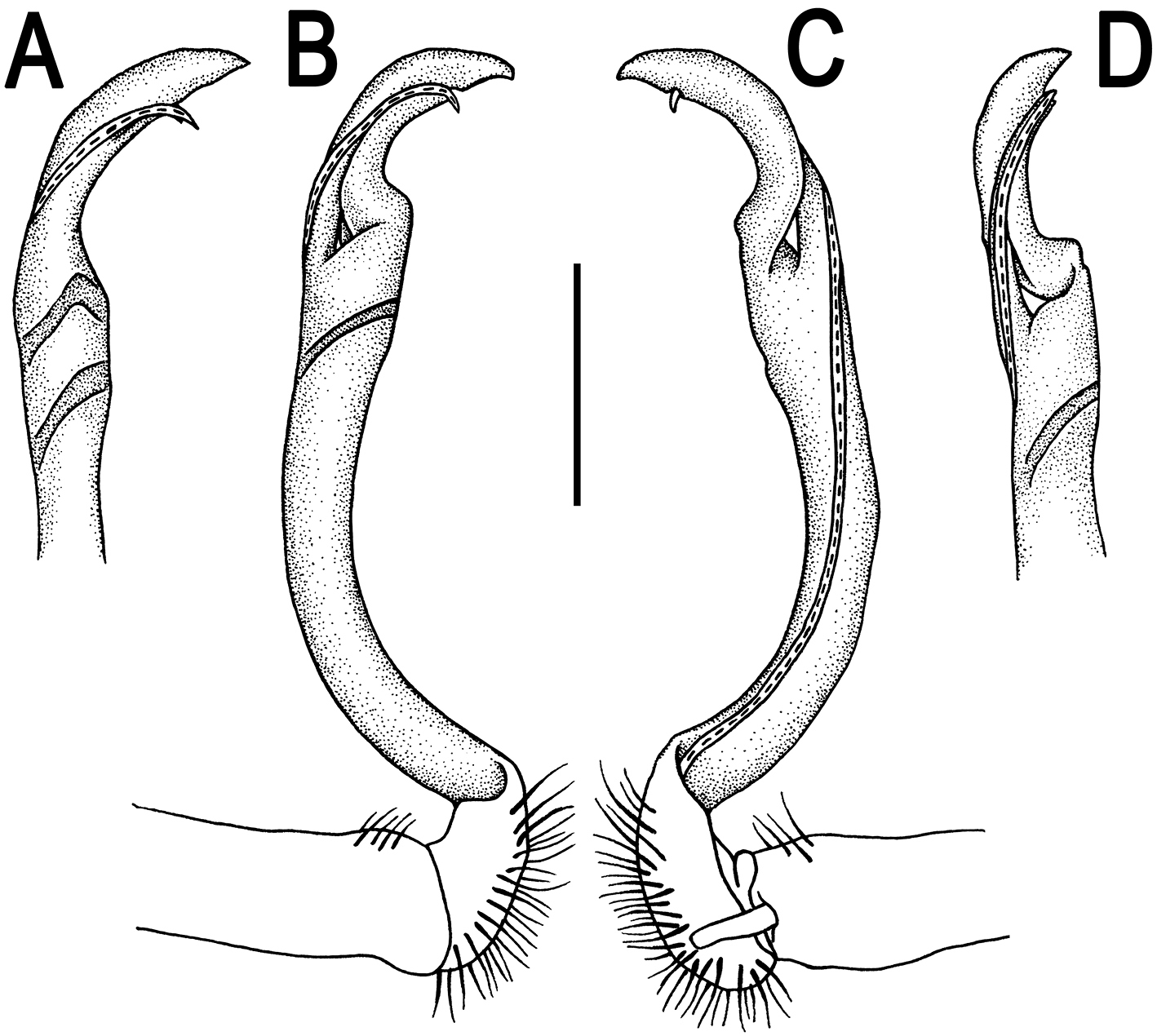
Orthomorpha gladiata sp. n., ♂ holotype. A–D right gonopod, suboral, lateral, mesal and subcaudal views, respectively. Scale bar: 0.5 mm.
The gonopod tip of Orthomorpha gladiata sp. n. is much like in Orthomorpha parasericata Likhitrakarn, Golovatch & Panha, 2010, from southern Thailand (
http://zoobank.org/D5E01463-1933-4A03-97C6-2D678FEAB8D2
http://species-id.net/wiki/Orthomorpha_sutchariti
Figs 13–15♂ (CUMZ), Laos, Champasak Province, Paksong District, Tadedu, 906 m a.s.l., 15°11'35"N, 106°06'07"E, 16.10.2013, leg. S. Panha and C. Sutcharit.
1 ♀ (CUMZ), same data, together with holotype.
To honour Dr. Chirasak Sutcharit, Professor at the Department of Biology of Chulalongorn University, Bangkok, who participated in collecting the type specimens and taught the first author the basics of taxonomy.
Differs in the colour pattern which has lighter caudal halves of the metaterga, the mostly strongly elevated and laterally bordered paraterga, and a trifid solenophore showing a subacuminate, longest, dorsal prong.
Length ca 42 mm (♂) or 44.5 mm (♀), width of midbody pro- and metazona 3.4 and 4.8 mm (♂), 3.5 and 5.6 mm (♀), respectively.
Coloration in alcohol, after one month of preservation, rather uniformly dark brown (Fig. 13A–G) with lighter caudal halves of metaterga, paraterga and epiproct contrasting light yellow, mid-dorsal regions of prozona strongly infuscate, blackish like most of metaterga; antennae and legs brown to light brown (Fig. 13A–I).
Orthomorpha sutchariti sp. n., ♂ holotype. A, B anterior part of body, dorsal and lateral views, respectively C, D segments 10–11, dorsal and lateral views, respectively E–G posterior part of body, lateral, dorsal and ventral views, respectively H, I sternal cones between coxae 4, subcaudal and sublateral views, respectively.
All other characters as in Orthomorpha paviei, except as follows.
Antennae (Fig. 13A, B) surpassing posterior end of body segment 3 (♂) or 2 (♀) when stretched dorsally. In width, head < collum < segment 3 = 4 < 2 < 5–15, gently and gradually tapering thereafter. Collum with three transverse rows of setae: 4+4 in anterior, 2+2 in intermediate, and 4+4 in posterior row; a very faint incision laterally near midway; caudal corner of paraterga very narrowly rounded, slightly upturned, but not drawn behind rear margin (Fig. 13A, B).
Tegument smooth and shining, metaterga smooth and delicately rugulose, leathery, posterior halves of metaterga rugose, with traces of tubercles/wrinkles; surface below paraterga finely microgranulate. Postcollum metaterga with two transverse rows of setae traceable at least as insertion points when setae broken off: 2+2 in anterior (pre-sulcus), 3+3 in posterior (post-sulcus) row. Axial line faint, but traceable both on pro- and metaterga. Paraterga very strongly developed (Fig. 13A–G), mostly slightly upturned, lying either faintly above (♂) or slightly below dorsum (♀), set at about upper 1/3 of midbody height, subhorizontal (♀), caudal corner very narrowly rounded, extended beyond rear tergal margin, increasingly well produced caudally in segments 16–19 (Fig. 13F, G); paraterga very thin in lateral view, blunt blades, modestly enlarged in pore-bearing segments, thinner in poreless ones. Calluses on paraterga 2 and 3 delimited by a sulcus only dorsally, on following paraterga both dorsally and ventrally. Paraterga 2 broad, anterior edge angular, lateral edge with one stronger and two smaller, but evident incisions in anterior 1/3; posterior edge clearly oblique (Fig. 13A, C, F). Anterior edges of paraterga broadly rounded and narrowly bordered, fused to callus. Transverse sulcus usually distinct (Fig. 13A, C, F), complete on metaterga 5–18, incomplete and nearly wanting on segments 4 and 19, shallow, not reaching bases of paraterga, rather faintly ribbed at bottom. Stricture between pro- and metazona narrow, clearly ribbed at bottom down to base of paraterga (Fig. 13A–F). Pleurosternal carinae complete crests with a sharp caudal tooth in segments 2–7 (♂) or 2–4 (♀), a very sharp, caudal tooth in segments 8–15 (♂) or 5–10 (♀), a small, mostly sharp tooth until segments 16–19 (♂) or 11–17 (♀) (Fig. 13B, D, E). Epiproct (Fig. 13F, G) conical, flattened dorsoventrally, with two evident apical papillae, both directed ventrocaudally and acute at tip; pre-apical papillae small, lying rather close to tip.
A pair of small, rounded, fully separated, sternal cones between ♂ coxae 4 (Fig. 13H, I). A paramedian pair of evident tubercles in front of gonopod aperture. Legs rather long and slender, faintly incrassate in ♂; midbody legs ca 1.2–1.4 (♂) or 0.9–1.1 (♀) times as long as body height, prefemora without modifications, ♂ tarsal brushes present until legs of segment 8.
Gonopods (Figs 14, 15) with slender and long coxae, the latter with several setae distoventrally. Femorite about 3 times as long as prefemoral portion. Femorite slender, slightly curved, postfemoral portion demarcated by an oblique lateral sulcus; tip of solenophore trifid, with dorsal lobule being subacuminate and longest, middle denticle spiniform, shortest; solenomere long and flagelliform.
Orthomorpha sutchariti sp. n., ♂ holotype, right gonopod. A, B mesal and lateral views, respectively C–F distal part, mesal, lateral, suboral and subcaudal views, respectively. Scale bar: 0.2 mm.
Orthomorpha sutchariti sp. n., ♂ holotype. A, B right gonopod, lateral and mesal views, respectively. Scale bar: 0.5 mm.
The colour pattern of Orthomorpha sutchariti sp. n. is similar to that observed in several species of the Oriental genus Antheromorpha Jeekel, 1968 (our personal observations), but the gonopod structure is typical of Orthomorpha spp. Due to the particularly short middle prong at the tip of the solenophore, this new species strongly resembles the numerous congeners within the former weberi-group (
A total of 34 species of millipedes have hitherto been reported from Laos (
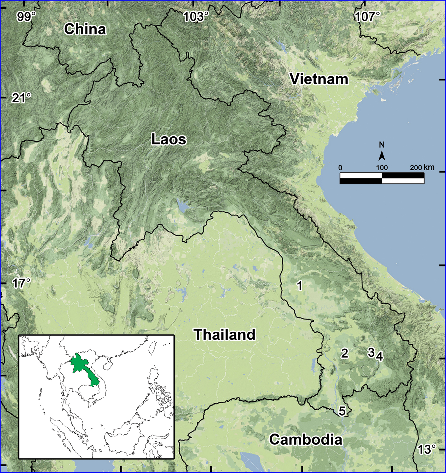
Distributions of the new records of Orthomorpha in Laos: 1 Ban Than Bang 2 Tadedu 3 Ban Lak No. 52 4 Ban Kasom 5 Khone Phapheng Waterfall.
This project was partly funded by Chulalongkorn University Graduate School Postdoctoral Project to NL, while most of the financial support was received from The Thailand Research Fund, The TRF Senior Research Scholar RTA 5580001 (2012–2015) to SP, and additional funding from the 100 Years Chulalongkorn University, Food and Water Research Cluster (CU56-FW04). We thank the members of the Animal Systematics Research Unit for their invaluable assistance in the field.