


(C) 2013 Yucheng Lin. This is an open access article distributed under the terms of the Creative Commons Attribution License 3.0 (CC-BY), which permits unrestricted use, distribution, and reproduction in any medium, provided the original author and source are credited.
For reference, use of the paginated PDF or printed version of this article is recommended.
Two new spider species of the family Mysmenidae Petrunkevitch, 1928 are reported from Southwestern China, i.e., Mysmena wawuensis sp. n. (male and female) from Sichuan and Trogloneta yuensis sp. n. (male) from Chongqing. Diagnoses and illustrations of the new species are provided.
Taxonomy, diagnosis, description, forest, etymology
Mysmenidae is a small family of minute araneoid spiders. Although the family Mysmenidae is distributed worldwide, it is one of the least-studied family-level groups among orb-weaving spiders, and its diversity is grossly undersampled due to their small size (0.7–3 mm) and cryptic life style (
The genus Mysmena was erected by
The genus Trogloneta was established and placed in the family Theridiidae by Simon in 1922 for a minute spider from caves in France, Trogloneta granulum (“Troglonata” was misspelled in the original description, see
At present, 9 Trogloneta species are known from America, Europe, Asia and some Atlantic islands (
In this paper we described two new species of genera Mysmena and Trogloneta from Wawu Mt., Sichuan and Jinyun Mt., Chongqing of Southwestern China, Mysmena wawuensis sp. n. and Trogloneta yuensis sp. n.
Specimens were examined and measured under an Olympus SZX7 stereomicroscope. Further details were studied under an Olympus BX43 compound microscope. All drawings were made using a drawing tube attached to Olympus BX43 compound microscope, and then inked on ink jet plotter paper. Photos were taken with a Canon EOS 60D wide zoom digital camera (8.5 megapixels). The images were montaged using Helicon Focus 3.10 software. Male pedipalpi and female genitalia were examined and illustrated after they were dissected and detached from the spiders’ bodies. Vulvae were removed and treated in lactic acid before illustration. To reveal the course of spermatic duct, the pedipalpal bulb was also treated in lactic acid and mounted in Hoyer’s Solution. Left pedipalp of male spiders was illustrated. All specimens are preserved in 85% ethanol solution.
All measurements are in millimeters. Leg measurements are given as: total length (femur, patella, tibia, metatarsus, and tarsus). The terminology mostly follows
Type species. Theridion leycoplagiatum Simon, 1879
urn:lsid:zoobank.org:act:FF5B96D7-39D7-4F3E-816D-521A57F1413C
http://species-id.net/wiki/Mysmena_wawuensis
Figs 1–7, 13Holotype: CHINA, Sichuan: Hongya County, Wawu Mt. National Forest Park, Gufuping, 29°40.114'N, 102°57.515'E, elevation ca 1929 m, 27 June 2012, by hand collection, Yucheng Lin leg., male (SCUM).
Paratypes: [same data as holotype] (SCUM), 2 females.
The specific name is taken from the type locality; adjective.
This new species is similar to Mysmena goudao Miller, Griswold & Yin, 2009 (see
Male (holotype). Somatic characters see Fig. 1A–C. Coloration: Prosoma brown centrally, dark marginally. Sternum black. Opisthosoma black, with tiny yellow speckles.
Measurement: Total length 0.60. Prosoma 0.36 long, 0.35 wide, 0.32 high. Opisthosoma 0.36 long, 0.32 wide, 0.39 high. Clypeus 0.12 high. Sternum 0.25 long, 0.21 wide. Length of legs [total length (femur + patella + tibia + metatarsus + tarsus)]: I 1.14 (0.36, 0.14, 0.25, 0.18, 0.21); II 0.97 (0.30, 0.13, 0.21, 0.14, 0.19); III 0.76 (0.21, 0.11, 0.13, 0.13, 0.18); IV 0.93 (0.29, 0.13, 0.20, 0.14, 0.17).
Prosoma (Fig. 1A, C): Carapace near round. Cephalic pars elevated, sharply vertical forward and slope backward. Ocular area at apex, dark. Eight eyes in two rows. AME black, others white. ALE and PLE contiguous. AME smallest, ALE largest. ARE slightly procurved, PRE straight. Chelicerae yellow, small, as long as endites (Fig. 1C).
Legs: Femora pale yellow, other segments yellow proximally, gray distally. Leg formula: I-II-IV-III. Leg I with a distal metatarsal clasping macroseta prolaterally on 1/3 position. Leg I and II with a subdistal sclerotized femoral spot ventrally. Patellae I–IV with a dorsal seta distally. Tibiae I–IV with a dorsal seta proximally, and with 3 trichobothria. Metatarsi I–IV with only one trichobothrium.
Opisthosoma (Fig. 1A–C): Globular dorsally. Spinnerets dark, the anteriors larger than the posteriors. Colulus indistinct. Anal tubercle grey.
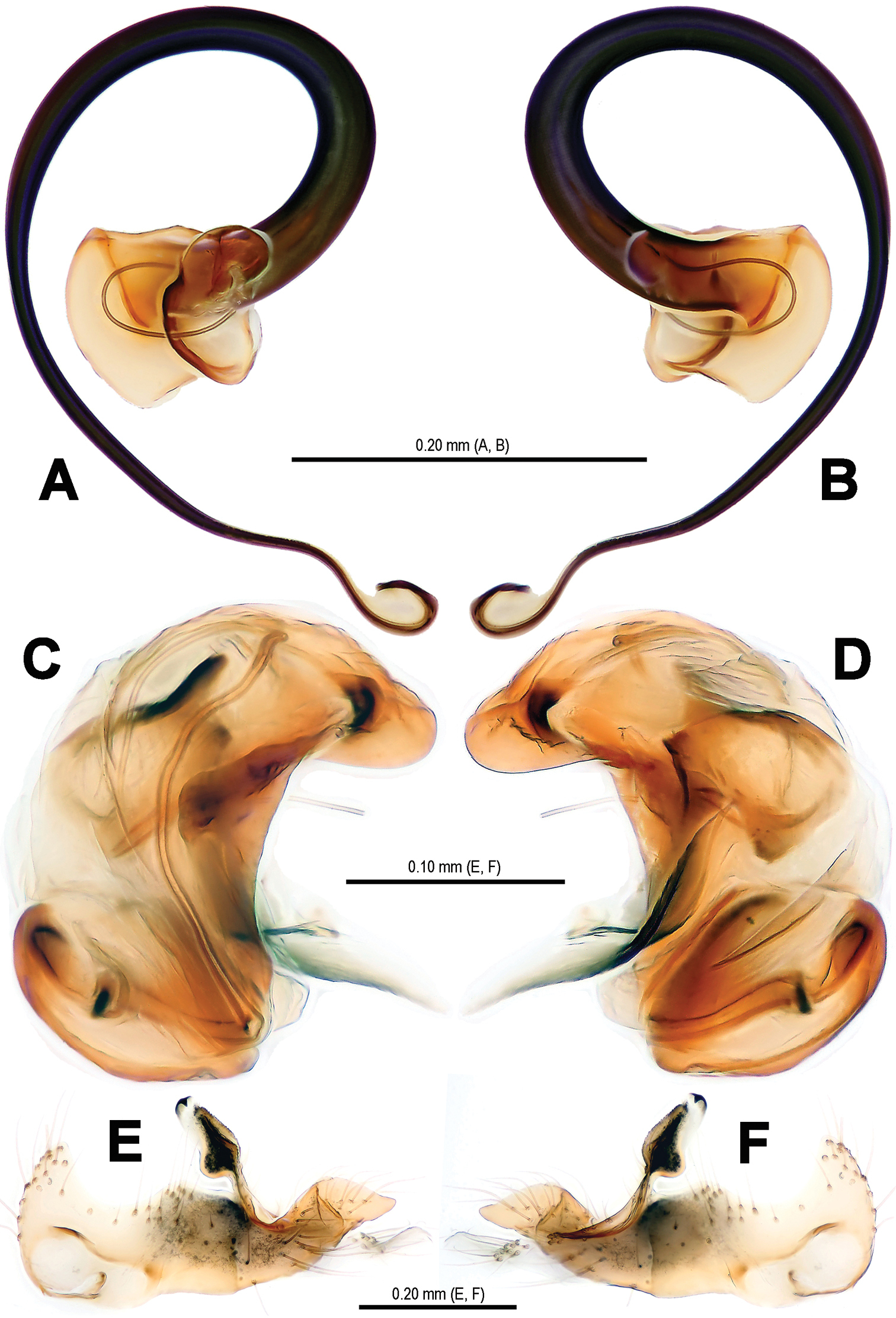
Pedipalp (Figs 2–3, 5–6): Femur long, with a subdistal macroseta ventrally (Figs 2A–B, 5A–B). Patella short, with a few setae. Tibia swollen, bowl-shaped, covered with long setae on distal margin ventrally and dorsally (Figs 3D–E, 6D–E). Cymbium membranous, wide, arisen from tibial margin ventrally (Fig. 6E), paracymbium attached with long setae along prolateral margin, a sclerotized cymbial process subdistally, a row of setae on cymbial fold subdistally and a primary cymbial conductor distally (Figs 3D–E, 6D–E). Tegulum rugose, translucent (Figs 2C, 3A–C). Spermatic duct visible through subtegulum (Figs 3A–C, 6A–C). Embolus long, thin and sparal (Figs 3C, 6C), coiling into four loops. Embolic end exceeded apex of cymbium (Figs 2C, 5A–C).
Female (one of paratypes). Somatic characters see Fig. 1D–F. Coloration: Same as in male.
Measurement: Total length 0.75. Prosoma 0.36 long, 0.32 wide, 0.30 high. Opisthosoma as in male, 0.54 long, 0.50 wide, 0.61 high. Clypeus 0.05 high, distinctly lower than in male. Sternum 0.23 long, 0.21 wide. Length of legs [total length (femur + patella + tibia + metatarsus + tarsus)]: I 1.05 (0.34, 0.14, 0.21, 0.16, 0.20); II 0.93 (0.29, 0.13, 0.18, 0.14, 0.19); III 0.77 (0.23, 0.11, 0.13, 0.13, 0.17); IV 0.99 (0.30, 0.13, 0.20, 0.16, 0.20).
Prosoma (Fig. 1D, F): Carapace near pear-shaped. Cephalic part lower than in male. Eyes arrangement, chelicerae and endites as in male.
Legs: Color, number of trichobothria same as in male, except for leg I without distal metatarsal clasping macroseta prolaterally. Sclerotized femoral spot present at leg I and II as in male. Leg formula: I-IV-II-III.
Opisthosoma (Fig. 1D–F): Globose dorsally. Spinnerets grey, the anteriors larger than the posteriors. Colulus small, pale.
Epigynum (Figs 4, 7): Large, weakly sclerotized, darkish. Epigynal area covered with short setae (Fig. 4B). A small, sclerotized scape stands on epigynal posteromargin mesially (Fig. 4B–C). Spermathecae short clubbed, weakly sclerotized, twisted, attached with membranous, rugose accessory bursae (Figs 4C, 7C). Fertilization ducts short, connected with spermathecae and accessory bursa. Copulatory ducts long, curved, weekly sclerotized, derives from inner side of spermathecae ventrally (Figs 4C, 7C).
Distribution. Known only from the type locality (Fig. 13).
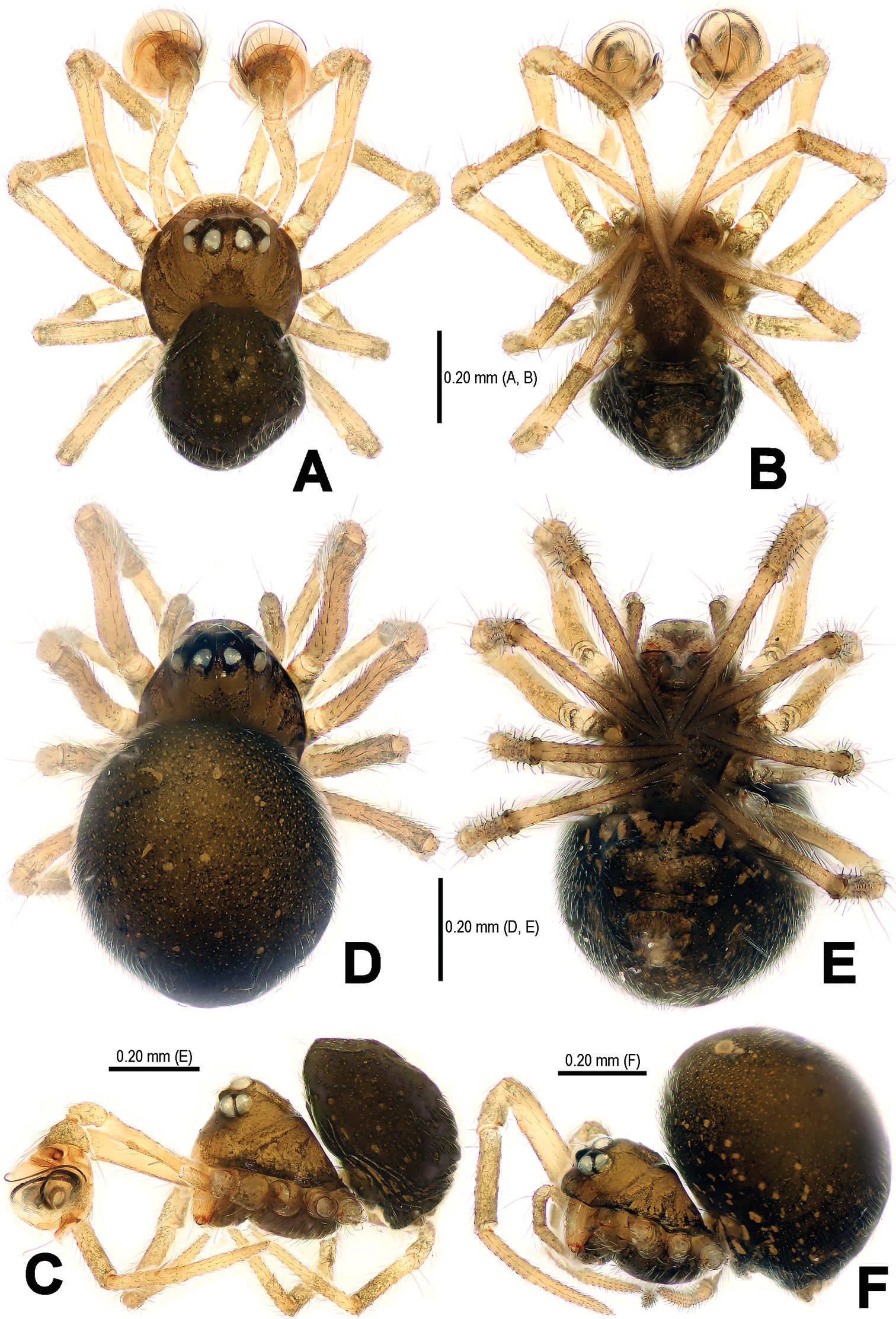
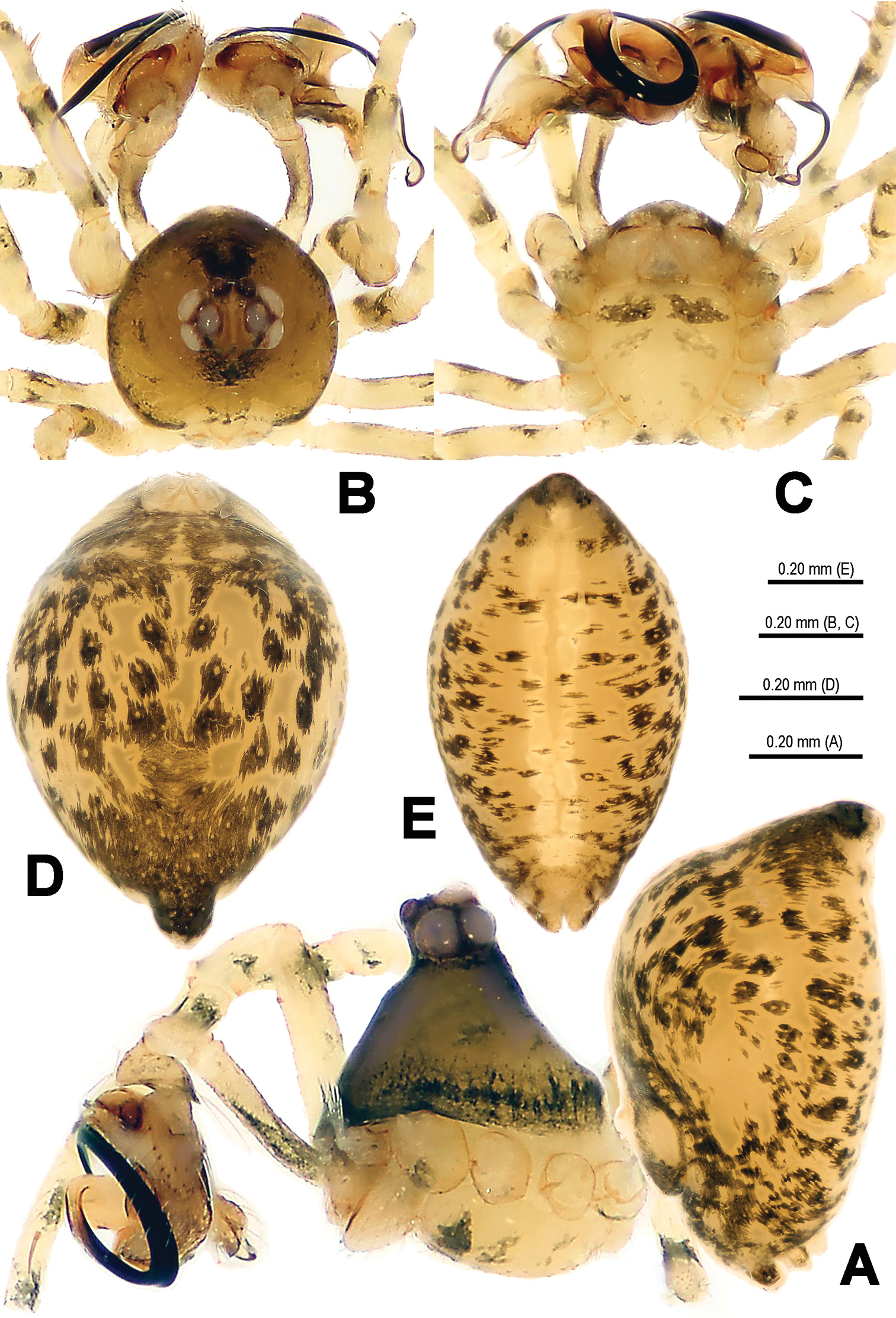
Mysmena wawuensis sp. n., male holotype (A–C) and female paratype (D–F). A–F Habitus. A, D dorsal view B, E ventral view C, F lateral view.
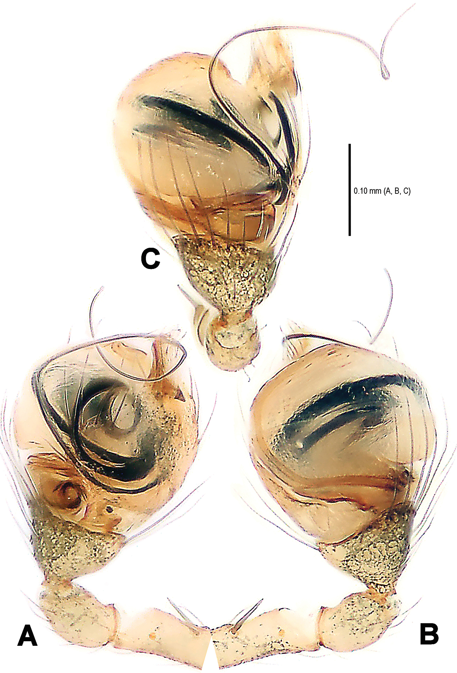
Mysmena wawuensis sp. n., male holotype. A–C Left pedipalp. A prolateral view B retrolateral view C dorsal view.
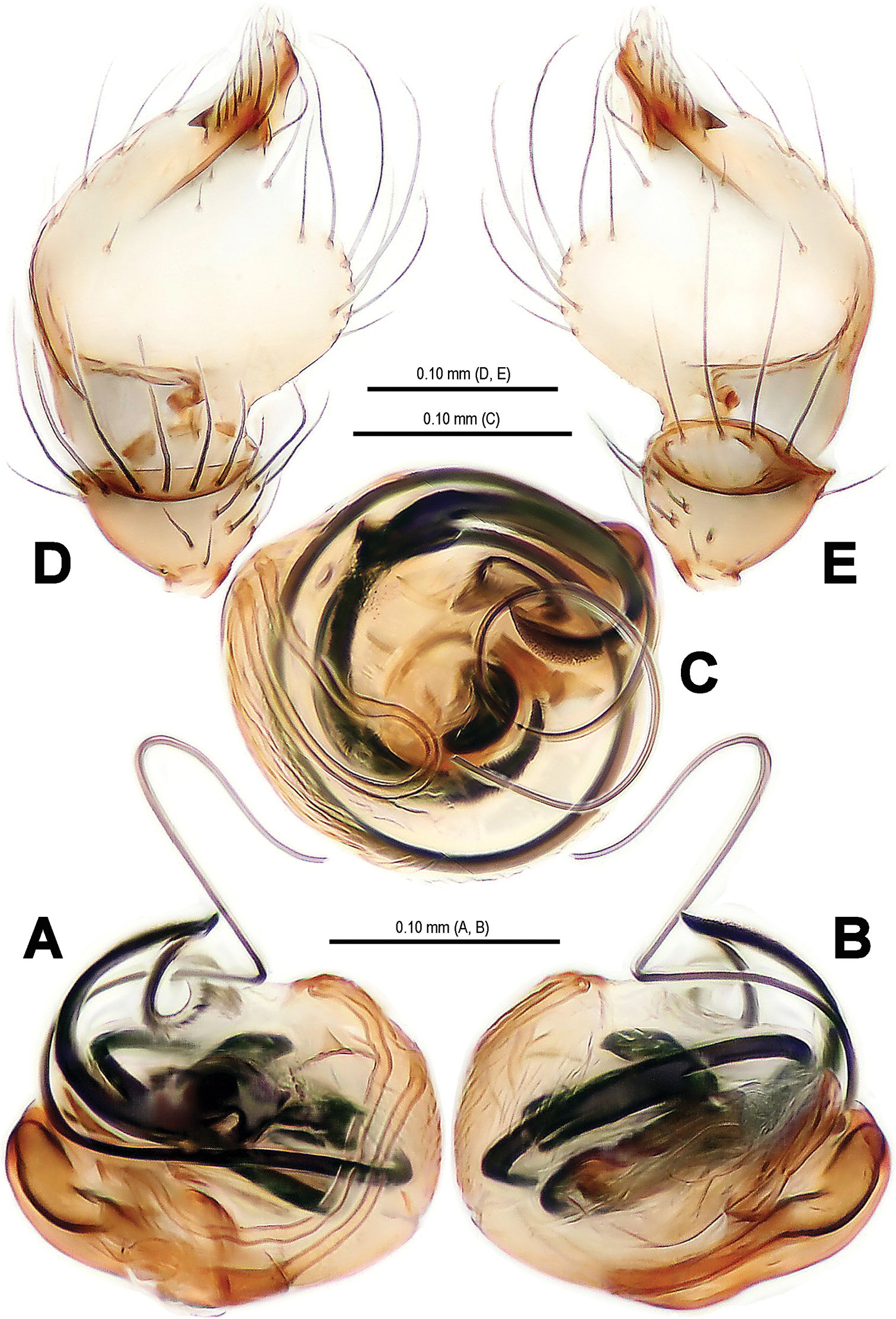
Mysmma wawuensis sp. n., male holotype. A–C Pedipalpal bulb D–E Cymbium. A ventral view B dorsal view C apical view D ventral view E dorsal view.
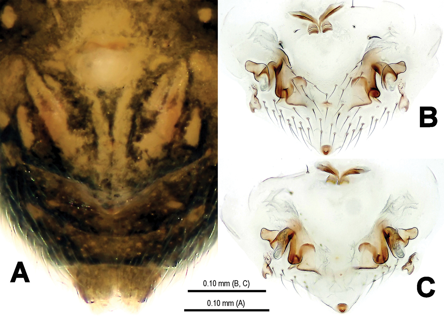
Mysmena wawuensis sp. n., female paratype. A Epigynum, ventral view B Epigynum (lactic acid-treated), ventral view C Vulva (cleared), dorsal view.

Mysmena wawuensis sp. n., male holotype. A–C Left pedipalp. A prolateral view B retrolateral view C dorsal view. Abbrs.: CyP cymbial process; E embolus; Pa patella; T tegulum; Ti tibia.
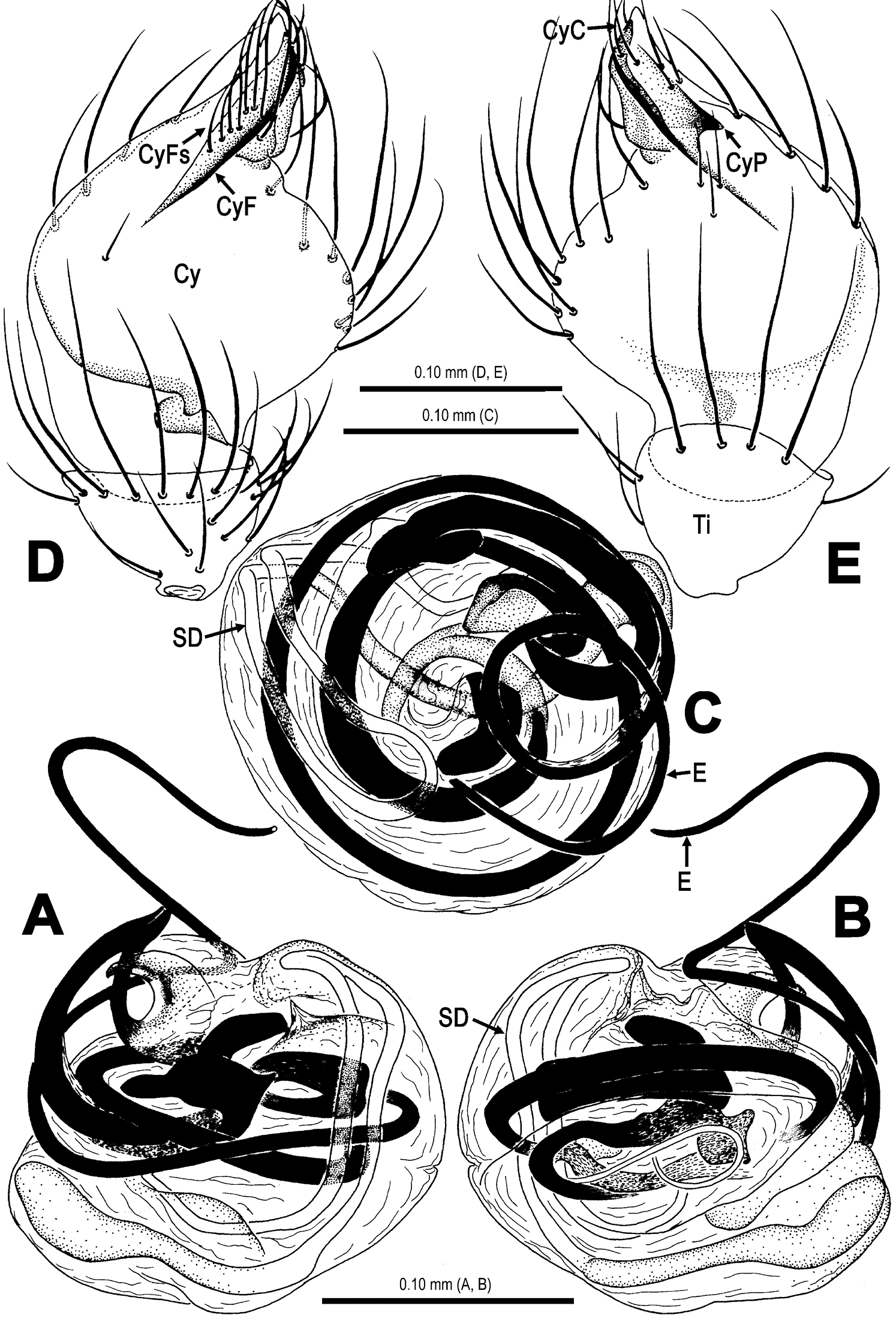
Mysmena wawuensis sp. n., male holotype. A–C Pedipalpal bulb, D–E Cymbium. A ventral view B dorsal view C apical view D ventral view E dorsal view. Abbrs.: Cy cymbium; CyC cymbial conductor; CyF cymbial fold; CyFs setae on cymbial fold; CyP cymbial process; E embolus; SD spermatic duct; Ti tibia.
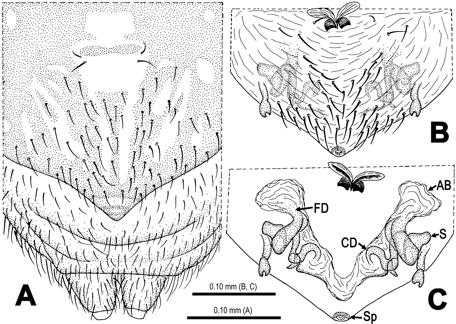
Mysmena wawuensis sp. n., female paratype. A Epigynum, ventral view B Epigynum (lactic acid-treated), ventral view C Vulva (cleared), dorsal view. Abbrs.: AB accessory bursa; CD copulatory duct; FD fertilization duct; S spermatheca; Sp scape.
Type species. Trogloneta granulum Simon, 1922
urn:lsid:zoobank.org:act:47B062D1-CCC8-4C2B-978B-6ABF9B135CDF
http://species-id.net/wiki/Trogloneta_yuensis
Figs 8–13Holotype: CHINA, Chongqing: Beibei District, Jinyun Mt., Guankou, 29°50.261'N, 106°23.811'E, elevation ca 531 m, 5 April 2010, by sieving, Zhisheng Zhang leg., male (SCUM).
The specific name is taken from the type locality; adjective. Yu is short name for Chongqing.
This new species has the following combinations of typical generic features: AME dark, smaller ALE (Fig. 8B); eyes at the apex (Fig. 8A); male leg I with a femoral spot and a metatarsal clasping spine; highly elevated and conical carapace (Fig. 8A); male pedipalp large (Fig. 8B–C). All indicating that this species belongs to the genus Trogloneta. This new species is similar to Trogloneta denticocleari Lin & Li, 2008 (see
Male (holotype). Somatic characters see Fig. 8A–E. Coloration: Prosoma yellow centrally, dark marginally. Clypeus black. Sternum yellow, with a pair of shoulder dark speckles. Opisthosoma yellow, with irregular dark spots.
Measurement: Total length 1.01. Prosoma 0.45 long, 0.45 wide, 0.59 high. Opisthosoma 0.54 long, 0.55 wide, 0.95 high. Clypeus 0.32 high. Sternum 0.31 long, 0.29 wide. Length of legs [total length (femur + patella + tibia + metatarsus + tarsus)]: I 1.42 (0.43, 0.17, 0.32, 0.29, 0.21); II 1.15 (0.38, 0.16, 0.23, 0.22, 0.16); III 0.96 (0.29, 0.13, 0.20, 0.18, 0.16); IV 1.15 (0.36, 0.14, 0.26, 0.22, 0.17).
Prosoma (Fig. 8A–C): Carapace near round. Cephalic pars sharply elevated, slope forward and backward. Ocular area at apex. Eight eyes in two rows. AME black, others white. AME smallest, ALE largest. ALE>PLE>PME>AME. ALE, PME and PLE contiguous. ARE procurved, PRE strongly procurved. Chelicerae pale, small, shorter than endites (Fig. 8A), fang furrow with 2 promaiginal and 1 retromarginal teeth.
Legs: Femora and other segments pale yellow mesially, but grey proximally and distally. Leg formula: I-II-IV-III. Leg I with a subdistal sclerotized femoral spot ventrally and a submesial metatarsal clasping macroseta prolaterally. Patellae I–IV with a dorsal seta distally. Tibiae I–IV with a dorsal seta proximally. Tibiae I, II and IV with 3 trichobothria, but 4 on tibia III. Metatarsi I–IV lack trichobothrium.
Opisthosoma (Fig. 8A, D–E): elliptic dorsally, fusiform posteriorly, triangular laterally, with a tubercle at rear. Spinnerets grey, the anteriors larger than the posteriors. Colulus small, tongue-shaped. Anal tubercle pale.
Pedipalp (Figs 9–12): Large, strongly sclerotized. Femur as 2.5 times long as patella (Fig. 9A, B). Patella short, with a few setae. Tibia wider than long, nearly cup-shaped, covered with a dorsal trichobothrium and a few marginal long setae ventrally (Figs 11A–B). Cymbium large (Figs 10E–F, 12E), membranous, paracymbium flattened, covered with dense long setae. A long cymbial process (aquiline distally, constricted proximally) arisen from inner side subdistal margin (Fig. 12E). Cymbial fold distinctly, with long setae. Distal primary cymbial conductor membranous, translucent, attaching with a cluster of setae (Fig. 12E). Tegulum smooth, sclerotized (Fig. 10C–D). Spermatic duct long, visible through subtegulum (Fig. 11C–D). A long, fingerlike median apophysis starts at the junction between tegulum and subtegulum (Figs 10D, 11D). Embolus long, arched, strongly sclerotized, gradually diminishing from base to end (Figs 9B, 12A–B). Embolic end unciform, with accessory membrane (Fig. 12A–B), hidden behind cymbial conductor (Figs 9B, 11B).
Female. Unknown.
Trogloneta yuensis sp. n., male holotype. A Habitus, lateral view B Prosoma, dorsal view C Ditto, ventral view D Opisthosoma, dorsal view E Ditto, posterior view.
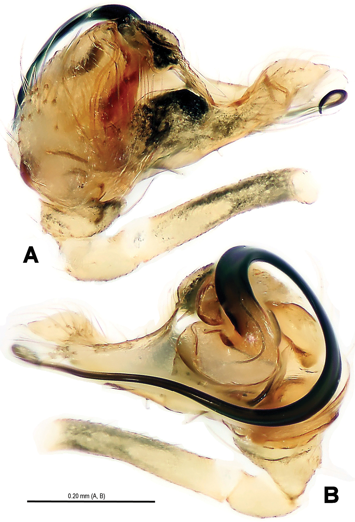
Trogloneta yuensis sp. n., male holotype. A Left pedipalp, retrolateral view B Ditto, prolateral view.
Trogloneta yuensis sp. n., male holotype. A Embolus, ventral view B Ditto, dorsal view C Pedipalpal bulb (excluding embolus), ventral view D Ditto, dorsal view E Cymbium, dorsal view F Ditto, ventral view.
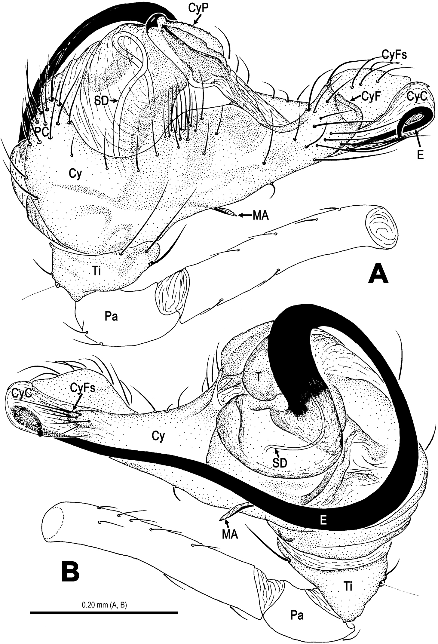
Trogloneta yuensis sp. n., male holotype. A Left pedipalp, retrolateral view B Ditto, prolateral view. Abbrs.: Cy cymbium; CyC cymbial conductor; CyF cymbial fold; CyFs setae on cymbial fold; CyP cymbial process; E embolus; MA median apophysis; Pa patella; PC paracymbium; SD spermatic duct; T tegulum; Ti tibia.
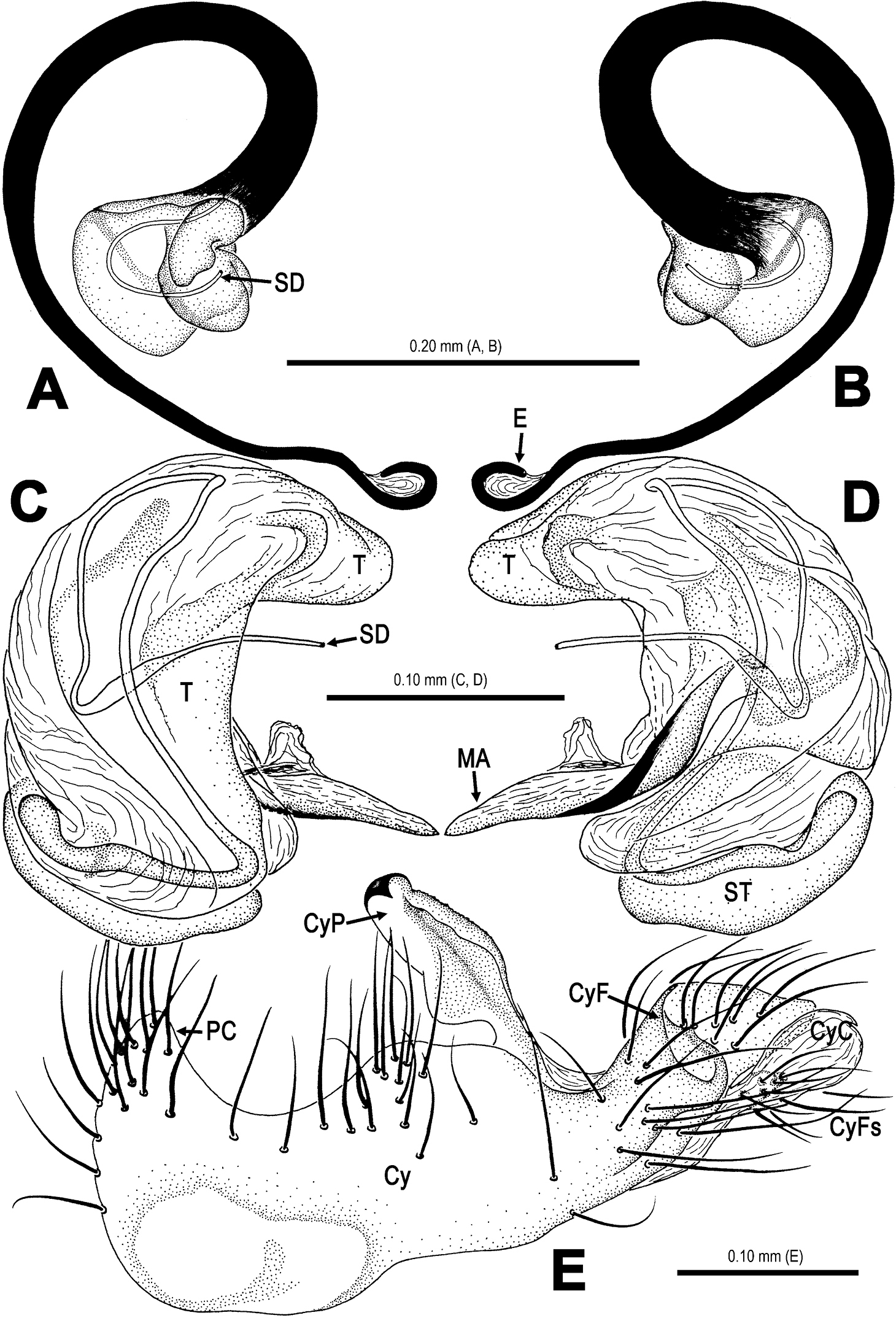
Trogloneta yuensis sp. n., male holotype. A–B Embolus. A ventral view B dorsal view C–D Pedipalpal bulb (excluding embolus) C ventral view D dorsal view E Cymbium, dorsal view. Abbrs.: Cy cymbium; CyC cymbial conductor; CyF cymbial fold; CyFs setae on cymbial fold; CyP cymbial process; E embolus; MA median apophysis; Pa patella; PC paracymbium; SD spermatic duct; ST subtegulum; T tegulum.
Known only from the type locality (Fig. 13).
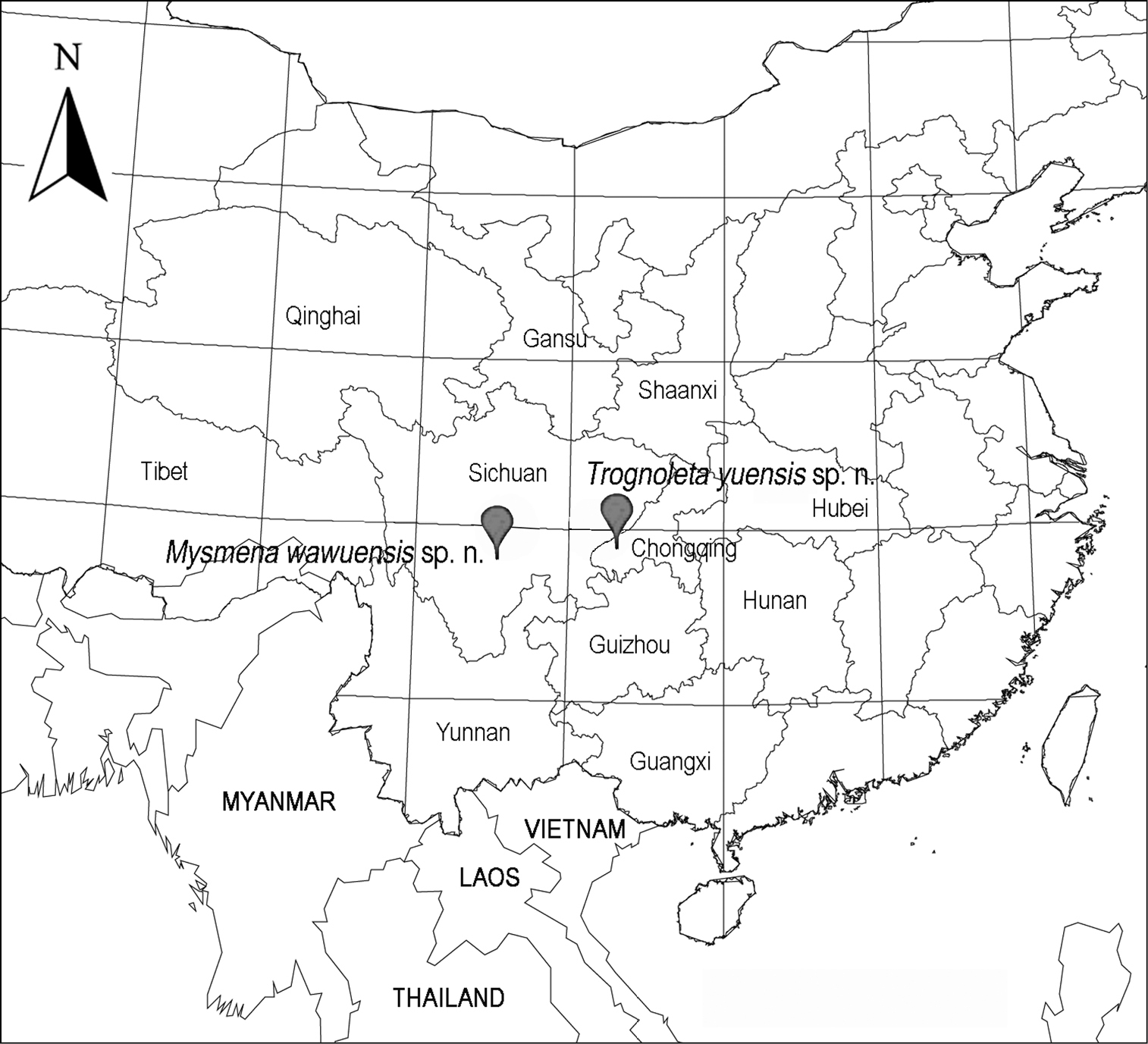
Distributional records of two new mysmenid species from China.
The manuscript benefited greatly from comments by Jeremy Miller (Naturalis Biodiversity Center, the Netherlands) and one anonymous reviewer. This study was supported by the National Natural Sciences Foundation of China (China National Funds for Distinguished Young Scientists-31025023 and NSFC-30870271, 31000946, 31272280), and by New Teacher Fund for Doctor Station of Ministry of Education of China (20100181120049).