Research Article |
|
Corresponding author: Pavel S. Nefediev ( p.nefediev@mail.ru ) Academic editor: Marzio Zapparoli
© 2018 Gyulli Sh. Farzalieva, Pavel S. Nefediev.
This is an open access article distributed under the terms of the Creative Commons Attribution License (CC BY 4.0), which permits unrestricted use, distribution, and reproduction in any medium, provided the original author and source are credited.
Citation:
Farzalieva GSh, Nefediev PS (2018) The first record of the anopsobiid genus Shikokuobius Shinohara, 1982 in continental Asia, with the description of a new species from the Altais, southwestern Siberia, Russia (Chilopoda, Lithobiomorpha, Anopsobiidae). ZooKeys 793: 15-28. https://doi.org/10.3897/zookeys.793.29221
|
Abstract
A new lithobiomorph species, Shikokuobius altaicus sp. n., is described from the Altai Mountains in southwestern Siberia, Russia. This is the first record of the genus Shikokuobius Shinohara, 1982 in continental Asia, all previous reports being from Japan. The distribution of Shikokuobius is mapped.
Keywords
Centipede, taxonomy, new species, Altai Mountains, Siberia, Russia
Introduction
The family Anopsobiidae Verhoeff, 1907 is currently known to be represented in the Northern Hemisphere by one small and eight monotypic genera: Yobius Chamberlin, 1945 (Utah, USA), Hedinobius Verhoeff, 1934 (Tien Shan, western China), Rhodobius Silvestri, 1932 (Europe), Anopsobiella Attems, 1938 (Vietnam), Shikokuobius Shinohara, 1982 (Japan), Ghilaroviella Zalesskaja, 1975 (Tajikistan), Dzhungaria Farzalieva, Zalesskaja et Edgecombe, 2004 (eastern Kazakhstan), Speleopsobius Shear, 2018 (southern Idaho, USA), as well as Buethobius Chamberlin, 1911, with 5 species from the USA (
A collection of lithobiomorph centipedes from the Republic of Altai, Russia, deposited in the Zoological Museum of the Lomonosov Moscow State University, has yielded a single male of a new anopsobiid species of Shikokuobius. Four additional specimens of that new species have also been freshly collected from the Altai Province, Russia.
Shikokuobius altaicus sp. n. is very similar to S. japonicus (Murakami, 1967) from Japan (Sakuragi, Honshu; Nakameguro and Shirogane; the DNA voucher specimen of
Material and methods
The material used in the present study was collected by S.I. Golovatch (Moscow, Russia) in the environs of Lake Teletskoye, Republic of Altai and by T.M. Krugova (Barnaul, Russia) with her team of volunteers in the Tigirek State Nature Reserve, Altai Province. Both sites are located in the Russian Altais, southwestern Siberia, Russia. Most of the material is currently deposited in the collection of the Zoological Museum of the Lomonosov Moscow State University, Moscow, Russia (
The total body length was measured from the fore margin of the cephalic plate to the posterior end of the postpedal tergite. Leg length was measured excluding the length of the claw. Lengths are given as the minimum and maximum values. All measurements are given in millimeters (mm).
The mouthparts, legs and body segments of the new species were cleared in 10% KOH and mounted in permanent slides in sandarac medium (
The terminology of the external anatomy follows
The following abbreviations are used in the text:
T, TT tergite, tergites;
C, CC coxa, coxae;
t trochanter;
P prefemur;
F femur.
Results
Taxonomy
Shikokuobius altaicus sp. n.
Type material
Holotype ♂ (
Paratypes
1 ♂ (
Non-type material
1 ♂ (
Name
The specific epithet refers to Altai, the locus typicus.
Diagnosis
A Shikokuobius species with the body 4–6 mm long, the antennae composed of 15–17 antennomeres, commonly 15; coxosternal teeth large, separated from each other by distances less than width at the base of a tooth; P, F and T of 15 leg relatively short and thick, 15 C with a prominent, acute, mesodistal process; the number of coxal pores varying from 1 to 2 on 12–15 CC (formula 1,1,1,1 in the male and 1,1,1,2 in the female); 15 t and 15 P with bifurcate ventral spines (seldom on legs 13 and 14); at least each leg 15 with a bifurcate spine at the distodorsal end of P; 1–10 tibiae with a distal spinose projection (Figs
Distribution
(Fig.
Description
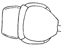
Holotype ♂. Body ca 4.0 mm long, ca 0.4 mm wide (in 70% alcohol); colour yellow. Tergites: almost smooth, with relatively long and sparse setae, as in Figs

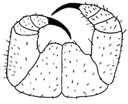
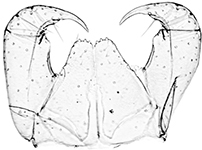
Shikokuobius altaicus sp. n., male paratype (1, 3, 6) and female paratype (2, 4, 5). 1 forcipulae, ventral view 2 head, ventral view 3 12–14 sternites and coxae, ventral view 4 dental margin of forcipular coxosternite 5 13–16 tergites, dorsal view 6 mandibula, ventrolateral view. Abbreviations: TO Tömösváry’s organ, CO coxal pore. Scale bars: 0.1 mm (1–5), 0.05 mm (6).
Tarsal articulation of legs 1–12 indistinct, tarsi distinctly longer than tibiae. 1–10 tibiae with a distal spinose projection, as in Figs


Shikokuobius altaicus sp. n., male holotype (20–22) and male paratype (23, 24). 20 left part of second maxilla, ventral view 21 ventral spine on prefemur 15, lateral view 22 ventral spine on trochanter 15, lateral view 23 leg 11, lateral view 24 leg 10, lateral view. Scale bars: 0.05 mm (20–22), 0.1 mm (23, 24).
Paratype ♂. Length 4.0 mm, width 0.4 mm. All other characters as in holotype, but coxal process on leg 15 broken off on both legs.
Non-type material ♂. Length 4.9 mm, width 0.5 mm. All other characters as in holotype (Figs
Paratype ♀♀. All characters as in ♂♂. The number of antennomeres in females unknown: one ♀ with antennae completely broken off, while another ♀ with damaged antennae, having 12+7 antennal articles. Coxal pores as in holotype, formula 1,1,1,2 (Figs

Shikokuobius altaicus sp. n., male holotype (25, 26, 28, 29) and female paratype (27). 25 left gonopod, ventral view 26 left mesodistal process on 15 coxa, ventral view 27 left gonopod, ventral view 28 left part of first maxilla 29 distodorsal process on tibia 3, lateral view. Scale bar: 0.05 mm.
Habitats
The new species was collected in the lowland Altais in small-leaved and mixed taiga forests at 450 to 920 m a.s.l. (Fig.
Remarks
The new species belongs to the genus Shikokuobius Shinohara, 1982 that shows the following synapomorphies: antenna with up to 18 articles, 3+3 coxosternal teeth; spiracles on leg-bearing segments 3, 5, 8, 10, 12 and 14; coxal pores on 12–15 legs; 15 C with a prominent, acute, mesodistal process; 15 t and 15 P with spines, ventrally bifurcated at their tips; at least 15 P with a bifurcate spine at distodorsal end (as some specimens with spines apparently broken off, so these are not visible).
S. altaicus sp. n. is similar to S. japonicus (Murakami, 1967), so far the single species in the genus Shikokuobius, with the above characters. The main differences between them are given in Table
Finally, S. altaicus sp. n. is also rather similar to Ghilaroviella valiachmedovi Zalesskaja, 1975, from the Tajikistan in showing the same body length, simple and plumose bristles on the second maxillae; the number of antennomeres, 1–2 coxal pores, 2+2 spurs and a simple ♀ gonopodal claw. However, S. altaicus sp. n. is well-distinguished from the latter species by: (1) 3+3 coxosternal teeth (vs. 2+2 in G. valiachmedovi); (2) coxal process well-developed only on leg 15 (vs. on legs 14 and 15 in G. valiachmedovi) and (3) the absence of small warts at the base of the ♀ gonopodal claw (vs. 2 small warts in G. valiachmedovi).


Shikokuobius altaicus sp. n., male paratype (36) and not-type male (37–43). 36 left part of first maxilla, ventral view 37 right part of first maxilla, ventral view 38 distal part of coxa 15, ventral view 39 spine on trochanter 15, lateral view 40 spine on prefemur 15, lateral view 41 trochanter and prefemur 15, lateral view 42 right gonopod, ventral view 43 distodorsal spine on prefemur 15, lateral view. Scale bars: 0.05 mm (36–40, 42, 43), 0.1 mm (41).


The main differences between S. japonicus (Murakami, 1967) and S. altaicus sp. n.
| S. japonicus (Murakami, 1967) | S. altaicus sp. n. | ||
|
sensu
|
sensu
|
||
| front body part |

|
|
 cephalic plate slightly elongate, width/length ratio 0.8; posterior margin of T1 slightly sinuate cephalic plate slightly elongate, width/length ratio 0.8; posterior margin of T1 slightly sinuate |
| cephalic plate equal in width and length; posterior margin of T1 straight | |||
| number of antennomeres | 18 | up to 18 | 15* |
| labrum |
 sides are smooth; pair of setae projecting across the labral midpiece absent** sides are smooth; pair of setae projecting across the labral midpiece absent** |
no data |
 sides with poorly-expressed fringes of bristles; pair of setae projecting across the labral midpiece present sides with poorly-expressed fringes of bristles; pair of setae projecting across the labral midpiece present |
| forcipular coxosternite |
|

|
 narrower, width/length ratio 1.2–1.3:1 narrower, width/length ratio 1.2–1.3:1 |
| approximately broad, width/length ratio 1.6–1.8:1 | |||
| dental margin of forcipulae coxosternite |

|

|
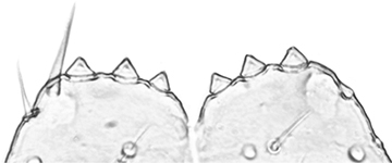 teeth relatively large, separated from each other by distances less than width at the base of a tooth teeth relatively large, separated from each other by distances less than width at the base of a tooth |
| teeth very small, separated from each other by distances more than width at the base of a tooth | |||
| leg 15 |

|
|
 P, F & T shorter and thicker P, F & T shorter and thicker |
| P, F & T elongate and thin | |||
Acknowledgments
The authors wish to thank S.I. Golovatch (Moscow, Russia) for kindly editing the English of an advanced draft. Special gratitude goes to S.L. Esyunin (Perm, Russia) for his constant guidance, encouragement and support of the first co-author. We are also very much obliged to A.A. Schileyko (Moscow, Russia) for the provision some of material from
References
- Attems C (1938) Die von Dr. C. Dawydoff in Französisch Indochina gesammelten Myriopoden. Mémoires du Muséum d’Histoire Naturelle (Paris) NS 6(2): 187–353.
- Bonato L, Edgecombe GD, Lewis JGE, Minelli A, Pereira LA, Shelley RM, Zapparoli M (2010) A common terminology for the external anatomy of centipedes (Chilopoda). ZooKeys 69: 17–51. https://doi.org/10.3897/zookeys.69.737
- Edgecombe GD, Giribet G (2003) Relationships of Henicopidae (Chilopoda: Lithobiomorpha): New molecular data, classification and biogeography. African Invertebrates 44: 13–38.
- Edgecombe GD, Giribet G (2004) Molecular phylogeny of Australasian anopsobiine centipedes (Chilopoda: Lithobiomorpha). Invertebrate Systematics 18: 235–49. https://doi.org/10.1071/IS03033
- Farzalieva GSh, Zalesskaja NT, Edgecombe GD (2004) A new genus and species of lithobiomorph centipede (Chilopoda: Lithobiomorpha: Anopsobiidae) from eastern Kazakhstan. Arthropoda Selecta 13(4): 219–224.
- Krasheninnikov AB (2011) Mounting technique of entomological preparations in sandarac medium. Eurasian entomological journal 10(3): 278–279.
- Murakami Y (1967) Postembryonic development of the common Myriapoda of Japan XXIV. A new species of the family Henicopidae. Zoological Magazine, Tokyo, 76: 7–12.
- Shear WA (2018) The centipede family Anopsobiidae new to North America, with the description of a new genus and species and notes on the Henicopidae of North America and the Anopsobiidae of the Northern Hemisphere (Chilopoda, Lithobiomorpha). Zootaxa 4422(2): 259–283. https://doi.org/10.11646/zootaxa.4422.2.6
- Shinohara K (1982) A new genus of centipede of the subfamily Anopsobiinae (Henicopidae, Chilopoda). Proceedings of the Japanese Society for Systematic Zoology 24: 41–46.
- Silvestri F (1909) Descrizioni preliminari di vari artropodi specialmente d’America. Rendiconti della R. Accademia dei Lincei. Classe di Scienze Fisiche Matematiche e Naturali 18: 267–271.
- Zalesskaja NT (1975) [New genera and species of Chilopoda (Lithobiomorpha) from Central Asia and Far East]. Zoologicheskii Zhurnal 54(9): 1316–1325. [in Russian]
- Zapparoli M, Edgecombe GD (2011) Order Lithobiomorpha. In: Minelli A (Ed.) Treatise on Zoology – Anatomy, Taxonomy, Biology – The Myriapoda, Volume 1.Brill, Leiden – Boston, 371–389.