


(C) 2012 Lu-Xi Liu. This is an open access article distributed under the terms of the Creative Commons Attribution License 3.0 (CC-BY), which permits unrestricted use, distribution, and reproduction in any medium, provided the original author and source are credited.
For reference, use of the paginated PDF or printed version of this article is recommended.
A review of species of the genus Neopsocopsis Badonnel, 1936 is presented. Four species are redescribed, viz. Neopsocopsis hirticornis (Reuter, 1893), Neopsocopsis quinquedentata (Li & Yang, 1988), Neopsocopsis profunda (Li, 1995), and Neopsocopsis flavida (Li, 1989), as well as the description of one new species, Neopsocopsis convexa sp. n. Seven new synonymies are proposed as follows: Pentablaste obconica Li syn. n. and Pentablaste clavata Li syn. n. of Neopsocopsis hirticornis, Pentablaste tetraedrica Li syn. n. of Neopsocopsis longiptera, Neoblaste schizopetala Li syn. n. and Neoblaste flavae Li syn. n. of Neopsocopsis profunda, Blastopsocidus pini Li syn. n. and Pentablaste lanceolata Li syn. n. of Neopsocopsis flavida. Neopsocopsis hirticornis (Reuter, 1893) is recorded from Japan and China for the first time, and Neopsocopsis longiptera Vishnyakova, 1986 is newly recorded from China. Illustrated keys to adult males and females are presented.
Psocodea, Psocoptera, Psocidae, Neopsocopsis, redescriptions, new synonymies, new species, new records, distribution, key
The psocopteran genus Neopsocopsis is a small group in the subfamily Amphigerontiinae, formerly characterized by head-covering glandular setae, female fore wing brachypterous, and male hypandrium with three lobes (one median lobe and two lateral lobes) and 2 internal apophyses (
In the present paper, one new species of Neopsocopsis is described, N. convexa sp. n., with redescriptions of four species: Neopsocopsis hirticornis (Reuter, 1893), Neopsocopsis quinquedentata (Li & Yang, 1988), Neopsocopsis profunda (Li, 1995), and Neopsocopsis flavida (Li, 1989). Seven new synonymies are proposed as follows: Pentablaste obconica Li, syn. n. and Pentablaste clavata Li, syn. n. of Neopsocopsis hirticornis, Pentablaste tetraedrica Li, syn. n. of Neopsocopsis longiptera, Neoblaste schizopetala Li, syn. n. and Neoblaste flavae Li, syn. n. of Neopsocopsis profunda, Blastopsocidus pini Li, syn. n. and Pentablaste lanceolata Li, syn. n. of Neopsocopsis flavida. Neopsocopsis hirticornis (Reuter, 1893) is recorded from Japan and China for the first time, and Neopsocopsis longiptera Vishnyakova, 1986 is newly recorded from China. Updated keys for adult males and females of world species in the genus is presented.
Material and methodsAll specimens treated in this paper were from Entomological Museum of China Agricultural University (CAU), Beijing, China, and Hokkaido University Insect Collection (SEHU), Sapporo, Japan. Specimen preparation and measurements were undertaken following
http://species-id.net/wiki/Neopsocopsis
Small to medium sized psocids. Antennae short, not reaching tip of fore wing. Wings membranous, usually hyaline with brownish tinge; fore wing normal in both sexes or brachypterous in female; fore wing Rs and M meeting at point, fused for short distance or connected by crossvein, areola postica pentagonous, first and second sections of Cu1a forming obtuse angle about 120°. Male abdomen with distal segments dark brown colored, 8th sternum broadly sclerotized and fused to hypandrium, with lateral margins overlapping clunium; epiproct round, dorsally with sclerotized projection at middle of anterior margin; hypandrium symmetrical and 5-lobed, with posteromedian lobe forming dorsal-curved structure, pair of lateral lobes carinate with outer surface covering denticles, and pair of internal lobes rod-like or expanded; phallosome free posteriorly, anteriorly fused or connected by membrane. Female subgenital plate with sclerotized arms forming flat V-shaped regions and expanded laterally, egg guide relatively long; ventral valve of gonapophyses distally tapering to slender tip, outer valve with well developed posterior lobe.
China; Finland; France; Germany; Hungary; Italy; Japan; Macedonia; Mongolia; Romania; Russia; Serbia; Spain; Sweden; Switzerland.
The genus Neopsocopsis is placed in the subfamily Amphigerontiinae mainly based on the following characters: male 8th sternum broadly sclerotized and fused to hypandrium, with lateral margins overlapping clunium (
One Indonesian genus, Javablaste Endang, Thornton & New, 2002, shared many generic characters of Neopsocopsis and was different from the latter by 1) female with normal fore wing, 2) subgenital plate with transverse sclerotized bar at mid line and 3) male hypandrium with lateral spinous lobes (
Pentablaste pentasticha (Li, 1990) is not included in the key as re-examination of the species and its possible relationship with Neoblaste were not possible for this study.
Key to adult males of Neopsocopsis| 1 | Internal lobes of hypandrium well developed, expanded and longer than posteromedian lobe (Fig. 3C) | 2 |
| – | Internal lobes of hypandrium not well developed, usually rod-like, equal length or shorter than posteromedian lobe (Fig. 4C) | 3 |
| 2 | Small in size, fore wing length about 2.5–3.0 mm; epiproct fully sclerotized, with tiny projection dorsally; lateral lobes of hypandrium small; phallosome with parameres absent. See Yoshizawa, 2010, Fig. 10 | Neopsocopsis sakishimensis |
| – | Large in size, fore wing length about 3.2–4.5 mm; epiproct with membranous regions medially (Fig. 3B), with large and sharp projection dorsally (Fig. 3AB); lateral lobes of hypandrium large (Fig. 3C); phallosome paired with parameres (Fig. 3D) | Neopsocopsis hirticornis (=Pentablaste obconica; =Pentablaste clavata) |
| 3 | Posteromedian lobe of hypandrium with distal margin almost straight, or concave with projection medially (Fig. 5C) | 4 |
| – | Posteromedian lobe of hypandrium with distal margin convex medially (Fig. 2C) | 6 |
| 4 | Posteromedian lobe of hypandrium with distal margin straight; lateral lobes of hypandrium with anterior part short. See Yoshizawa, 2010, Fig. 12 | Neopsocopsis longiptera (=Pentablaste tetraedrica) |
| – | Posteromedian lobe of hypandrium with distal margin concave, and with projection medially; lateral lobes of hypandrium with anterior part long and curved anteromedially (Fig. 4C) | 5 |
| 5 | Epiproct with sharp projection dorsally (Fig. 4B); internal lobes of hypandrium not forked distally (Fig. 4C) | Neopsocopsis quinquedentata |
| – | Epiproct with large and round projection dorsally (Fig. 5B); internal lobes of hypandrium forked distally (Fig. 5C) | Neopsocopsis profunda (= Neoblaste schizopetala; = Neoblaste flavae) |
| 6 | Clunium with posterior margin sharply convex medially (Fig. 2B); internal lobes of hypandrium distally forked (Fig. 2C) | Neopsocopsis convexa sp. n. |
| – | Clunium with posterior margin smoothly convex medially (Fig. 6B); internal lobes of hypandrium tortuous forming right-angle and distally not forked (Fig. 6C) | Neopsocopsis flavida (=Blastopsocidus pini; =Pentablaste lanceolata) |
| 1 | Subgenital plate with egg guide not sclerotized | 2 |
| – | Subgenital plate with egg guide sclerotized wholly (Fig. 2E) or at basal 1/3–1/2 (Fig. 5E) | 4 |
| 2 | Fore wing pale brown with anterior part dark colored; outer valve of gonapophyses with posterior lobe narrowing to internal tip. See |
Neopsocopsis minuscula |
| – | Fore wing pale brown wholly; outer valve of gonapophyses with posterior lobe broad at internal tip | 3 |
| 3 | Subgenital plate with large membranous region anteromedially, pigment arms with small lateral expansions, widely separated; dorsal valve of gonapophyses slender. See |
Neopsocopsis longicaudata |
| – | Subgenital plate with small membranous region anteromedially, pigment arms with large lateral expansions close to each other; dorsal valve of gonapophyses broad. See |
Neopsocopsis auctachila |
| 4 | Subgenital plate with egg guide sclerotized wholly (Fig. 2E) | Neopsocopsis convexa sp. n. |
| – | Subgenital plate with egg guide sclerotized at basal 1/3–1/2 | 5 |
| 5 | Subgenital plate with distal margin of sclerotized regions straight (Fig. 5E) | 6 |
| – | Subgenital plate with distal margin of sclerotized regions not straight (Fig. 3E) | 7 |
| 6 | Subgenital plate with egg guide sclerotized at basal 1/3 (Fig. 5E); internal plate with round plate surrounding spermathecal opening (Fig. 5G) | Neopsocopsis profunda (= Neoblaste schizopetala; = Neoblaste flavae) |
| – | Subgenital plate with egg guide sclerotized at basal 1/2; internal plate without round plate surrounding spermathecal opening. See Li, 2002, pp. 1385, Fig. 1244 | Neopsocopsis jinxiuica |
| 7 | Egg guide with distal margin of sclerotized regions sharply convex (Fig. 6E) | 8 |
| – | Egg guide with distal margin of sclerotized regions concave medially, forming fork-like structure (Fig. 3E) | 9 |
| 8 | Small in size, forewing length about 3.1–3.7 mm; outer valve of gonapophyses with posterior lobe broad at internal tip (Fig. 6F) | Neopsocopsis flavida (=Blastopsocidus pini; =Pentablaste lanceolata) |
| – | Large in size, forewing length about 4.1 mm; outer valve of gonapophyses with posterior lobe narrowing to internal tip. See |
Neopsocopsis lushannensis |
| 9 | Small in size, forewing length about 2.5–3.0 mm; egg guide with short neck region; subgenital plate with large membranous region anteromedially. See |
Neopsocopsis sakishimensis |
| – | Large in size, forewing length more than 3.0 mm; egg guide with relatively long neck region; subgenital plate with small membranous region anteromedially | 10 |
| 10 | Egg guide with fork-like sclerotized regions slightly concave; internal plate with two small sclerotized regions laterally. See |
Neopsocopsis longiptera (= Pentablaste tetraedrica) |
| – | Egg guide with fork-like sclerotized regions strongly concave (Fig. 3E); internal plate with sclerotized regions marginally, paired with strong processes directed laterally (Fig. 3G) | Neopsocopsis hirticornis (=Pentablaste obconica; =Pentablaste clavata) |
urn:lsid:zoobank.org:act:DB945C8B-2D6F-4367-B9C7-15332C71F172
http://species-id.net/wiki/Neopsocopsis_convexa
Figures 1A , 2Holotype ♂: China, Yunnan Prov., Lüchun Co., Huanglianshan Natural Reserve, 5.v.2011 (LX Liu). Paratypes. China: 1♀, same date as holotype; 1♂1♀, same locality and collector as holotype, 6.v.2011; 1♀, Yunnan Prov., Jinping Co., Fenshuiling Natural Reserve, 9.v.2011 (LX Liu); 1♂1♀, same locality and collector, 10.v.2011; 2♂, Yunnan Prov., Pingbian Co., Daweishan Natural Reserve, 16.v.2006 (JX Cui); 1♂, same locality, 12.v.2006 (Y Tang); 3♂, same locality, 24.v.2009 (XS Yang); 1♂, Gansu Prov., Wenxian Co., Qiujiaba Reg., 26.vii.2011 (SP Liu).
The specific name refers to the characteristic convex-shaped posteromedian lobe of the hypandrium.
Medium sized psocids. Fore wing hyaline with brownish coloration; Rs and M fused for very short distance, meeting at point or connected by crossvein. Male: 8th sternum strongly sclerotized and fused to hypandrium; epiproct swollen with tiny projection at middle of anterior margin; hypandrium 5-lobed with posteromedian lobe convex distaromedially, internal lobes rod-like and distally forked. Female: subgenital plate with egg guide distally round, slightly sclerotized, pigment arms forming flat V-shaped regions and expanded laterally.
Male. Head creamy brown; compound eyes grayish black, ocelli black with grayish black ocellar field; antennae and labrum brown; maxillary palpi brown with distal segments dark colored. Thorax brown with dark brown markings on mesonotum; legs brown, with tarsi and distal part of tibia dark brown. Abdominal segments mostly creamy white, with apical regions dark brown. Fore wing (Fig. 1A) hyaline with brownish tinge, pterostigma dark brown with dark brown band along proximal margin; veins brown, except for Rs fork and M-Cu1a fusion hyaline. Venation: Rs and M fused for very short distance, meeting at point or connected by crossvein; distal margin of discoidal cell straight; first and second sections of Cu1a almost equal length, diverging at angle about 120°. Hind wing hyaline with brownish coloration; veins brown.
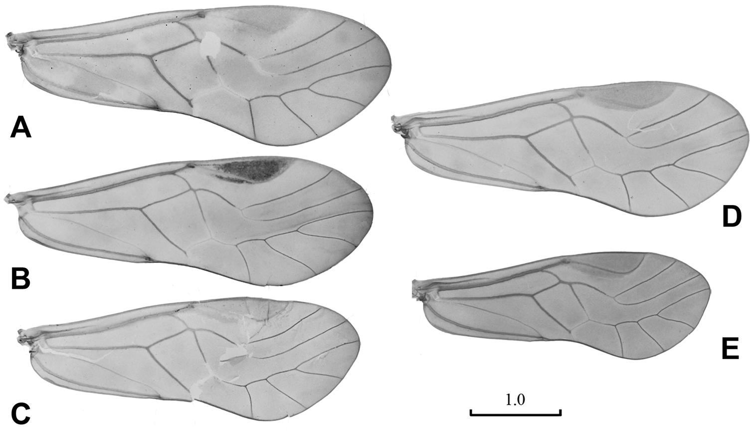
Male wings. A Neopsocopsis convexa sp. n. B Neopsocopsis hirticornis C Neopsocopsis quinquedentata D Neopsocopsis profunda E Neopsocopsis flavida. Scales in mm.
Abdomen. Male genitalia: 8th sternum strongly sclerotized and fused to hypandrium. Clunium (Fig. 2A) with posterior margin sharply convex medially. Epiproct (Fig. 2AB) swollen, unsclerotized medially, with tiny projection at middle of anterior margin. Paraproct (Fig. 2A) round and broadened distally. Hypandrium 5-lobed, lateral lobes carinate with outer surface covering denticles; posteromedian lobe forming dorsal-curved structure, with distal margin convex medially, basally with small membranous regions; internal lobes rod-like and distally forked. Phallosome (Fig. 2D) free posteriorly, distally broadened and paired with parameres.
Female genitalia: Subgenital plate (Fig. 2E) with egg guide round distally, invaginated basally and slightly sclerotized; pigment arms forming flat V-shaped regions and expanded laterally. Gonapophyses (Fig. 2F) with ventral valve distally tapering to slender tip; dorsal valve broad with pointed distal process; outer valve oval, with posterior lobe slender and well pointed. Internal plate (Fig. 2G) brown around spermathecal opening and marginally, with rod-like dark brown sclerotization anteriorly.
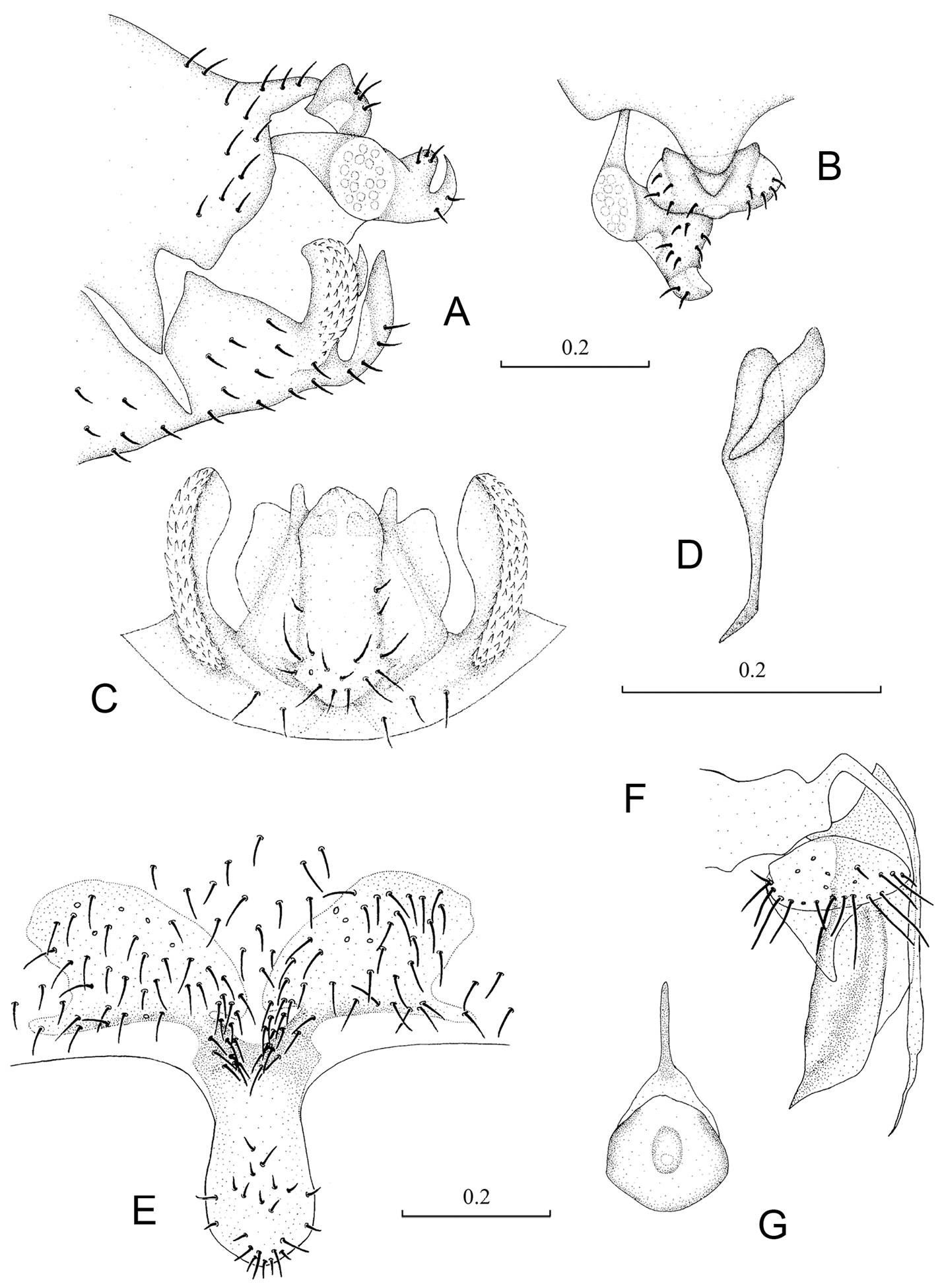
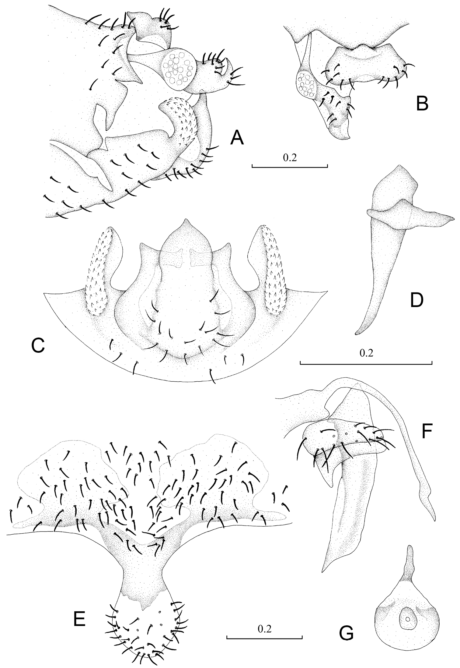
Terminalia of Neopsocopsis convexa sp. n.. A terminalia, lateral view B terminalia, dorsal view C hypandrium, posterior view D phallosome, lateral view E subgenital plate, ventral view F gonapophyses G internal plate, ventral view. Scales in mm. AB, CD, E–G to common scale.
Male: Body length 2.5–3.2 mm; fore wing length 3.9–4.3 mm; hind wing length 2.9–3.6 mm. Female: Body length 3.2–3.9 mm; fore wing length 3.7–4.4 mm; hind wing length 2.6–3.2 mm.
China (Gansu, Yunnan).
The new species appears to be closely related to Neopsocopsis hirticornis (Reuter, 1893), Neopsocopsis sakishimensis (Yoshizawa, 2010) and Neopsocopsis flavida (Li, 1989) based on similarity of the hypandrium with posteromedian lobe convex distaromedially. However, it can be easily separated from them by the larger body size, by the structure of male clunium, and by the shape and sclerotized pattern of the female subgenital plate. The new species is distinguished from all the other Neopsocopsis species by the characteristic shape of the internal lobes of hypandrium.
http://species-id.net/wiki/Neopsocopsis_hirticornis
Figures 1B , 3Pentablaste obconica – Holotype ♂: China, Shanxi Prov., Wenshui Co., Guandishan Reg., 2.viii.1981 (FS Li); Pentablaste clavata – Holotype ♂: China, Hebei Prov., Pingquan Co., Guangtoushan Reg., 2.vii.1986 (FS Li).Other material examined.China: 1♂, Nei Mongol Aut. Reg., Alax Left. B., Helanshan Natural Reserve, 6.viii.2010 (YL Tian); 1♂, same locality, 13.viii.2010 (SG Liang); 2♂, Shanxi Prov., Wenshui Co., Guandishan Reg., 3.viii.1981 (CK Yang); 1♀, same locality, 3.viii.1981 (FS Li); 1♀, Hebei Prov., Pingquan Co., Guangtoushan Reg., 2.vii.1986 (FS Li); 1♂, Beijing M., Xiangshan Reg., 12.v.1962 (FS Li). Japan: 1♀, Kanagawa Pref., Yokohama C., Serigatani, 7.iv.2011 (Y Hoshino); 2♀, same locality and collector, 12.iv.2011.
Male. Head creamy brown, with dark brown markings; compound eyes grayish black, ocelli black with grayish black ocellar field; antennae, labrum and maxillary palpi brown. Thorax brown with dark brown spots; legs brown, with band of dark brown marking on femur, tarsi and distal part of tibia dark brown. Fore wing (Fig. 1B) hyaline with brownish tinge, pterostigma dark brown with dark brown band along proximal margin; veins brown, except for Rs fork and M-Cu1a fusion hyaline. Venation: Rs and M fused for very short distance; distal margin of discoidal cell straight; first and second sections of Cu1a almost equal length, diverging at angle about 120°. Hind wing hyaline; veins brown.
Abdomen. Male genitalia: 8th sternum strongly sclerotized and fused to hypandrium. Clunium (Fig. 3A) with posterior margin convex medially and invaginated bilaterally. Epiproct (Fig. 3AB) swollen, unsclerotized medially, with sharp projection at middle of anterior margin. Paraproct (Fig. 3A) round and broadened distally. Hypandrium (Fig. 3C) 5-lobed, lateral lobes carinate with outer surface covering denticles; posteromedian lobe forming dorsal-curved structure, with distal margin smoothly round, basally with fan-shaped membranous regions; internal lobes well developed, with distal part crescent-like and directed dorsolaterally. Phallosome (Fig. 3D) free posteriorly, distally broadened and paired with parameres.
Female genitalia: Subgenital plate (Fig. 3E) with egg guide round distally, basally invaginated; pigment arms forming flat V-shaped regions and expanded laterally, posteriorly forked. Gonapophyses (Fig. 3F) with ventral valve distally tapering to slender tip; dorsal valve broad with pointed distal process; outer valve oval, with posterior lobe broad and well pointed. Internal plate (Fig. 3G) with brown coloration around spermathecal opening and marginally, with rod-like dark brown sclerotization anteriorly.
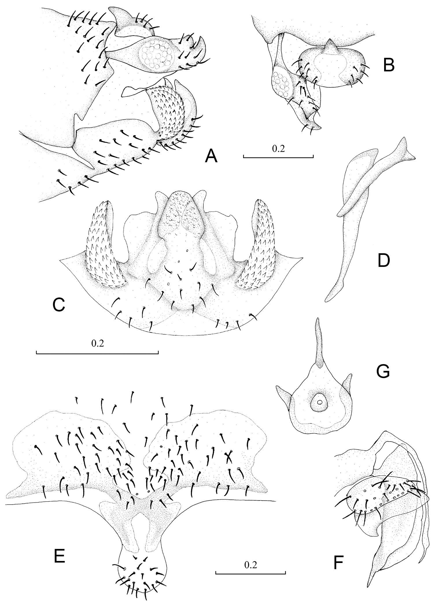
Terminalia of Neopsocopsis hirticornis. A terminalia, lateral view B terminalia, dorsal view C hypandrium, posterior view D phallosome, lateral view E subgenital plate, ventral view F gonapophyses G internal plate, ventral view. Scales in mm. AB, CD, E–G to common scale.
Male: Body length 2.4–2.9 mm; fore wing length 3.2–4.5 mm; hind wing length 2.5–3.3 mm. Female: Body length 3.0–3.5 mm; fore wing length 3.7–4.5 mm; hind wing length 3.0–3.6 mm.
China (Beijing, Gansu, Hebei, Hubei, Hunan, Jilin, Nei Mongol, Ningxia, Shanxi, Zhejiang: new distributional record); Finland; France; Germany; Hungary; Italy; Japan (new distributional record) ; Macedonia; Mongolia; Romania; Russia; Serbia; Spain; Sweden; Switzerland.
Pentablaste clavata was described by
http://species-id.net/wiki/Neopsocopsis_longiptera
Pentablaste tetraedrica – Holotype ♂: China, Hebei Prov., Pingquan Co., Guangtoushan Reg., 2.vii.1986 (FS Li). Other material examined.China: 2♂3♀, same locality and collector, 2.vii.1986; Japan: 1♂1♀, Fukuoka Pref., Hisayama C., Yamada, 5.vi.1994 (K Yoshizawa).
China (Hebei: new distributional record); Russia; Japan.
Pentablaste tetraedrica was described based on 3 males and 3 females from China. The species is distinguished from other Chinese species based on the character of the hypandrium posteromedian lobe lacking apically horn-like processes (Li, 2002). Neopsocopsis longiptera was described based on the specimens from the Russian Far East and differed from Neopsocopsis hirticornis (Reuter, 1893) in having a macropterous female and a larger male IO/D (Vishnyakova, 1986). After reexamining the two specimens, we found the main characters of the wings and genitalia are nearly identical. Thus we consider Pentablaste tetraedrica to be a new synonym of Neopsocopsis longiptera.
http://species-id.net/wiki/Neopsocopsis_quinquedentata
Figures 1C , 4Holotype ♂: China, Guizhou Prov., Jiangkou Co., Fanjingshan Natural Reserve, 27.vii.1983 (FS Li). Other material examined. China: 1♂, Guizhou Prov., Leishan Co., Leigongshan Natural Reserve, 14.iv.2005 (Y Tang).
Male. Head creamy brown; compound eyes grayish black, ocelli black with grayish black ocellar field; antennae and labrum brown, maxillary palpi brown with apical segment lighter. Thorax brown with dark brown spots; legs brown, with band of dark brown marking on femur, tarsi and distal part of tibia dark colored. Fore wing (Fig. 1C) hyaline with light brownish tinge, pterostigma brown; veins brown, except for Rs fork and M-Cu1a fusion hyaline. Venation: Rs and M fused for very short distance; distal margin of discoidal cell straight; first section of Cu1a shorter than the second section, diverging at angle about 120°. Hind wing hyaline; veins brown.
Abdomen. Male genitalia: 8th sternum strongly sclerotized and fused to hypandrium. Clunium (Fig. 4A) with posterior margin sharply convex medially and with tiny projection bilaterally. Epiproct (Fig. 4AB) swollen, unsclerotized medially, with moderate projection at middle of anterior margin. Paraproct (Fig. 4A) round and broadened distally. Hypandrium (Fig. 4C) 5-lobed, lateral lobes carinate with anterior part long and curved anteromedially, outer surface covering denticles; posteromedian lobe forming dorsal-curved structure, with distal margin almost straight and with tiny projection medially, basally with small membranous regions; internal lobes not well developed, much smaller than posteromedian lobe and distally bud-like. Phallosome (Fig. 4D) free posteriorly, distally broadened and paired with parameres.
Female unknown.
Terminalia of Neopsocopsis quinquedentata. A terminalia, lateral view B terminalia, dorsal view C hypandrium, posterior view D phallosome, lateral view Scales in mm. AB, CD to common scale.
Male: Body length 1.9–2.3 mm; fore wing length 4.0–4.1 mm; hind wing length 2.9–3.1 mm.
China (Guizhou).
Neopsocopsis quinquedentata was described based on one male from Guizhou, with the character of fore wing Rs-M fusion. It can be separated from other species by the hypandrial posteromedian lobe with projection medially, and by the characteristic structures of internal lobes and parameres.
http://species-id.net/wiki/Neopsocopsis_profunda
Figures 1D , 5Neoblaste profunda – Holotype ♂: China, Zhejiang Prov., Qingyuan Co., Baishanzu Natural Reserve, 3.x.1993 (H Wu). Neoblaste schizopetala – Holotype ♂: China, Chongqing M., Fengdu Co., Shiping Reg., 5.x.1994 (FS Li). Neoblaste flavae – Holotype ♀: China, Zhejiang Prov., Qingyuan Co., Baishanzu Natural Reserve, 27.x.1993 (H Wu). Other material examined. China: 1♀3♂, Zhejiang Prov., Qingyuan Co., Baishanzu Natural Reserve, 20.xi.1993 (H Wu); 1♀, same locality and collector, 3.x.1993; 2♀, same locality and collector, 27.x.1993; 1♂, Chongqing M., Fengdu Co., Guicheng Reg., 4.x.1994 (FS Li); 1♀, Chongqing M., Fengdu Co., Shiping Reg., 5.x.1994 (FS Li); 1♂, Hubei Prov., Xingshan Co., Longmenhe Reg., 12.ix.1994 (FS Li); 1♀, Henan Prov., Luanchuan Co., Longyuwan Reg., 7.viii.2008 (WH Li).
Male.Head yellowish, with brown markings; compound eyes grayish black, ocelli black with grayish black ocellar field; antennae and labrum brown, maxillary palpi brown with distal segments dark colored. Thorax brown with dark brown spots; legs pale brown. Fore wing (Fig. 1D) hyaline with yellowish tinge, pterostigma and veins brown, except for Rs fork and M-Cu1a fusion hyaline. Venation: Rs and M connected by short crossvein or meeting at point; distal margin of discoidal cell straight; first and second sections of Cu1a almost equal length, diverging at angle about 120°. Hind wing hyaline; veins brown.
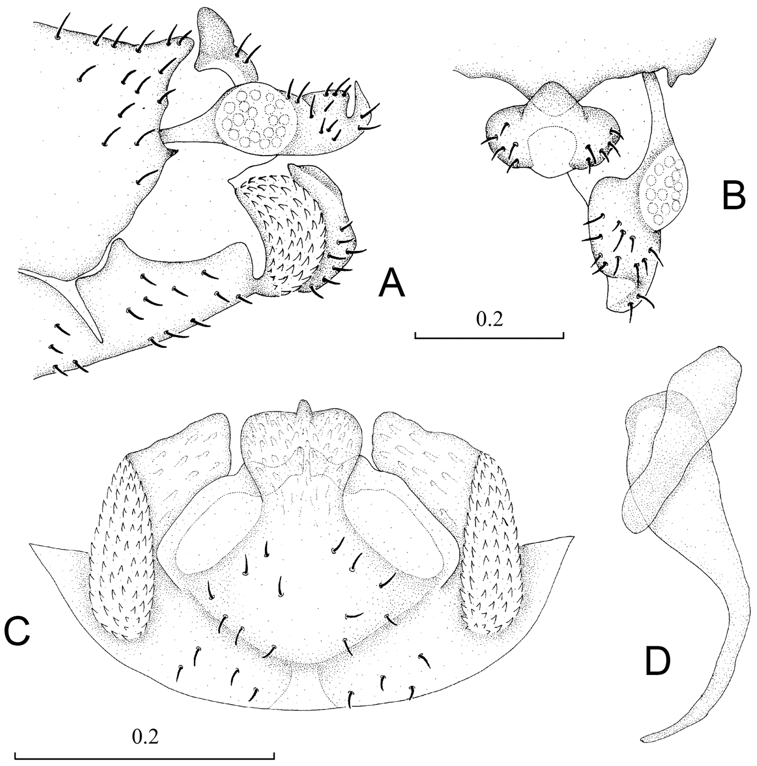
Abdomen. Male genitalia: 8th sternum strongly sclerotized and fused to hypandrium. Clunium (Fig. 5A) with posterior margin convex medially and with slight invagination bilaterally. Epiproct (Fig. 5AB) swollen, unsclerotized medially, with round projection at middle of anterior margin. Paraproct (Fig. 5A) round and broadened distally. Hypandrium (Fig. 5C) 5-lobed, lateral lobes carintae with anterior part long and curved anteromedially, outer surface covering denticles; posteromedian lobe forming dorsal-curved structure, with distal margin concave and with tiny projection medially, basally with small membranous regions; internal lobes rod-like and distally forked. Phallosome (Fig. 5D) free posteriorly, distally broadened and paired with parameres.
Female genitalia: Subgenital plate (Fig. 5E) with egg guide round with slightly narrowed margins distally, basally invaginated and sclerotized at basal 1/3; pigment arms forming flat V-shaped regions and expanded laterally. Gonapophyses (Fig. 5F) with ventral valve distally tapering to slender tip; dorsal valve broad with pointed distal process; outer valve oval, with posterior lobe broad and well pointed. Internal plate (Fig. 5G) with brown coloration around spermathecal opening and marginally, with rod-like dark brown sclerotization anteriorly.

Terminalia of Neopsocopsis profunda. A terminalia, lateral view B terminalia, dorsal view C hypandrium, posterior view D phallosome, lateral view E subgenital plate, ventral view F gonapophyses G internal plate, ventral view. Scales in mm. AB, CD, E–G to common scale.
Male: Body length 3.0–3.2 mm; fore wing length 3.7–4.1 mm; hind wing length 2.9–3.1 mm. Female: Body length 3.1–3.5 mm; fore wing length 3.8–4.6 mm; hind wing length 2.8–3.4 mm.
China (Chongqing, Henan, Hubei, Zhejiang).
Neoblaste profunda was described by
http://species-id.net/wiki/Neopsocopsis_flavida
Figures 1E , 6Blastopsocidus flavidus – Holotype ♂: China, Guizhou Prov., Guiyang C., Huaxi D., 9.vi.1981 (FS Li).Blastopsocidus pini – Holotype ♂: China, Guizhou Prov., Guiyang C., Bagongli Reg., 21.viii.1988 (FS Li).Pentablaste lanceolata – Holotype ♂: Guizhou Prov., Guiyang C., Bagongli Reg., 21.viii.1988 (FS Li).Other material examined.China: 13♀22♂, Guizhou Prov., Guiyang C., Huaxi D., 9.vi.1981 (FS Li); 1♂, same locality and collector, 27.v.1981; 1♂, same locality and collector, 28.v.1981; 7♀3♂, Guizhou Prov., Guiyang C., Bagongli Reg., 21.viii.1988 (FS Li); 1♂, Guizhou Prov., Leishan Co., Leigongshan Natural Reserve, 16.iv.2005 (Y Tang); 1♂, Fujian Prov., Nanping C., Wuyishan Reg., 4.vii.2009 (XS Yang); 4♀6♂, Hunan Prov., Hengyang C., Nanyue D., 20.vi.1963 (CK Yang); 1♂, Shanxi Prov., Wutai Co., Wutaishan Reg., 24.vii.1981 (FS Li); 1♂, Guangxi Prov., Longzhou Co., Nonggang Natural Reserve, 21.v.1982 (CK Yang); 1♀, Jiangxi Prov., Jiujiang C., Lushan Reg., 6.ix.1959 (CK Yang); 1♀1♂, Shaanxi Prov., Pingxiang C., Wugongshan Reg., 22.vii.1962 (CK Yang); 3♀3♂, Anhui Prov., Huangshan C., Huangshan Reg., 18.vii.1977 (FS Li).
Male. Head brownish, with dark brown markings; compound eyes grayish black, ocelli black with grayish black ocellar field; antennae and labrum brown, maxillary palpi brown with distal segments dark colored. Thorax brown with dark brown spots dorsally; legs pale brown, with tarsi dark brown. Fore wing (Fig. 1E) hyaline with yellowish tinge, pterostigma and veins brown, except for Rs fork and M-Cu1a fusion hyaline. Venation: Rs and M fused for short distance; distal margin of discoidal cell straight; first section of Cu1a little longer than the second section, diverging at angle about 120°. Hind wing hyaline; veins brown.
Abdomen. Male genitalia: 8th sternum strongly sclerotized and fused to hypandrium. Clunium (Fig. 6A) with posterior margin convex medially. Epiproct (Fig. 6AB) swollen, unsclerotized medially, with round projection at middle of anterior margin. Paraproct broad. Hypandrium (Fig. 6C) 5-lobed, lateral lobes carinate with outer surface covering denticles; posteromedian lobe forming dorsal-curved structure, with distal margin convex tapering to point; internal lobes tortuous forming right-angle and distally not forked. Phallosome (Fig. 6D) free posteriorly, distally broadened and paired with parameres.
Female genitalia: Subgenital plate (Fig. 6E) with egg guide round distally, basally invaginated and sclerotized at basal 1/3; pigment arms forming flat V-shaped regions and expanded laterally. Gonapophyses (Fig. 6F) with ventral valve distally tapering to slender tip; dorsal valve long and narrow with pointed distal process; outer valve oval, with posterior lobe narrow and well pointed. Internal plate (Fig. 6G) with brown coloration around spermathecal opening and marginally, with rod-like dark brown sclerotization anteriorly.
Terminalia of Neopsocopsis flavida. A terminalia, lateral view B terminalia, dorsal view C hypandrium, posterior view D phallosome, lateral view E subgenital plate, ventral view F gonapophyses G internal plate, ventral view. Scales in mm. AB, CD, E–G to common scale.
Male: Body length 2.1–2.9 mm; fore wing length 2.6–3.6 mm; hind wing length 2.0–2.7 mm. Female: Body length 2.6–3.3 mm; fore wing length 3.1–3.8 mm; hind wing length 2.4–2.9 mm.
China (Anhui, Fujian, Guangxi, Guizhou, Hunan, Jiangxi, Shaanxi, Shanxi).
These three species were very similar according to
We would like to thank Xiu-Shuai Yang, Yan-Lin Tian, Si-Pei Liu, Jian-Xin Cui , Wei-Hai Li, Yi Tang and Shao-Guang Liang for their help collecting field materials, and not least thanks are due to Professor Xin-Li Wang for giving experimental equipment assistance. We are also much indebted to Professor Ding Yang and Associate Professor Xing-Yue Liu for their kind help. KY thanks Yasuo Hoshino for supplying the specimens of Neopsocopsis hirticornis which were collected from Japan for the first time. This research was supported by the National Natural Science Foundation of China (No. 31071962) and Foundation of the Ministry of Agriculture of China (No. 201103022)