


(C) 2012 Hao-Sen Li. This is an open access article distributed under the terms of the Creative Commons Attribution License 3.0 (CC-BY), which permits unrestricted use, distribution, and reproduction in any medium, provided the original author and source are credited.
For reference, use of the paginated PDF or printed version of this article is recommended.
Eriophyoid mites from Qinghai Province, northwestern China were studied herein. Up to now, only six species have been reported from Qinghai Province. In field surveys, 17 eriophyoid mite species were collected, among which nine species were found new to science. The new species and their host plants are listed as follows: Acaphyllisa tuberculumae sp. n. on Populus sp. (Salicaceae); Proiectus xiningensis sp. n. on Pinus sp. (Pinaceae); Phyllocoptes beishaniensis sp. n. on Spiraea mongolica Maxim. (Rosaceae); Tetra pruniana sp. n. on Prunus tomentosa Thunb. (Rosaceae) Rupr. (Berberidaceae); Tetra pyriana sp. n. on Pyrus calleryana Decne. (Rosaceae); Tetra simonia sp. n. on Populus simonii Carr. (Salicaceae); Diptacus berberinus sp. n. on Berberis amurensis Rupr. (Berberidaceae); Diptacus mengdaensis sp. n. on Lonicera elisae Franch. (Caprifoliaceae); Rhyncaphytoptus spinus sp. n. on Lonicera rupicola Hook. f. et Thoms. (Caprifoliaceae). Aculops ulmi Hong & Xue, 2005 was re-described.
Eriophyoid mites, Qinghai, taxonomy, new species
Qinghai Province (89°35'E–103°04'E, 13°39'N–39°19'N), located in the northwest of the People’s Republic of China, is a part of the Qinghai-Tibet Plateau, with an average elevation of over 3000m (1650m–6860m). The average temperature ranges from 0.4°C to 7.4°C. (
Up to now, six species of eriophyoid mites from Qinghai have been reported. They are Aceria paramacrodonis Kuang, 1988 on Lycium sp. (Solanaceae), Aceria qinghaiensis Kuang, 1997 on Salix babylonica L. (Salicaceae), Aculodes salicis Kuang, 1997 on Salix babylonica L. (Salicaceae), Aculops xiningensis Kuang, 2000 on Malus pumila P. Mill. (Rosaceae), Aculus huangzhongensis Kuang, 2000 on Syringa oblata Lindl. (Oleaceae) and Tetraspinus syringae Lin & Kuang, 2001 on Syringa oblata Lindl. (Oleaceae). In July 2007, field surveys were conducted in Qinghai Province, northwestern China. Twenty-five eriophyoid mite samples were collected and 17 eriophyoid mite species were identified, among which nine species were found new to science. No species reported earlier from Qinghai were collected in this survey. In total, there are 23 species of the Eriophyoidea from Qinghai Province, belonging to two families and 12 genera. A list of eriophyoid mites from Qinghai Province is given (Table 1).
List of eriophyoid mites and their hosts in Qinghai Province.
| Family | Subfamily | Tribe | Species | Host |
|---|---|---|---|---|
| Eriophyidae | Eriophyinae | Aceriini | Aceria paramacrodonis Kuang, 1988 | Lycium sp. (Solanaceae) |
| Aceria qinghaiensis Kuang, 1997 | Salix babylonica L. (Salicaceae) | |||
| Phyllocoptinae | Acaricalini | Acaphyllisa tuberculumae sp. n. | Populus sp. (Salicaceae) | |
| Phyllocoptini | Proiectus xiningensis sp. n. | Pinus sp. (Pinaceae) | ||
| Phyllocoptes beishaniensis sp. n. | Spiraea mongolica Maxim. (Rosaceae) | |||
| Phyllocoptes asperatae Song, Xue & Hong, 2006 | Picea meyeri Rehd. Et Wils. (Pinaceae) | |||
| Phyllocoptes dangchangi Song, Xue & Hong, 2006 | Picea sp. (Pinaceae) | |||
| Phyllocoptes gansunensis Kuang & Luo, 1998 | Potentilla parvifolia Fisch. ap. Lehm. (Rosaceae) | |||
| Phyllocoptruta platycladusa Xue, Song, Amrine & Hong, 2007 | Juniperus chinensis L. (Cupressaceae) | |||
| Anthocoptini | Aculus changbais Xue, Song & Hong, 2008 | Salix chaenomeloides Kimura ( Salicaceae) | ||
| Aculus huangzhongensis Kuang, 2000 | Syringa oblata Lindl. (Oleaceae) | |||
| Aculodes salicis Kuang, 1997 | Salix babylonica L. (Rosaceae) | |||
| Aculops umli Hong & Xue, 2005 | Ulmus sp. (Ulmaceae) | |||
| Aculops xiningensis Kuang, 2000 | Malus pumila P. Mill. (Rosaceae) | |||
| Tetraspinus syringae Lin & Kuang, 2001 | Syringa oblata Lindl. (Oleaceae) | |||
| Tetra pinnatifidae Xue, Song & Hong, 2006 | Prunus armeniaca Linn. (Rosaceae) | |||
| Tetra pruniana sp. n. | Prunus tomentosa Thunb. (Rosaceae) | |||
| Tetra pyriana sp. n. | Pyrus calleryana Decne. (Rosaceae) | |||
| Pyrus betulifolia Bunge. (Rosaceae) | ||||
| Tetra simonia sp. n. | Populus simonii Carr. (Salicaceae) | |||
| Diptilomiopidae | Diptilomiopinae | Diptacus berberinus sp. n. | Berberis amurensis Rupr. (Berberidaceae) | |
| Diptacus mengdaensis sp. n. | Lonicera elisae Franch. (Caprifoliaceae) | |||
| Rhyncaphytoptinae | Rhyncaphytoptus spinus sp. n. | Lonicera rupicola Hook. f. et Thoms. (Caprifoliaceae) | ||
| Rhyncaphytoptus ulmi Xin & Dong, 1981 | Ulmus sp. (Ulmaceae) |
In the field, eriophyoid mites were collected by the aid of a hand-lens (30X) from the lower surface of host plant leaves. Eriophyoid mites, together with host plants, were immersed in 75% alcohol and kept in vials. Each vial was marked with the collection data, such as specimen number, collection date, host plant, mite color, location, collector, and mite relationship to host plant. The collection data were also recorded in the collection notebook for further use. The host plants were kept in a plant specimen folder in a dry environment.
The morphological terminology used here follows that of
Subfamily Eriophyinae Nalepa, 1898
Tribe Aceriini Amrine & Stasny, 1994
Genus Aceria Keifer, 1944
http://species-id.net/wiki/Aceria_paramacrodonis
Lycium sp. (Solanaceae).
Leaf gall; mites produce pocket galls on the lower side of leaves.
China (Gansu, Ningxia, Qinghai, Shandong).
http://species-id.net/wiki/Aceria_qinghaiensis
Salix babylonica L. (Salicaceae).
The mites produce pockets on the lower surface of the leaves.
China (Gansu, Qinghai).
Tribe Acaricalini Amrine & Stasny, 1994
Genus Acaphyllisa Keifer, 1978
urn:lsid:zoobank.org:act:90E42ADF-5C3A-449D-A4C1-BA012B3D09F1
http://species-id.net/wiki/Acaphyllisa_tuberculumae
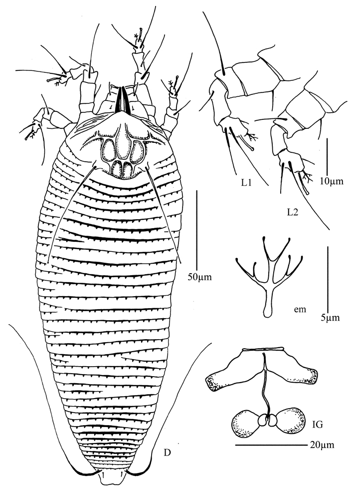
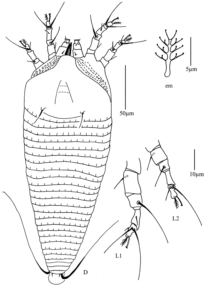
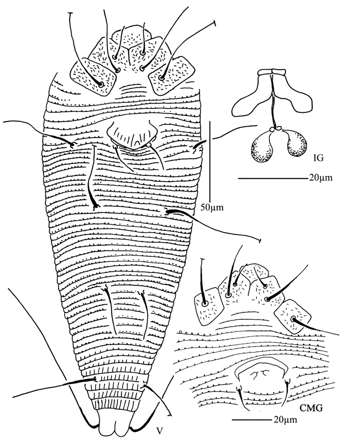
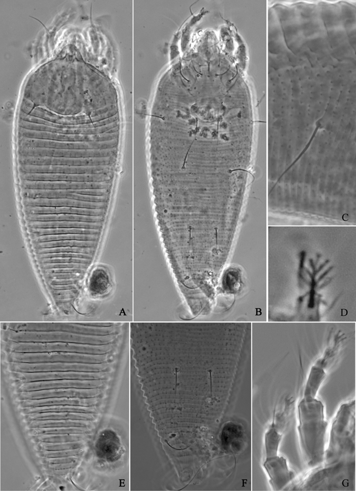
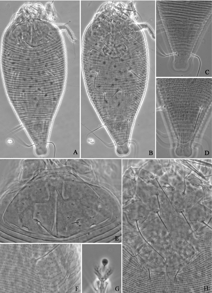
Figures 1–3Female. (n = 8) Body fusiform, light yellow, 171 (171–195), 72 (70–75) wide. Gnathosoma 21 (20–21), projecting obliquely down, suboral plate present, pedipalp coxal seta (ep) 4 (4–5), dorsal pedipalp genual seta (d) 7 (7–8), cheliceral stylets 16 (16–18). Prodorsal shield 49 (45–49), 53 (53–60) wide, subtriangular; frontal lobe 6 (5–8); median, admedian and submedian lines present, median line ending at basal 1/2 of prodorsal shield, admedian lines connected at basal 1/2 and 2/3 of prodorsal shield, forming three cells on each side of the median line. Scapular tubercles ahead of rear shield margin, 2 (2–3), 18 (18–19) apart, scapular setae (sc) 8 (6–8), projecting centrad. Coxigenital region with 10 smooth annuli. Coxisternal plates with granules, anterolateral setae on coxisternum I (1b) 7 (7–8), 14 (14–15) apart, proximal setae on coxisternum I (1a) 19 (19–21), 11 (10–11) apart, proximal setae on coxisternum II (2a) 51 (51–53), 26 (26–28) apart, tubercles 1b and 1a 8 (8–9) apart, tubercles 1a and 2a 8 (8–9) apart. Prosternal apodeme combined, 7 (6–7). Leg I 36 (35–36), femur 10 (9–10), basiventral femoral seta (bv) 8 (8–10); genu 5 (5–6), antaxial genual seta (l") 29 (29–31); tibia 8 (7–8), paraxial tibial seta (l’) 7 (7–8), located at 1/4 from dorsal base; tarsus 8 (7–8), seta ft’ 17 (17–19), seta ft" 22 (22–23), seta u’ 5 (5–6); tarsal empodium (em) 6 (6–7), divided, 2-rayed on each side, tarsal solenidion (ω) 7 (6–7), knobbed. Leg II 28 (28–33), femur 10 (9–10), basiventral femoral seta (bv) 10 (9–10); genu 5 (4–5), antaxial genual seta (l") 6 (6–8); tibia 6 (6–7); tarsus 7 (6–7), seta ft’ 5 (5–7), seta ft" 21 (21–22), seta u’ 5 (5–6); tarsal empodium (em) 6 (5–6), divided, 2-rayed on each side, tarsal solenidion (ω) 6 (6–7), knobbed. Opisthosoma dorsally with 55 (55–57) annuli, with round microtubercles, ventrally with 77 (74–77) annuli, with round microtubercles. Setae c2 31 (28–31) on ventral annulus 14 (14–16), 56 (54–56) apart; setae d 55 (55–60) on ventral annulus 34 (33–34), 38 (33–38) apart; setae e 17 (16–17) on ventral annulus 52 (52–53), 20 (18–20) apart; setae f 28 (25–28) on ventral annulus 71 (69–71), 26 (25–26) apart. Setae h1 2 (2–3), h2 70 (70–75). Female genitalia 18 (17–18), 24 (24–25) wide, coverflap smooth, setae 3a 33 (33–35), 14 (14–15) apart.
Male. (n = 1) Body fusiform, light yellow, 150, 57 wide. Gnathosoma 17, projecting obliquely down, suboral plate present, pedipalp coxal seta (ep) 5, dorsal pedipalp genual seta (d) 5, cheliceral stylets 13. Prodorsal shield has the same design as female, 44, 48 wide, subtriangular; frontal lobe 5. Scapular tubercles ahead of rear shield margin, 3, 18 apart, scapular setae (sc) 6, projecting centrad. Coxigenital region with 10 smooth annuli. Coxisternal plates with granules, anterolateral setae on coxisternum I (1b) 7, 12 apart, proximal setae on coxisternum I (1a) 18, 10 apart, proximal setae on coxisternum II (2a) 45, 23 apart, tubercles 1b and 1a 7 apart, tubercles 1a and 2a 7 apart. Prosternal apodeme combined, 5. Legs with usual series of setae. Leg I 29, femur 9, basiventral femoral seta (bv) 8; genu 5, antaxial genual seta (l") 31; tibia 7, paraxial tibial seta (l’) 6, located at 1/4 from dorsal base; tarsus 6, seta ft’ 17), seta ft" 18, seta u’ 4; tarsal empodium (em) 6, divided, 2-rayed on each side, tarsal solenidion (ω) 6, knobbed. Leg II 28, femur 10, basiventral femoral seta (bv) 8; genu 4, antaxial genual seta (l") 8; tibia 6; tarsus 6, seta ft’ 5, seta ft" 18, seta u’ 4; tarsal empodium (em) 5, divided, 2-rayed on each side, tarsal solenidion (ω) 6, knobbed. Opisthosoma dorsally with 51 annuli, with round microtubercles, ventrally with 66 annuli, with round microtubercles. Setae c2 21 on ventral annulus 12, 42 apart; setae d 42 on ventral annulus 27, 28 apart; setae e 16 on ventral annulus 43, 16 apart; setae f 25 on ventral annulus 61, 21 apart. Setae h1 2, h2 60. Male genitalia forming a “Y” like structure in the middle, 19 wide, setae 3a 22, 15 apart.
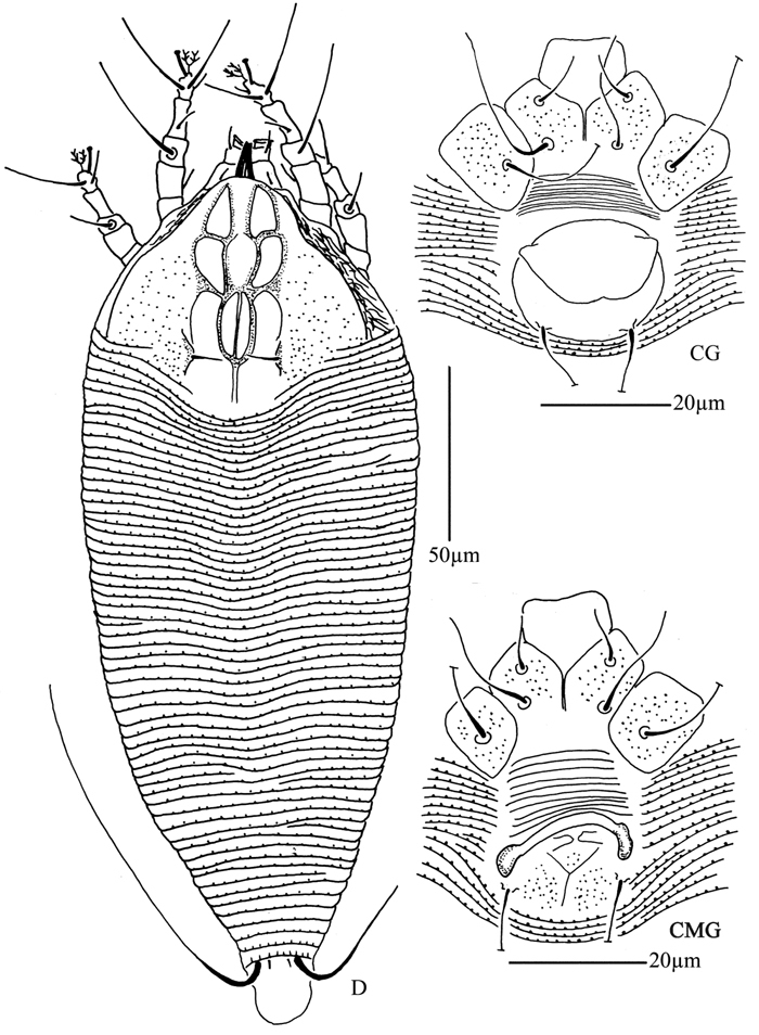
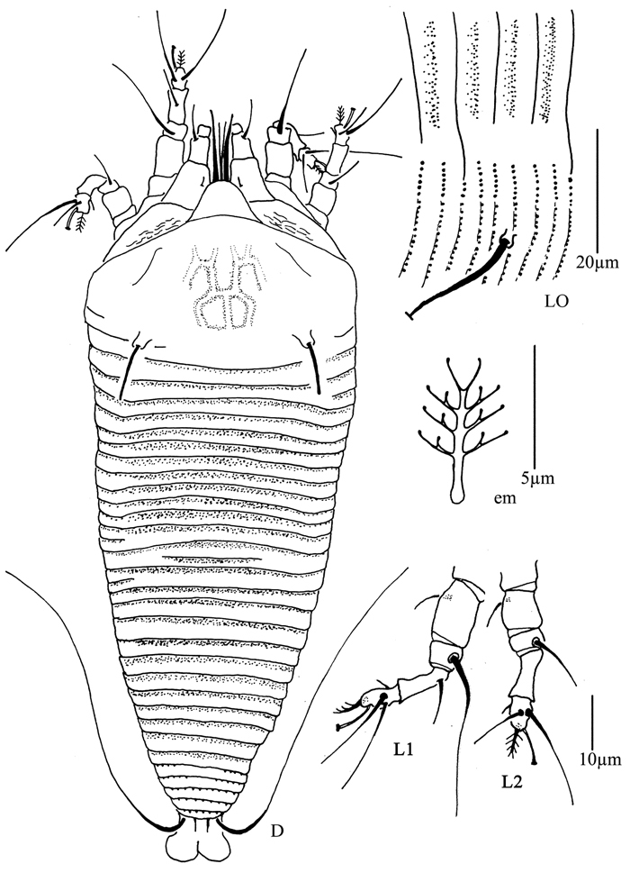
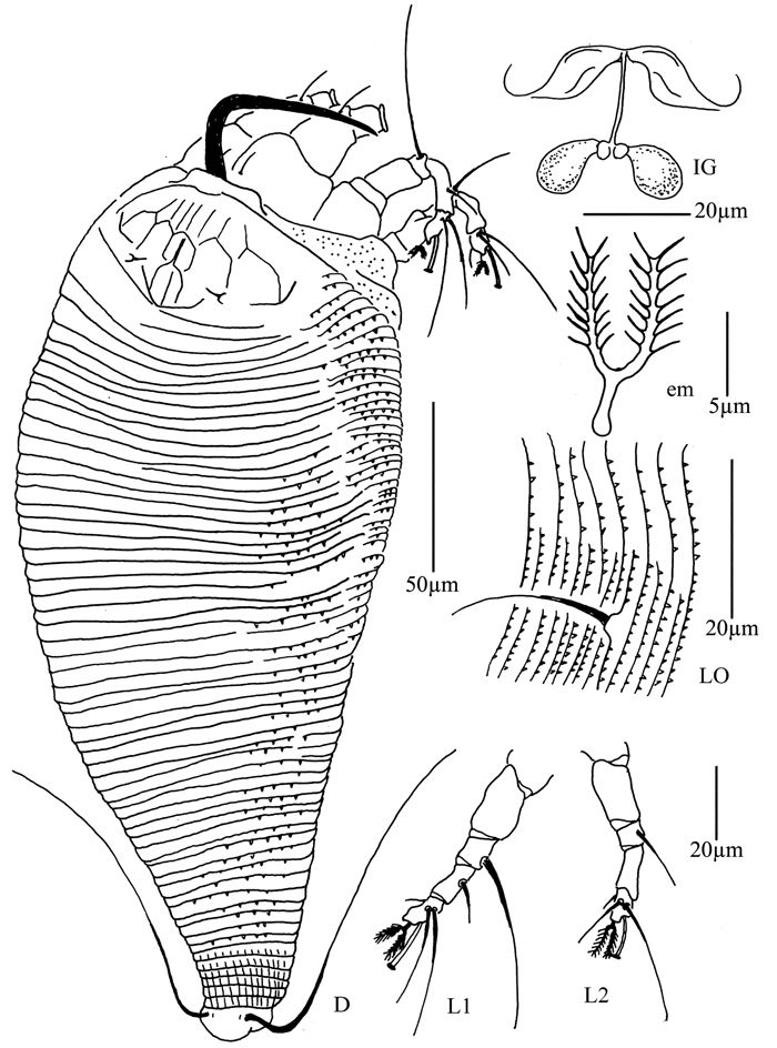
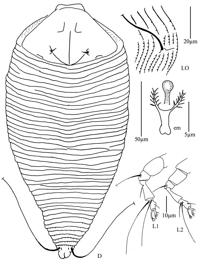
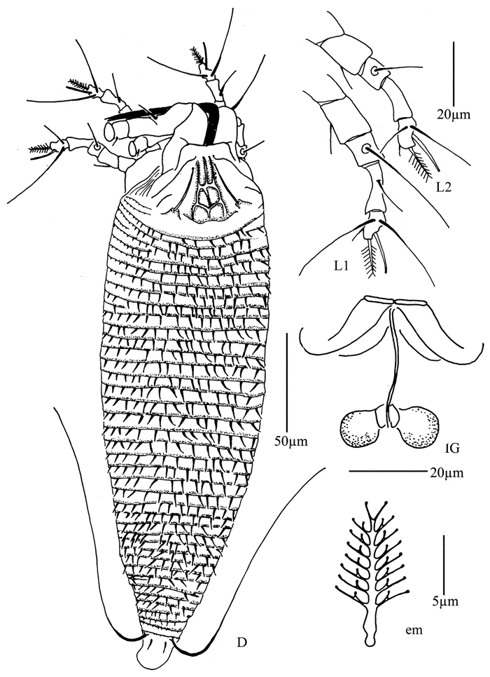
Acaphyllisa tuberculumae sp. n.: D dorsal view of female CG coxae and female genitalia CMG coxae and male genitalia.
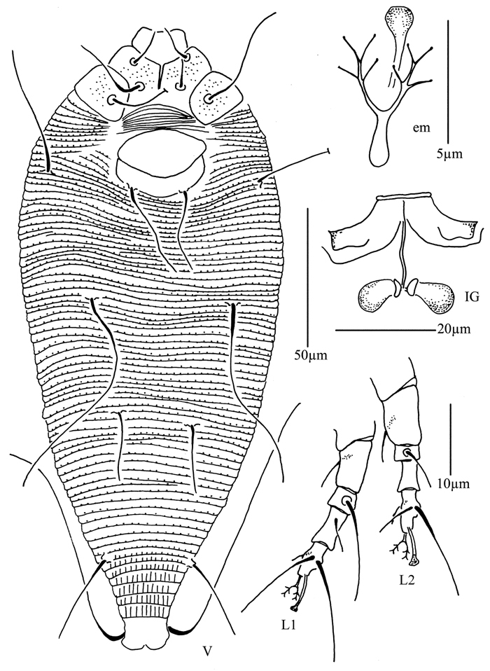
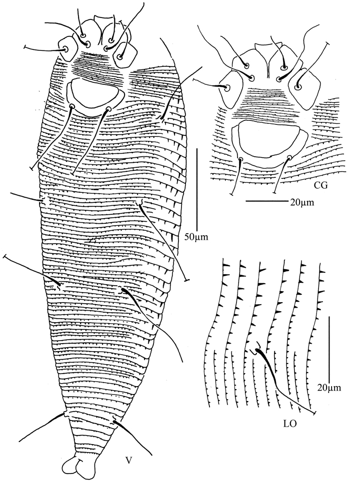
Acaphyllisa tuberculumae sp. n.: V ventral view of female em empodium IG female internal genitalia L1 leg I L2 leg II.
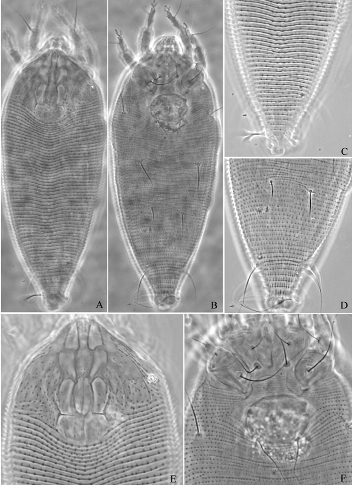
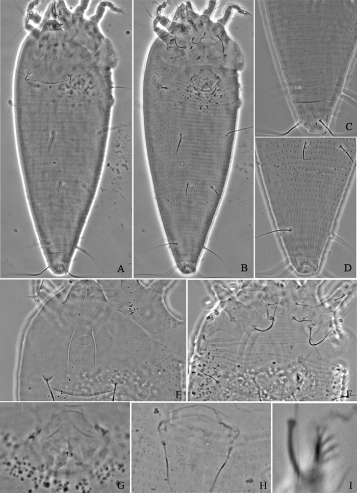
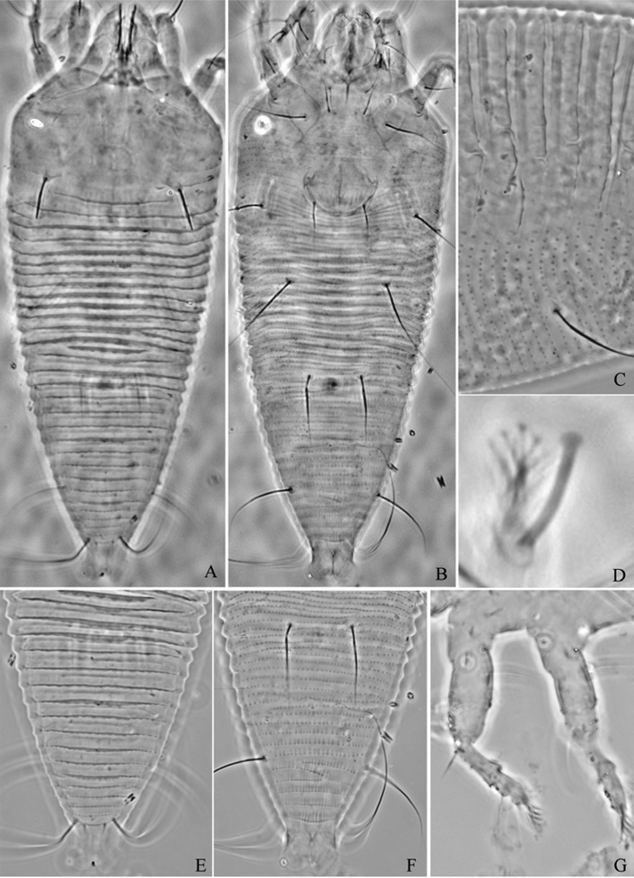
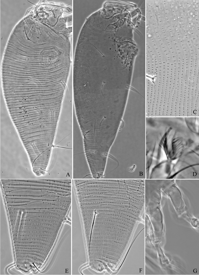
Acaphyllisa tuberculumae sp. n.: A dorsal view of female B ventral view of female C dorsal view of female posterior part D ventral view of female posterior part E prodorsal shield F coxae and female genitalia.
Holotype, female (slide number NJAUEri789B, marked Holotype), from Populus sp. (Salicaceae), Xining City, Qinghai Province, P. R. China, 36°38'18"N, 101°45'27"E, elevation 2241m, 21 July 2007, coll. Xiao-Feng Xue. Paratypes, 7 females and 1 male (slide number NJAUEri789B), with the same data as holotype.
Vagrant on leaf lower surface. No damage to the host was observed.
The specific designation tuberculumae is from the character of dorsal opisthosomal microtubercles, “tuberculum” in Latin; masculine in gender.
This species is similar to Acaphyllisa populi Xue & Hong, 2006, but can be differentiated from the latter by prodorsal shield with six cells in the middle (prodorsal shield without cells in Acaphyllisa populi), opisthosoma dorsally with round microtubercles (opisthosoma dorsally with elliptical microtubercles only on ridges in Acaphyllisa populi), female genitalia coverflap smooth (female genital coverflap with 10 longitudinal ridges in Acaphyllisa populi).
Genus Proiectus Huang, 2001
urn:lsid:zoobank.org:act:21DA68FE-18B9-4305-B088-20BF3A865A3C
http://species-id.net/wiki/Proiectus_xiningensis
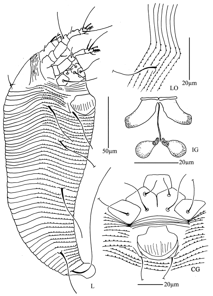
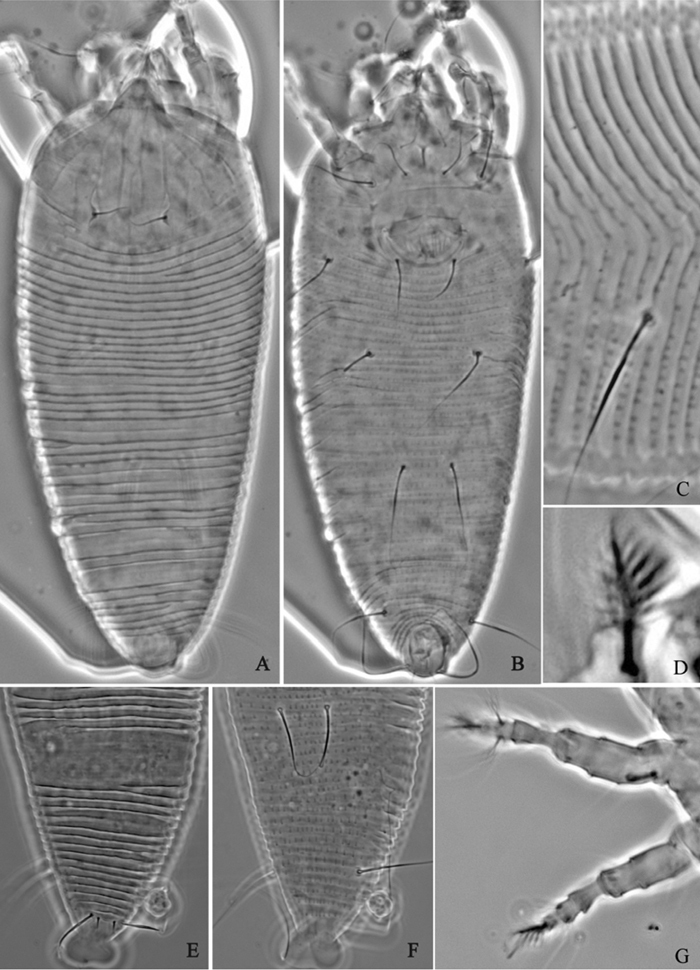
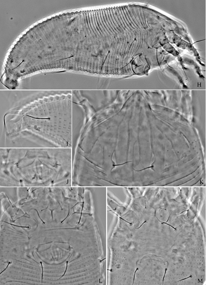
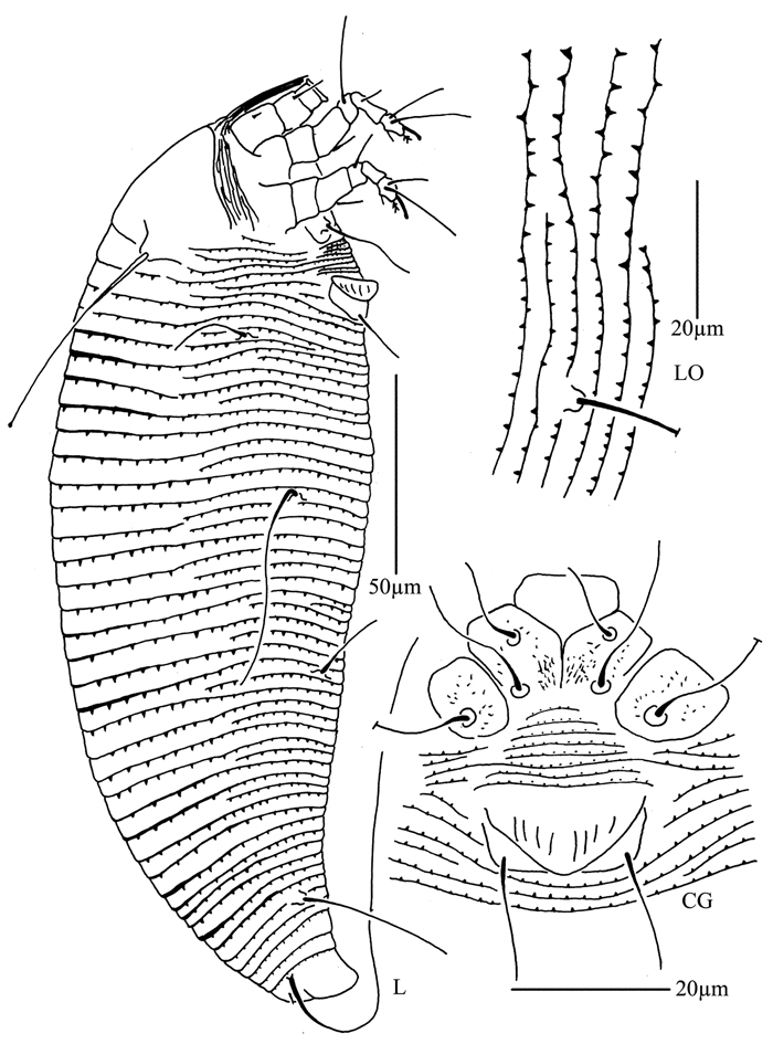
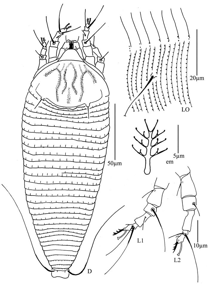
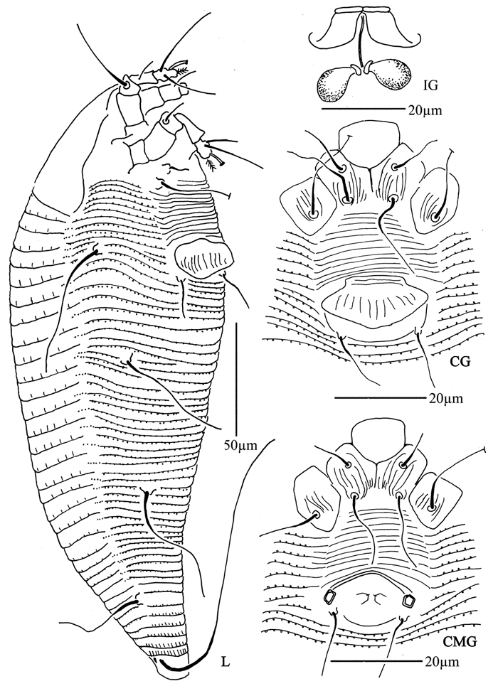
Figures 4–7Female. (n = 5) Body fusiform, light yellow, 248 (223–308), 100 (100–110) wide, 90 (90–91) thick. Gnathosoma 33 (33–34), projecting obliquely down, suboral plate present, pedipalp coxal seta (ep) 3 (3–5), dorsal pedipalp genual seta (d) 15 (11–15), cheliceral stylets 33 (33–34). Prodorsal shield 73 (65–73), 100 (100–110) wide, subtriangular, with a large projection on each lateral margin 7 (6–7); frontal lobe broad 24 (22–24); median, admedian and submedian lines obscure, median and admedian lines connected at base. Scapular tubercles ahead of rear shield margin, 2 (2–3), 30 (27–30) apart, scapular setae (sc) 8 (6–8), projecting centrad. Coxigenital region with 17 (14–17) annuli, with round microtubercles, with deep seam under coxisternal plate II. Coxisternal plates with short lines, anterolateral setae on coxisternum I (1b) 10 (10–11), 18 (18–19) apart, proximal setae on coxisternum I (1a) 22 (20–22), 13 (12–13) apart, proximal setae on coxisternum II (2a) 52 (50–52), 32 (32–33) apart, tubercles 1b and 1a 11 (10–11) apart, tubercles 1a and 2a 10 (10–11) apart. Prosternal apodeme separated, 4 (4–5). Leg I 43 (40–45), femur 9 (8–10), basiventral femoral seta (bv) 14 (12–14); genu 9 (8–10), antaxial genual seta (l") 28 (28–34); tibia 15 (13–15), paraxial tibial seta (l’) 5 (5–6), located at 1/2 from dorsal base; tarsus 10 (9–10), seta ft’ 17 (17–18), seta ft" 30 (30–32), seta u’ 5 (5–6); tarsal empodium (em) 8 (7–8), simple, 5-rayed, tarsal solenidion (ω) 10 (9–10), knobbed. Leg II 40 (40–41), femur 13 (13–16), basiventral femoral seta (bv) 13 (10–13); genu 6 (6–8), antaxial genual seta (l") 8 (7–8); tibia 10 (10–12); tarsus 8 (8–9), seta ft’ 9 (8–9), seta ft" 27 (27–28), seta u’ 5 (5–6); tarsal empodium (em) 8 (7–8), simple, 5-rayed, tarsal solenidion (ω) 10 (9–10), knobbed. Opisthosoma dorsally with 41 (39–41) annuli, with weak filamentous microtubercles, ventrally with 92 (92–100) annuli, with round microtubercles. Setae c2 21 (21–22) on ventral annulus 15 (15–20), 70 (70–73) apart; setae d 100 (80–100) on ventral annulus 34 (34–39), 37 (37–46) apart; setae e 55 (55–65) on ventral annulus 56 (56–61), 21 (21–22) apart; setae f 33 (30–33) on ventral annulus 84 (84–90), 28 (28–29) apart. Setae h1 7 (6–7), h2 65 (55–65). Female genitalia 28 (28–30), 33 (33–34) wide, coverflap with 19 longitudinal ridges, setae 3a 10 (10–12), 23 (21–22) apart.
Male. (n = 1) Body fusiform, light yellow, 250, 90 wide. Gnathosoma 34, projecting obliquely down, suboral plate present, pedipalp coxal seta (ep) 3, dorsal pedipalp genual seta (d) 11, cheliceral stylets 50. Prodorsal shield has the same design as female, 70, 90 wide, subtriangular; with a large projection on each lateral margin, 6; frontal lobe broad, 21. Scapular tubercles ahead of rear shield margin, 2, 23 apart, scapular setae (sc) 6, projecting centrad. Coxigenital region with 16 annuli, with round microtubercles, with deep seam under coxisternal plate II. Coxisternal plates with short lines, anterolateral setae on coxisternum I (1b) 10, 15 apart, proximal setae on coxisternum I (1a) 15, 12 apart, proximal setae on coxisternum II (2a) 52, 29 apart, tubercles 1b and 1a 10 apart, tubercles 1a and 2a 11 apart. Prosternal apodeme separated, 5. Leg I 40, femur 10, basiventral femoral seta (bv) 13; genu 8, antaxial genual seta (l") 31; tibia 13, paraxial tibial seta (l’) 5, located at 1/2 from dorsal base; tarsus 8, seta ft’ 17, seta ft" 30, seta u’ 5; tarsal empodium (em) 7, simple, 5-rayed, tarsal solenidion (ω) 9, knobbed. Leg II 37, femur 15, basiventral femoral seta (bv) 12; genu 6, antaxial genual seta (l") 7; tibia 10; tarsus 8), seta ft’ 7, seta ft" 25, seta u’ 5; tarsal empodium (em) 7, simple, 5-rayed, tarsal solenidion (ω) 9, knobbed. Opisthosoma dorsally with 41 annuli, with weak filamentous microtubercles, ventrally with 94 annuli, with round microtubercles. Setae c2 21 on ventral annulus 18, 60 apart; setae d 80 on ventral annulus 36, 38 apart; setae e 60 on ventral annulus 58, 20 apart; setae f 27 on ventral annulus 88, 25 apart. Setae h1 6, h2 65. Male genitalia 25 wide, setae 3a 12, 22 apart.
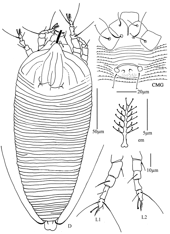
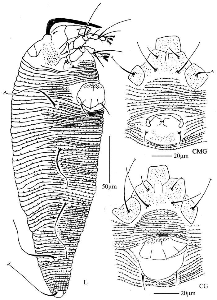
Proiectus xiningensis sp. n.: D dorsal view of female CMG coxae and male genitalia CG coxae and female genitalia.
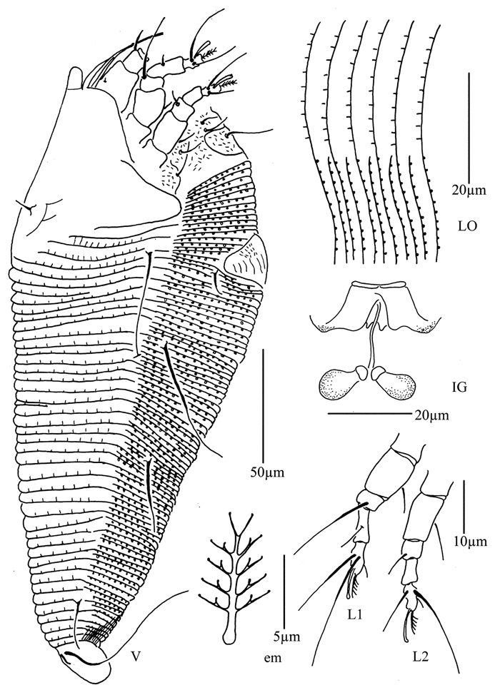
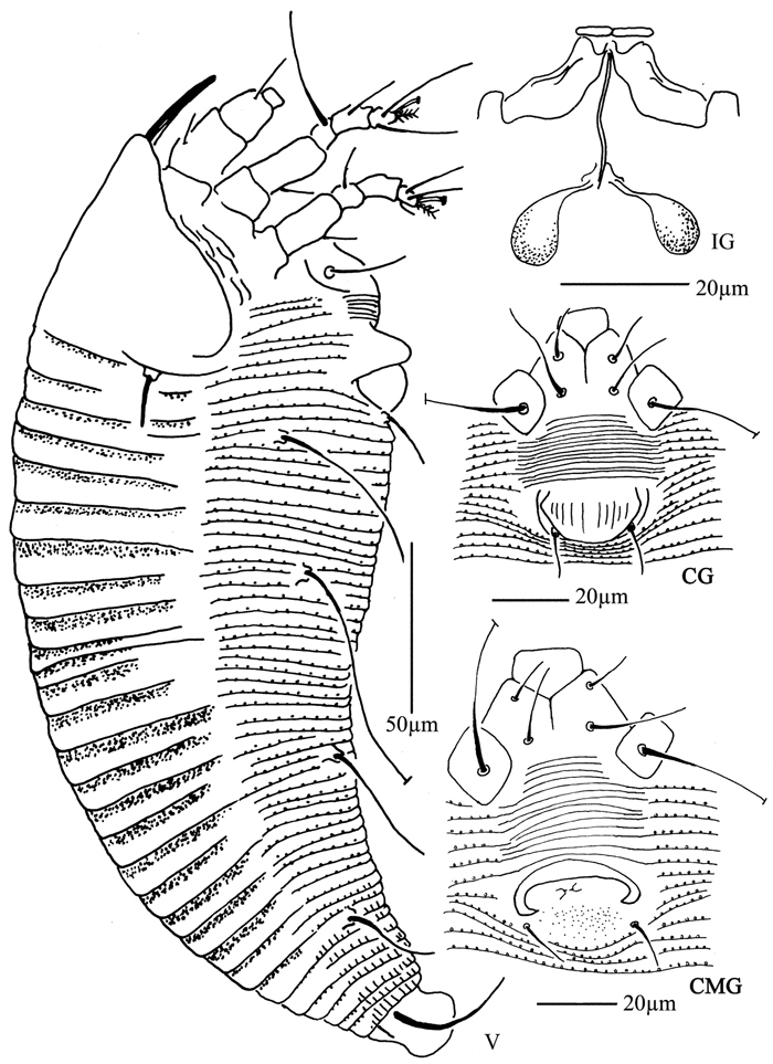
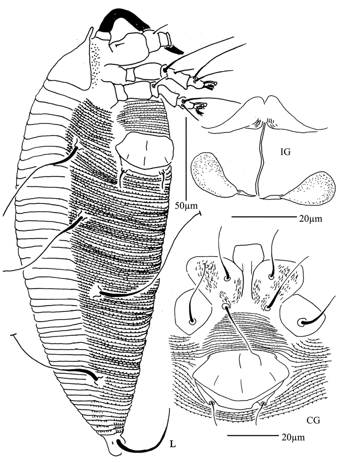
Proiectus xiningensis sp. n.: L lateral view of female LO lateral microtubercles IG female internal genitalia em empodium L1 leg I L2 leg II.
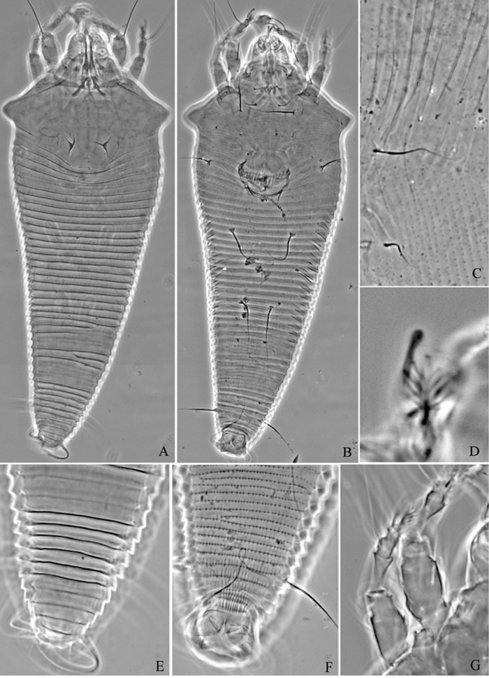
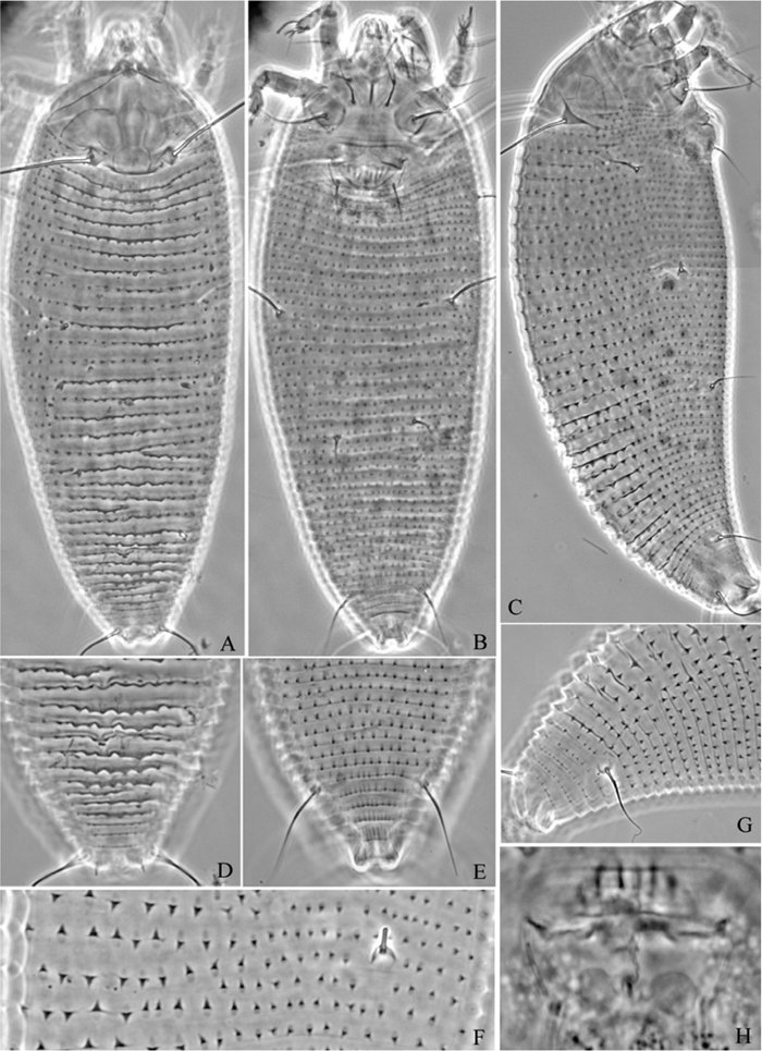
Proiectus xiningensis sp. n.: A dorsal view of female B ventral view of female C lateral microtubercles D empodium E dorsal view of female posterior part F ventral view of female posterior part G leg I and leg II.
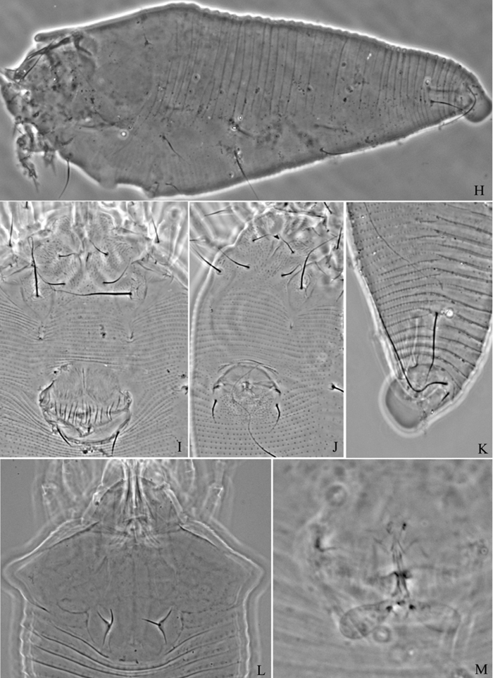
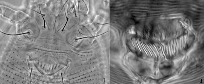
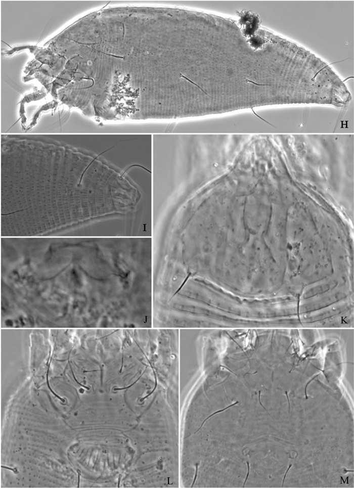
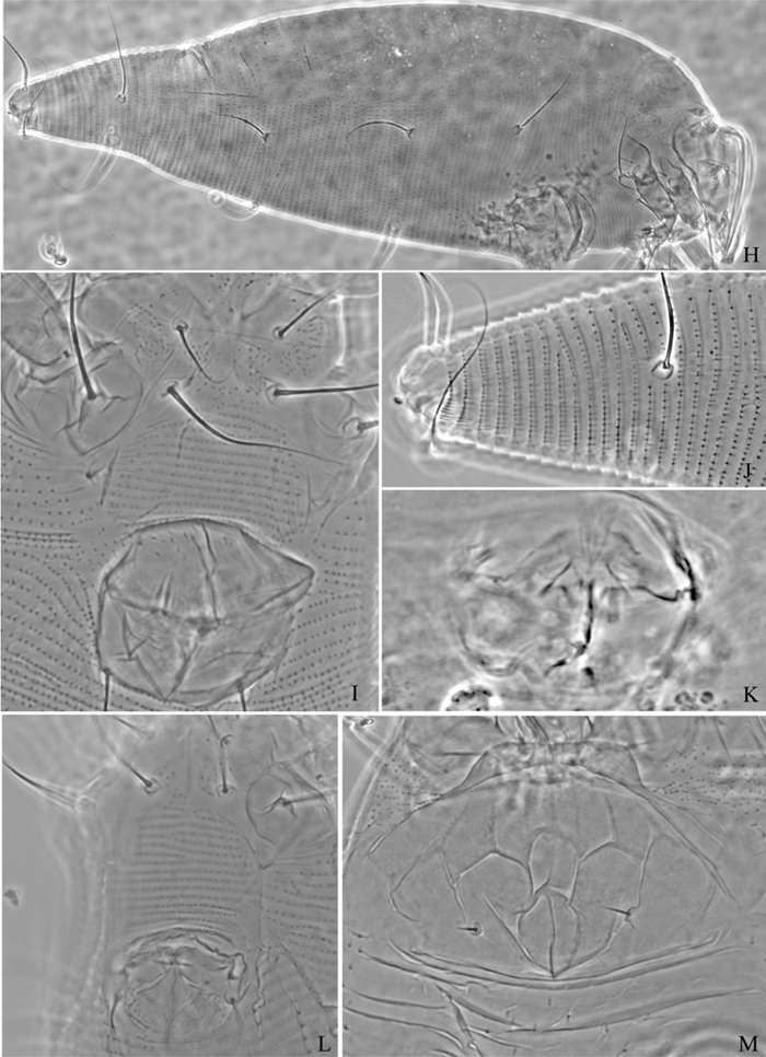
Proiectus xiningensis sp. n.: H lateral view of female I coxae and female genitalia J coxae and male genitalia K lateral view of female posterior part L prodorsal shield M female internal genitalia.
Holotype, female (slide number NJAUEri790, marked Holotype), from Pinus sp. (Pinaceae), Xining City, Qinghai Province, P. R. China, 36°38'18"N, 101°45'27"E, elevation 2241m, 21 July 2007, coll. Xiao-Feng Xue. Paratypes, 4 females and 1 male (slide number NJAUEri790), with the same data as holotype.
Vagrant on terminal part of the needles. No damage to the host was observed.
Etymology. The specific designation xiningensis is from the place name Xining City, where this new species was collected; feminine in gender.
This species is similar to Proiectus thunbergis Xue, Song, Amrine & Hong, 2007, but can be differentiated from the latter by median and admedian lines of prodorsal shield simple (median and admedian lines with granules in Proiectus thunbergis), opisthosoma dorsally annuli with weak filamentous microtubercles (opisthosoma dorsally annuli with round microtubercles in Proiectus thunbergis), tarsal empodium (em) 5-rayed (4-rayed in Proiectus thunbergis).
urn:lsid:zoobank.org:act:0ABE4028-A29D-4B4A-B7FE-133603A01C99
http://species-id.net/wiki/Phyllocoptes_beishaniensis
Figures 8–11Female. (n = 13) Body fusiform, light yellow, 168 (160–178), 63 (66–67) wide, 61 (61–68) thick. Gnathosoma 20 (20–25), projecting obliquely down, suboral plate present, pedipalp coxal seta (ep) 6 (5–6), dorsal pedipalp genual seta (d) 6 (6–7), cheliceral stylets 16 (16–20). Prodorsal shield 48 (46–48), 65 (60–65) wide, subtriangular; frontal lobe 10 (9–10); median, admedian and submedian lines present, median line ending at basal 1/2 of prodorsal shield and connected with admedian lines at basal 1/4. Scapular tubercles ahead of rear shield margin, 2 (2–3), 22 (21–22) apart, scapular setae (sc) 10 (8–11), projecting centrad. Coxigenital region with 5 (4–5) smooth annuli. Coxisternal plates smooth, anterolateral setae on coxisternum I (1b) 8 (7–8), 13 (13–14) apart, proximal setae on coxisternum I (1a) 21 (17–21), 10 (10–11) apart, proximal setae on coxisternum II (2a) 42 (42–45), 30 (30–31) apart, tubercles 1b and 1a 9 (9–10) apart, tubercles 1a and 2a 10 (10–11) apart. Prosternal apodeme combined 6 (6–7). Leg I 33 (33–34), femur 11 (10–11), basiventral femoral seta (bv) 12 (12–13); genu 6 (5–6), antaxial genual seta (l") 23 (22–23); tibia 8 (8–9), paraxial tibial seta (l’) 7 (6–7), located at 1/3 from dorsal base; tarsus 6 (6–7), seta ft’ 18 (18–19), seta ft" 22 (22–23), seta u’ 5 (5–6); tarsal empodium (em) 8 (8–9), simple, 6-rayed, tarsal solenidion (ω) 7 (6–7), knobbed. Leg II 31 (29–31), femur 10 (10–11), basiventral femoral seta (bv) 10 (9–10); genu 6 (5–6), antaxial genual seta (l") 10 (8–10); tibia 6 (6–7); tarsus 6 (6–7), seta ft’ 7 (6–7), seta ft" 21 (21–22), seta u’ 5 (5–6); tarsal empodium (em) 8 (8–9), simple, 6-rayed, tarsal solenidion (ω) 8 (8–9), knobbed. Opisthosoma dorsally with 45 (45–53) annuli, smooth; ventrally with 52 (51–52) annuli, with round microtubercles. Setae c2 30 (29–30) on ventral annulus 10 (9–13), 55 (54–55) apart; setae d 60 (55–60) on ventral annulus 20 (19–20), 30 (30–31) apart; setae e 40 (40–45) on ventral annulus 31 (30–31), 15 (15–16) apart; setae f 25 (25–27) on ventral annulus 46 (45–46), 24 (24–25) apart. Setae h1 5 (4–5), h2 80 (80–85). Female genitalia 17 (17–18), 22 (22–23) wide, coverflap with 10 longitudinal ridges, setae 3a 56 (55–56), 16 (16–17) apart.
Male. (n = 9) Body fusiform, light yellow, 169–195, 56–67 wide. Gnathosoma 19–22, projecting obliquely down, suboral plate present, pedipalp coxal seta (ep) 4–5, dorsal pedipalp genual seta (d) 6–7, cheliceral stylets 17–18. Prodorsal shield has the same design as female, 42–50, 49–56 wide, subtriangular; frontal lobe 8–9. Scapular tubercles ahead of rear shield margin, 2–3, 18–19 apart, scapular setae (sc) 8–9, projecting centrad. Coxigenital region with 5 smooth annuli. Coxisternal plates smooth, anterolateral setae on coxisternum I (1b) 5–6, 12–15 apart, proximal setae on coxisternum I (1a) 13–14, 10–11 apart, proximal setae on coxisternum II (2a) 22–27, 27–31 apart, tubercles 1b and 1a 8–9 apart, tubercles 1a and 2a 9–10 apart. Prosternal apodeme combined, 5–7. Leg I 27–32, femur 10–11, basiventral femoral seta (bv) 11–12; genu 5–6, antaxial genual seta (l") 21–22; tibia 6–7, paraxial tibial seta (l’) 5–6, located at 1/3 from dorsal base; tarsus 6–7, seta ft’ 18–19, seta ft" 21–22, seta u’ 5–6; tarsal empodium (em) 7–9, simple, 6-rayed, tarsal solenidion (ω) 6–7, knobbed. Leg II 27–30, femur 9–10, basiventral femoral seta (bv) 9–10; genu 5–6, antaxial genual seta (l") 7–9; tibia 5–6; tarsus 5–6, seta ft’ 5–6, seta ft" 17–19, seta u’ 5–6; tarsal empodium (em) 7–8, simple, 6-rayed, tarsal solenidion (ω) 7–8, knobbed. Opisthosoma dorsally with 52 annuli, smooth, ventrally with 58 annuli, with round microtubercles. Setae c2 21–23 on ventral annulus 11, 46–53 apart; setae d 29–30 on ventral annulus 21, 25–28 apart; setae e 25–28 on ventral annulus 35, 12–13 apart; setae f 20 (20–23) on ventral annulus 54, 20–21 apart. Setae h1 4–5, h2 42–45. Male genitalia 19–21 wide, setae 3a 13–14, 17–19 apart.
Phyllocoptes beishaniensis sp. n.: D dorsal view of female CMG coxae and male genitalia em empodium L1 leg I L2 leg II.
Phyllocoptes beishaniensis sp. n.: L lateral view of female LO lateral microtubercles IG female internal genitalia CG coxae and female genitalia
Phyllocoptes beishaniensis sp. n.: A dorsal view of female B ventral view of female C lateral microtubercles D empodium E dorsal view of female posterior part F ventral view of female posterior part G leg I and leg II.
Phyllocoptes beishaniensis sp. n.: H lateral view of female I lateral view of female posterior part J female internal genitalia K prodorsal shield L coxae and female genitalia M coxae and male genitalia.
Holotype, female (slide number NJAUEri815, marked Holotype), from Spiraea mongolica Maxim. (Rosaceae), Beishan National Forest Park, Huzhu County, Qinghai Province, P. R. China, 36°53'35"N, 102°25'56"E, elevation 2610m, 22 July 2007, coll. Xiao-Feng Xue. Paratypes, 7 females and 2 males (slide number NJAUEri815), with the same data as holotype.
Vagrant on leaf lower surface.
The specific designation beishaniensis is from the place name Beishan National Forest Park, where the new species were collected; feminine in gender.
This species is similar to Phyllocoptes adalius (Keifer, 1939) from Rosa sp., but can be differentiated from the latter by prodorsal shield front lobe stout (prodorsal shield front lobe pointed in Phyllocoptes adalius), dorsal opisthosoma annuli smooth (opisthosoma annuli entirely covered with spinuliferous microtubercles in Phyllocoptes adalius), Coxisternal plates smooth (coxisternal plates with short lines in Phyllocoptes adalius).
http://species-id.net/wiki/Phyllocoptes_asperatae
4 females and 2 males (slide number NJAUEri810), from a new host, Picea meyeri Rehd. Et Wils. (Pinaceae), Beishan National Forest Park, Huzhu County, Qinghai Province, P. R. China, 36°53'35"N, 102°25'56"E, elevation 2610m, 22 July 2007, coll. Xiao-Feng Xue.
Picea asperata Mast. (Pinaceae); Picea meyeri Rehd. Et Wils. (Pinaceae)
Vagrant on leaf surface. No damage to the host was observed.
China (Shaanxi, Qinghai).
http://species-id.net/wiki/Phyllocoptes_dangchangi
11 females and 1 male (slide number NJAUEri811), from Picea sp. (Pinaceae), Beishan National Forest Park, Huzhu County, Qinghai Province, P. R. China, 36°53'35"N, 102°25'56"E, elevation 2610m, 22 July 2007, coll. Xiao-Feng Xue.
Picea asperata Mast. (Pinaceae); Picea sp. (Pinaceae).
Vagrant on terminal part of the needles. No damage to the host was observed.
China (Gansu, Qinghai).
http://species-id.net/wiki/Phyllocoptes_gansuensis
8 females and 2 males (slide number NJAUEri824), from a new host, Potentilla parvifolia Fisch. ap. Lehm. (Rosaceae), Beishan National Forest Park, Huzhu County, Qinghai Province, P. R. China, 36°53'35"N, 102°25'56"E, elevation 2610m, 22 July 2007, coll. Xiao-Feng Xue.
Potentilla glabra Lodd. (Rosaceae); Potentilla parvifolica Fisch. et. Lehm. (Rosaceae).
Vagrant on leaf lower surface. No damage to the host was observed.
China (Gansu, Qinghai).
http://species-id.net/wiki/Phyllocoptruta_platyclada
8 females (slide number NJAUEri814), from a new host, Juniperus chinensis L. (Cupressaceae), Beishan National Forest Park, Huzhu County, Qinghai Province, P. R. China, 36°53'35"N, 102°25'56"E, elevation 2610m, 22 July 2007, coll. Xiao-Feng Xue.
Platycladus orientalis (Linn.) Franco (Cupressaceae); Juniperus chinensis L. (Cupressaceae).
Vagrant on terminal part of the needles. No damage to the host was observed.
China (Shaanxi, Qinghai).
Genus Aculus Keifer, 1959
http://species-id.net/wiki/Aculus_changbais
8 females and 1 male (slide number NJAUEri778), from a new host, Salix chaenomeloides Kimura (Salicaceae), Mengda Natural Reserve, Xunhua County, Qinghai Province, P. R. China, 35°47'38"N, 102°40'40"E, elevation 2523m, 19 July 2007, coll. Xiao-Feng Xue.
Salix gracilistyla Miq. (Salicaceae); Salix chaenomeloides Kimura (Salicaceae).
Vagrant on leaf lower surface. No damage to the host was observed.
China (Jilin, Qinghai).
http://species-id.net/wiki/Aculus_huangzhongensis
Syringa oblata Lindl. (Oleaceae).
Vagrant on leaf surface. No damage to the host was observed.
China (Qinghai).
http://species-id.net/wiki/Aculodes_salicis
Salix babylonica L. (Salicaceae).
Forming galls on the leaf of the host.
China (Qinghai), Poland.
http://species-id.net/wiki/Aculops_ulmi
Figures 12–15Female. (n = 10) Body fusiform, light yellow, 192 (192–230), 70 (62–72) wide, 80 (80–81) thick. Gnathosoma 23 (23–25), projecting obliquely down, suboral plate present, pedipalp coxal seta (ep) 3 (2–4), dorsal pedipalp genual seta (d) 6 (5–6), cheliceral stylets 22 (22–25). Prodorsal shield 33 (33–34), 48 (48–51) wide, subtriangular; median, admedian and submedian lines present, median line ending at basal 1/4 of prodorsal shield, median and admedian lines connected at basal 1/4 of prodorsal shield, admedian and submedian lines connected at basal 2/3 of prodorsal shield, forming two cells on both sides of median line. Scapular tubercles on rear shield margin, 4 (4–5), 26 (26–28) apart, scapular setae (sc) 55 (55–60), projecting posteriorly, knobbed at the end. Coxigenital region with 7 (6–7) annuli, with triangular microtubercles. Coxisternal plates with short lines and granules, anterolateral setae on coxisternum I (1b) 13 (10–13), 14 (14–15) apart, proximal setae on coxisternum I (1a) 30 (27–30), 11 (11–13) apart, proximal setae on coxisternum II (2a) 54 (54–57), 26 (26–28) apart, tubercles 1b and 1a 7 (7–8) apart, tubercles 1a and 2a 9 (9–10) apart. Prosternal apodeme combined, 7 (5–11). Leg I 31 (31–35), femur 10 (9–12), basiventral femoral seta (bv) 12 (11–14); genu 5 (5–6), antaxial genual seta (l") 22 (22–29); tibia 7 (7–9), paraxial tibial seta (l’) 6 (6–8), located at 1/3 from dorsal base; tarsus 8 (8–11), seta ft’ 16 (16–18), seta ft" 20 (20–25), seta u’ 6 (5–6); tarsal empodium (em) 6 (6–7), simple, 2-rayed, tarsal solenidion (ω) 9 (8–10), slightly knobbed. Leg II 28 (28–31), femur 9 (9–11), basiventral femoral seta (bv) 12 (11–12); genu 5 (5–6), antaxial genual seta (l") 9 (9–13); tibia 5 (5–7); tarsus 9 (9–10), seta ft’ 7 (6–7), seta ft" 18 (18–20), seta u’ 5 (4–5); tarsal empodium (em) 6 (6–7), simple, 2-rayed, tarsal solenidion (ω) 8 (8–11), slightly knobbed. Opisthosoma dorsally with 35 (22–38) annuli, with triangular microtubercles, ventrally with 55 (55–56) annuli, with triangular microtubercles. Setae c2 16 (14–16) on ventral annulus 10 (10–11), 61 (61–69) apart; setae d 57 (55–65) on ventral annulus 21 (21–22), 50 (48–50) apart; setae e 14 (12–19) on ventral annulus 33 (32–34), 23 (23–24) apart; setae f 27 (26–30) on ventral annulus 51 (49–53), 21 (19–21) apart. Setae h1 3 (3–4), h2 90 (85–90). Female genitalia 11 (11–14), 23 (22–23) wide, coverflap with 8 longitudinal ridges, setae 3a 17 (17–22), 17 (16–17) apart.
Male. Unknown.
Aculops ulmi Hong & Xue: D dorsal view of female L1 leg I L2 leg II em empodium IG female internal genitalia.
Aculops ulmi Hong & Xue: L lateral view of female LO lateral microtubercles CG coxae and female genitalia.
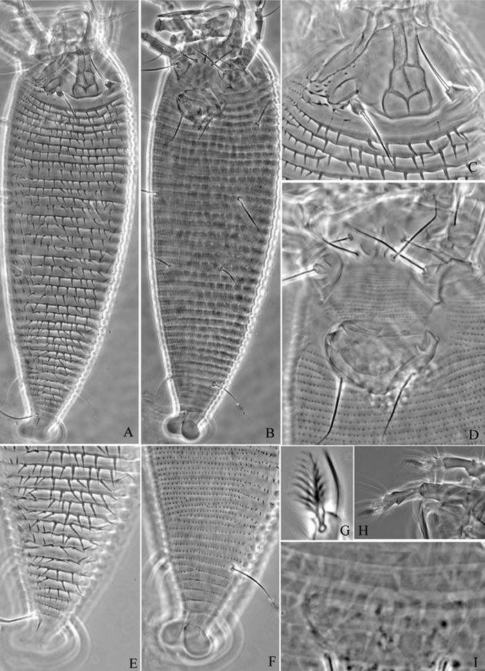
Aculops ulmi Hong & Xue: A dorsal view of female B ventral view of female C lateral view of female D dorsal view of female posterior part E ventral view of female posterior part F lateral microtubercles G lateral view of female posterior part H female internal genitalia.
Aculops ulmi Hong & Xue: I female genitalia J female genitalia from holotype.
6 females (slide number NJAUEri792) from Ulmus sp. (Ulmaceae), Xining, Qinghai Province, P. R. China, 36°38'18"N, 101°45'27"E, elevation 2241m, 21 July 2007, coll. Xiao-Feng Xue.
Vagrant on leaf lower surface. No damage to the host was observed.
China (Hebei, Qinghai).
Instead of the original description: female genitalia coverflap smooth, female genitalia coverflap is with 8 longitudinal ridges in this redescription.
http://species-id.net/wiki/Aculops_xiningensis
Malus pumila P. Mill. (Rosaceae).
Vagrant on leaf lower surface. No damage to the host was observed.
China (Qinghai).
http://species-id.net/wiki/Tetraspinus_syringae
Syringa oblata Lindl. (Oleaceae).
Vagrant on leaf lower surface. No damage to the host was observed.
China (Qinghai).
http://species-id.net/wiki/Tetra_pinnatifidae
8 females and 2 males (slide number NJAUEri793) from a new host, Prunus armeniaca Linn. (Rosaceae), Xining, Qinghai Province, P. R. China, 36°38'18"N, 101°45'27"E, elevation 2241m, 21 July 2007, coll. Xiao-Feng Xue.
Crataegus pinnatifida Bunge (Rosaceae); Prunus armeniaca Linn. (Rosaceae).
Vagrant on leaf lower surface. No damage to the host was observed.
China (Shaanxi, Qinghai).
urn:lsid:zoobank.org:act:D0B42704-F828-4F47-9265-47B3EF8E603A
http://species-id.net/wiki/Tetra_pruniana
Figures 16–18Female. (n = 9) Body fusiform, light yellow, 215 (210–225), 77 (75–78) wide. Gnathosoma 21 (21–23), projecting obliquely down, suboral plate present, pedipalp coxal seta (ep) 4 (4–5), dorsal pedipalp genual seta (d) 7 (7–8), cheliceral stylets 16 (16–18). Prodorsal shield 54 (54–55), 75 (75–78) wide, subtriangular; frontal lobe 12 (11–12); median and submedian lines absent, admedian lines connected by two weak transverse lines. Scapular tubercles near rear shield margin, 3 (3–4), 34 (32–34) apart, scapular setae (sc) 14 (13–15), projecting posteriorly. Rear shield with wave-like margin. Coxigenital region with 9 annuli. Coxisternal plates with short lines, anterolateral setae on coxisternum I (1b) 11 (10–13), 14 (13–14) apart, proximal setae on coxisternum I (1a) 20 (20–25), 10 (10–11) apart, proximal setae on coxisternum II (2a) 50 (45–50), 28 (28–29) apart, tubercles 1b and 1a 7 (7–8) apart, tubercles 1a and 2a 10 (10–12) apart. Prosternal apodeme combined, 8 (6–8). Leg I 34 (32–35), femur 12 (12–13), basiventral femoral seta (bv) 12 (12–14); genu 7 (6–7), antaxial genual seta (l") 20 (19–20); tibia 10 (9–10), paraxial tibial seta (l’) 6 (5–6), located at 1/3 from dorsal base; tarsus 7 (7–8), seta ft’ 19 (19–21), seta ft" 25 (24–25), seta u’ 5 (4–5); tarsal empodium (em) 7 (6–7), simple, 4-rayed, tarsal solenidion (ω) 6 (6–7), knobbed. Leg II 27 (27–31), femur 10 (10–11), basiventral femoral seta (bv) 12 (12–13); genu 5 (5–6), antaxial genual seta (l") 8 (7–8); tibia 6 (6–7); tarsus 6 (6–7), seta ft’ 6 (6–7), seta ft" 25 (23–25), seta u’ 5 (5–6); tarsal empodium (em) 6 (5–6), simple, 4-rayed, tarsal solenidion (ω) 6 (6–7), knobbed. Opisthosoma dorsally with 25 (24–25) annuli, with weak filamentous microtubercles, ventrally with 53 (53–59) annuli, with round microtubercles. Setae c2 25 (24–26) on ventral annulus 9 (9–11), 58 (58–59) apart; setae d 60 (57–65) on ventral annulus 22 (22–24), 33 (32–33) apart; setae e 18 (17–20) on ventral annulus 37 (37–40), 18 (17–18) apart; setae f 29 (29–33) on ventral annulus 50 (50–56), 25 (22–25) apart. Setae h1 3 (2–3), h2 85 (80–95). Female genitalia 15 (15–17), 25 (24–25) wide, coverflap with 10 longitudinal ridges, setae 3a 18 (16–19), 17 (17–18) apart.
Male. (n = 6) Body fusiform, light yellow, 170–186, 73–74 wide. Gnathosoma 16–17, projecting obliquely downwards, suboral plate present, pedipalp coxal seta (ep) 4–5, dorsal pedipalp genual seta (d) 7–8, cheliceral stylets 14–16. Prodorsal shield has the same design as female, 47–50, 72–73 wide, subtriangular; frontal lobe 10–12. Scapular tubercles on rear shield margin, 3–4, 30–33 apart, scapular setae (sc) 7–9, projecting posteriorly. Coxigenital region with 9 smooth annuli. Coxisternal plates with short lines, anterolateral setae on coxisternum I (1b) 10–11, 13–14 apart, proximal setae on coxisternum I (1a) 21–22, 10–11 apart, proximal setae on coxisternum II (2a) 45–48, 28–30 apart, tubercles 1b and 1a 7–8 apart, tubercles 1a and 2a 9–10 apart. Prosternal apodeme combined, 7–8. Leg I 34–35, femur 10–12, basiventral femoral seta (bv) 9–10; genu 6–7, antaxial genual seta (l") 19–22; tibia 9–10, paraxial tibial seta (l’) 6–7, located at 1/3 from dorsal base; tarsus 7–8, seta ft’ 15–17, seta ft" 21–24, seta u’ 4–5; tarsal empodium (em) 7–8, simple, 4-rayed, tarsal solenidion (ω) 6–7, knobbed. Leg II 28–29, femur 9–10, basiventral femoral seta (bv) 10–11; genu 5–6, antaxial genual seta (l") 6–7; tibia 7–8; tarsus 6–7, seta ft’ 6–7, seta ft" 22–25, seta u’ 5–6; tarsal empodium (em) 6–7, simple, 4-rayed, tarsal solenidion (ω) 7–8, knobbed. Opisthosoma dorsally with 26–27 annuli, with weak filamentous microtubercles, ventrally with 53–54 annuli, with round microtubercles. Setae c2 18–20 on ventral annulus 10–11, 55–65 apart; setae d 32–35 on ventral annulus 20–22, 30–45 apart; setae e 18–20 on ventral annulus 33–35, 18–30 apart; setae f 25–28 on ventral annulus 50–51, 20–22 apart. Setae h1 2–3, h2 80–90. Male genitalia 13–14, 23–28 wide, setae 3a 17–18, 20–21 apart.
Tetra pruniana sp. n.: D dorsal view of female em empodium L1 leg I L2 leg II.
Tetra pruniana sp. n.: V ventral view of female IG female internal genitalia CMG coxae and male genitalia.
Tetra prunianasp. n.: A dorsal view of female B ventral view of female C dorsal view of female posterior part D ventral view of female posterior part E prodorsal shield F coxae and female genitalia G female internal genitalia H male genitalia I empodium.
Holotype, female (slide number NJAUEri808, marked Holotype), from Prunus tomentosa Thunb. (Rosaceae), Xining City, Qinghai Province, P. R. China, 36°38'18"N, 101°45'27"E, elevation 2241m, 21 July 2007, coll. Xiao-Feng Xue. Paratypes, 2 females and 4 males (slide number NJAUEri808), with the same data as holotype.
Vagrant on leaf lower surface. No damage to the host was observed.
The specific designation pruniana is from the generic name of host plant, Prunus; feminine in gender.
This species is similar to Tetra pinnatifidae Xue, Song & Hong, 2006a, but can be differentiated from the latter by median and submedian lines absent (median and submedian lines present in Tetra pinnatifidae), admedian lines connected by two weak transverse lines (admedian lines separated in Tetra pinnatifidae).
urn:lsid:zoobank.org:act:99B1B2A9-DB3F-466C-8900-951BB3B35485
http://species-id.net/wiki/Tetra_pyriana
Figures 19–22Female. (n = 11) Body fusiform, light yellow, 180 (169–185), 73 (72–76) wide, 65 (64–65) thick. Gnathosoma 20 (20–23), projecting obliquely down, pedipalp coxal seta (ep) 4 (3–4), dorsal pedipalp genual seta (d) 6 (5–7), cheliceral stylets 20 (20–21). Prodorsal shield 47 (47–49), 68 (68–72) wide, subtriangular; frontal lobe 12 (11–13); median, admedian and submedian lines present, median line obscure and ending at basal 1/3 of prodorsal shield. Scapular tubercles on rear shield margin, 4 (3–4), 36 (33–36) apart, scapular setae (sc) 12 (12–13), projecting posteriorly. Coxigenital region with 13 (12–13) smooth annuli. Coxisternal plates with short lines, anterolateral setae on coxisternum I (1b) 7 (7–9), 12 (11–12) apart, proximal setae on coxisternum I (1a) 21 (20–21), 10 (9–10) apart, proximal setae on coxisternum II (2a) 40 (40–45), 27 (25–27) apart, tubercles 1b and 1a 6 (6–7) apart, tubercles 1a and 2a 10 (9–10) apart. Prosternal apodeme combined, 7 (6–7). Leg I 32 (30–32), femur 12 (11–12), basiventral femoral seta (bv) 11 (10–11); genu 6 (5–6), antaxial genual seta (l") 20 (20–21); tibia 7 (7–8), paraxial tibial seta (l’) 5 (5–6), located at 1/3 from dorsal base; tarsus 8 (7–8), seta ft’ 19 (17–19), seta ft" 25 (21–25), seta u’ 5 (4–5); tarsal empodium (em) 6 (6–7), simple, 4-rayed, tarsal solenidion (ω) 6 (6–7), knobbed. Leg II 28 (27–28), femur 11 (10–11), basiventral femoral seta (bv) 8 (8–11); genu 5 (4–5), antaxial genual seta (l") 8 (7–8); tibia 7 (6–7); tarsus 7 (6–7), seta ft’ 5 (5–6), seta ft" 20 (19–20), seta u’ 4 (4–5); tarsal empodium (em) 6 (6–7), simple, 4-rayed, tarsal solenidion (ω) 5 (5–6), knobbed. Opisthosoma dorsally with 32 (30–32) annuli, with filamentous microtubercles, ventrally with 58 (58–60) annuli, with round microtubercles. Setae c2 25 (24–30) on ventral annulus 11 (11–13), 57 (54–57) apart; setae d 45 (45–50) on ventral annulus 22 (22–24), 32 (32–33) apart; setae e 18 (17–18) on ventral annulus 37 (37–41), 17 (16–17) apart; setae f 29 (28–30) on ventral annulus 53 (53–54), 25 (24–25) apart. Setae h1 3 (2–3), h2 85 (80–90). Female genitalia 15 (14–16), 22 (22–23) wide, coverflap with 11 (10–12) longitudinal ridges, setae 3a 22 (19–24), 14 (14–15) apart.
Male. (n = 2) Body fusiform, light yellow, 150–176 63–65 wide. Gnathosoma 21–22, projecting obliquely down, pedipalp coxal seta (ep) 2–3, dorsal pedipalp genual seta (d) 6–7, cheliceral stylets 19–20. Prodorsal shield has the same design as female, 42–47, 63–65 wide, subtriangular; frontal lobe 10–11. Scapular tubercles on rear shield margin, 3–4, 30–31 apart, scapular setae (sc) 11–13, projecting posteriorly. Coxigenital region with 12–13 smooth annuli. Coxisternal plates with short lines, anterolateral setae on coxisternum I (1b) 7–8, 12–13 apart, proximal setae on coxisternum I (1a) 18–20, 9–10 apart, proximal setae on coxisternum II (2a) 45–46, 24–26 apart, tubercles 1b and 1a 6–7 apart, tubercles 1a and 2a 7–9 apart. Prosternal apodeme combined, 5–6. Leg I 29–32, femur 7–9, basiventral femoral seta (bv) 9–12; genu 5–6, antaxial genual seta (l") 20–23; tibia 6–8, paraxial tibial seta (l’) 5–6, located at 1/3 from dorsal base; tarsus 7–8, seta ft’ 17–18, seta ft" 21–23, seta u’ 3–4; tarsal empodium (em) 6–7, simple, 4-rayed, tarsal solenidion (ω) 5–6, knobbed. Leg II 25–27, femur 7–9, basiventral femoral seta (bv) 9–10; genu 4–5, antaxial genual seta (l") 6–8; tibia 7–8; tarsus 6–7, seta ft’ 2–3, seta ft" 20–21, seta u’ 2–3; tarsal empodium (em) 6–7, simple, 4-rayed, tarsal solenidion (ω) 6–7, knobbed. Opisthosoma dorsally with 29–31 annuli, with filamentous microtubercles, ventrally with 60–61 annuli, with round microtubercles. Setae c2 24–25 on ventral annulus 13–14, 50–51 apart; setae d 36–37 on ventral annulus 24–25, 29–30 apart; setae e 15–16 on ventral annulus 41–42, 16–17 apart; setae f 28–29 on ventral annulus 55–56, 20–21 apart. Setae h1 2–3, h2 85–90. Male genitalia 13–14, 19–20 wide, setae 3a 17–18, 16–17 apart.
Tetra pyriana sp. n.: D dorsal view of female LO lateral microtubercles em empodium L1 leg I L2 leg II.
Tetra pyriana sp. n.: L lateral view of female IG female internal genitalia CG coxae and female genitalia CMG coxae and male genitalia.
Tetra pyriana sp. n.: A dorsal view of female B ventral view of female C lateral microtubercles D empodium E dorsal view of female posterior part F ventral view of female posterior part G leg I and leg II.
Tetra pyriana sp. n.: H lateral view of female I lateral view of female posterior part J female internal genitalia K prodorsal shield L coxae and female genitalia M coxae and male genitalia.
Holotype, female (slide number NJAUEri796, marked Holotype), from Pyrus calleryana Decne. (Rosaceae), Xining, Qinghai Province, P. R. China, 36°38'18"N, 101°45'27"E, elevation 2241m, 21 July 2007, coll. Xiao-Feng Xue. Paratypes, 10 females and 2 males (slide number NJAUEri796), with the same data as holotype.
13 females (slide number NJAUEri807), from Pyrus betulifolia Bunge. (Rosaceae), Xining, Qinghai Province, P. R. China, 36°38'18"N, 101°45'27"E, elevation 2241m, 21 July 2007, coll. Xiao-Feng Xue; 14 females and 1 male (slide number NJAUEri806), from Pyrus calleryana Decne. (Rosaceae), Xining, Qinghai Province, P. R. China, 36°38'18"N, 101°45'27"E, elevation 2241m, 21 July 2007, coll. Xiao-Feng Xue.
Vagrant on leaf lower surface. No damage to the host was observed.
The specific designation pyriana is from the generic name of host plant, Pyrus; feminine in gender.
This species is similar to Tetra pinnatifidae Xue, Song & Hong, 2006a, but can be differentiated from the latter by median line weak and ending at basal 1/3 of prodorsal shield (median normal and does not disappear at basal 1/3 of prodorsal shield in Tetra pinnatifidae), coxigenital region with smooth annuli (coxigenital region annuli with round microtubercles in Tetra pinnatifidae).
urn:lsid:zoobank.org:act:11D2D601-440B-4A7D-AD50-18F7B91B3A33
http://species-id.net/wiki/Tetra_simonia
Figures 23–26Female. (n = 14) Body fusiform, light yellow, 196 (196–224), 72 (72–77) wide, 78 (78) thick. Gnathosoma 22 (20–22), projecting obliquely down, pedipalp coxal seta (ep) 3 (3–4), dorsal pedipalp genual seta (d) 12 (10–12), cheliceral stylets 19 (19–20). Prodorsal shield 46 (42–46), 72 (72–77) wide, subtriangular; frontal lobe 11 (11–12); median, admedian and submedian lines robust and connected, forming an “M” shape in the middle and two “H” shapes anterior to the “M”. Scapular tubercles on rear shield margin, 3 (3–4), 46 (44–46) apart, scapular setae (sc) 14 (14–15), projecting posteriorly. Coxigenital region with 14 (14–15) smooth annuli. Coxisternal plates smooth, anterolateral setae on coxisternum I (1b) 11 (11–12), 14 (14–19) apart, proximal setae on coxisternum I (1a) 17 (17–23), 12 (12–13) apart, proximal setae on coxisternum II (2a) 53 (53–58), 28 (27–31) apart, tubercles 1b and 1a 8 (8–9) apart, tubercles 1a and 2a 9 (9–10) apart. Prosternal apodeme combined, 6 (6–8). Leg I 34 (34–37), femur 12 (11–12), basiventral femoral seta (bv) 12 (12–13); genu 8 (8–9), antaxial genual seta (l") 24 (22–24); tibia 13 (11–13), paraxial tibial seta (l’) 7 (6–7), located at 1/3 from dorsal base; tarsus 7 (7–8), seta ft’ 22 (21–22), seta ft" 23 (22–23), seta u’ 6 (6–7); tarsal empodium (em) 6 (6–7), simple, 4-rayed, tarsal solenidion (ω) 6 (6–7), knobbed. Leg II 30 (30–34), femur 11 (11–12), basiventral femoral seta (bv) 13 (12–13); genu 6 (6–7), antaxial genual seta (l") 11 (11–13); tibia 9 (7–9); tarsus 7 (6–7), seta ft’ 5 (5–6), seta ft" 22 (22–24), seta u’ 6 (6–7); tarsal empodium (em) 6 (6–7), simple, 4-rayed, tarsal solenidion (ω) 6 (6–7), knobbed. Opisthosoma dorsally with 31 (30–31) annuli, with dark shading on rear annular margins, ventrally with 58 (58–65) annuli, with round microtubercles. Setae c2 36 (36–42) on ventral annulus 11 (11–13), 52 (52–56) apart; setae d 77 (71–77) on ventral annulus 21 (21–23), 33 (33–38) apart; setae e 23 (20–23) on ventral annulus 37 (37–39), 18 (18–19) apart; setae f 40 (36–40) on ventral annulus 51 (51–54), 30 (30–32) apart. Setae h1 5 (4–5), h2 150 (135–150). Female genitalia 20 (18–22), 25 (24–25) wide, coverflap with 10 (10–13) longitudinal ridges, setae 3a 17 (17–18), 18 (17–18) apart.
Male. (n = 1) Body fusiform, light yellow, 188, 74 wide. Gnathosoma 21, projecting obliquely downwards, pedipalp coxal seta (ep) 3, dorsal pedipalp genual seta (d) 11, cheliceral stylets 19. Prodorsal shield has the same design as female, 42, 74 wide, subtriangular; frontal lobe 11. Scapular tubercles on rear shield margin, 44 apart, scapular setae (sc) 13, projecting posteriorly. Coxigenital region with 14 smooth annuli. Coxisternal plates smooth, anterolateral setae on coxisternum I (1b) 13), 14 apart, proximal setae on coxisternum I (1a) 23, 12 apart, proximal setae on coxisternum II (2a) 49, 30 apart, tubercles 1b and 1a apart 8, tubercles 1a and 2a 10 apart. Prosternal apodeme combined, 6. Leg I 35, femur 11, basiventral femoral seta (bv) 12; genu 8, antaxial genual seta (l") 26; tibia 12, paraxial tibial seta (l’) 6, located at 1/3 from dorsal base; tarsus 7, seta ft’ 21, seta ft" 24, seta u’ 6; tarsal empodium (em) 6, simple, 4-rayed, tarsal solenidion (ω) 6, knobbed. Leg II 32, femur 10, basiventral femoral seta (bv) 14; genu 6, antaxial genual seta (l") 11; tibia 8; tarsus 8, seta ft’ 5, seta ft" 18, seta u’ 6; tarsal empodium (em) 6, simple, 4-rayed, tarsal solenidion (ω) 6, knobbed. Opisthosoma dorsally with 31 annuli, with dark shading on rear annular margins, ventrally with 67 annuli, with round microtubercles. Setae c2 42 on ventral annulus 13, 58 apart; setae d 70 on ventral annulus 27, 40 apart; setae e 25 on ventral annulus 47, 20 apart; setae f 40 on ventral annulus 63, 30 apart. Setae h1 5, h2 130. Male genitalia 15, 25 wide, setae 3a 18, 19 apart.
Tetra simonia sp. n.: D dorsal view of female LO lateral microtubercles em empodium L1 leg I L2 leg II.
Tetra simonia sp. n.: L lateral view of female IG female internal genitalia CG coxae and female genitalia CMG coxae and male genitalia.
Tetra simonia sp. n.: A dorsal view of female B ventral view of female C lateral microtubercles D empodium E dorsal view of female posterior part F ventral view of female posterior part G leg I and leg II.
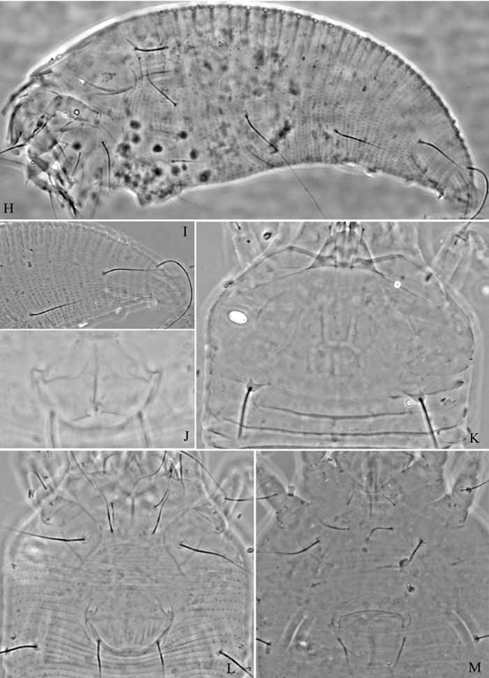
Tetra simonia sp. n.: H lateral view of female I lateral view of female posterior part J female internal genitalia K prodorsal shield L coxae and female genitalia M coxae and male genitalia.
Holotype, female (slide number NJAUEri799, marked Holotype), from Populus simonii Carr. (Salicaceae), Xining, Qinghai Province, P. R. China, 36°38'18"N, 101°45'27"E, elevation 2241m, 21 July 2007, coll. Xiao-Feng Xue. Paratypes, 13 females and 1 male (slide number NJAUEri799), with the same data as holotype.
4 females (slide number NJAUEri789A), from Populus simonii Carr. (Salicaceae), Xining, Qinghai Province, P. R. China, 36°38'18"N, 101°45'27"E, elevation 2241m, 21 July 2007, coll. Xiao-Feng Xue. 4 females and 2 males (slide number NJAUEri823), from Populus simonii Carr. (Salicaceae), Beishan National Forest Park, Huzhu County, Qinghai Province, P. R. China, 36°53'35"N, 102°25'56"E, elevation 2610m, 22 July 2007, coll. Xiao-Feng Xue.
Vagrant on leaf lower surface. No damage to the host was observed.
The specific designation simonia is from the species name of host plant, simonii; feminine in gender.
This species is similar to Tetra smilaxis Xue, Song & Hong, 2006a, but can be differentiated from the latter by opisthosoma with dark shading on rear annular margins (dark shading absent in Tetra smilaxis), prodorsal shield as wide as opisthosoma (opisthosoma wider than prodorsal shield in Tetra smilaxis), admedian lines connected at basal 1/3 but separate at basal 2/3 of prodorsal shield (admedian lines connected at basal 1/3 and basal 2/3 of prodorsal shield in Tetra smilaxis).
Subfamily Diptilomiopinae Keifer, 1944
Genus Diptacus Keifer, 1951
urn:lsid:zoobank.org:act:3EF374D2-74CD-46A0-833C-A80FC1EAE1E2
http://species-id.net/wiki/Diptacus_berberinus
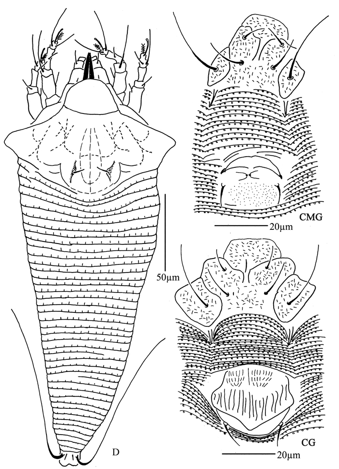
Figures 27–30Female. (n = 9) Body fusiform, light yellow, 283 (280–360), 110 (102–110) wide, 115 (114–115) thick. Gnathosoma 26 (25–27), projecting downwards, pedipalp coxal seta (ep) 6 (5–6), dorsal pedipalp genual seta (d) 12 (11–12), cheliceral stylets 65 (65–66). Prodorsal shield 40 (40–46), 80 (77–80) wide, with wide and broad frontal lobe, 7 (7–8); median, admedian and submedian lines present, admedian lines connected at the basal 1/3 and 2/3 of prodorsal shield, forming 3 cells on each side, submedian lines connected with the median and admedian at the basal 2/3 of prodorsal shield, forming the cell-like pattern at anterior shield margin. Scapular tubercles ahead of rear shield margin, 3 (2–3), 30 (27–30) apart, scapular setae (sc) 5 (4–5), projecting centrad to forward. Coxigenital region with 13 (13–15) annuli, with triangular microtubercles. Coxisternal plate I with granules, coxisternal plate II smooth, anterolateral setae on coxisternum I (1b) 20 (18–20), 17 (17–18) apart, proximal setae on coxisternum I (1a) 43 (43–45), 19 (17–19) apart, proximal setae on coxisternum II (2a) 70 (70–80), 49 (43–56) apart, tubercles 1b and 1a 12 (12–13) apart, tubercles 1a and 2a 17 (14–17) apart. Prosternal apodeme separated, 5 (5–6). Leg I 65 (60–65), femur 20 (20–22), basiventral femoral seta (bv) absent; genu 8 (8–9), antaxial genual seta (l") 48 (48–52); tibia 18 (17–19), paraxial tibial seta (l’) 11 (10–11), located at 1/2 from dorsal base; tarsus 11 (11–12), seta ft’ 30 (29–30), seta ft" 40 (37–40), seta u’ 7 (6–7); tarsal empodium (em) 11 (10–11), divided, 7-rayed on each side, tarsal solenidion (ω) 10 (10–14), knobbed. Leg II 55 (54–55), femur 20 (19–20), basiventral femoral seta (bv) absent; genu 8 (7–8), antaxial genual seta (l") 18 (15–18); tibia 17 (16–17); tarsus 11 (10–11), seta ft’ 11 (10–11), seta ft" 44 (44–50), seta u’ 8 (7–8); tarsal empodium (em) 11 (10–11), divided, 7-rayed on each side, tarsal solenidion (ω) 10 (10–11), knobbed. Opisthosoma dorsally with 59 (54–62) annuli, smooth, ventrally with 106 (101–106) annuli, with triangular microtubercles. Setae c2 115 (110–115) on ventral annulus 18 (18–20), 74 (74–75) apart; setae d 100 (100–120) on ventral annulus 40 (37–40), 56 (49–56) apart; setae e 60 (55–60) on ventral annulus 65 (60–65), 31 (29–35) apart; setae f 65 (60–70) on ventral annulus 93 (87–93), 35 (35–37) apart. Setae h1 2 (1–2), h2 103 (95–165). Female genitalia 35 (31–40), 35 (34–42) wide, coverflap with short lines on base, and 4 longitudinal ridges in 2 ranks, 1 ridge near the base and 3 ridges at distal margin, setae 3a 12 (11–15), 24 (24–25) apart.
Male. (n = 1) Body fusiform, light yellow, 269, 86 wide. Gnathosoma 60, projecting downwards, pedipalp coxal seta (ep) 5, dorsal pedipalp genual seta (d) 11, cheliceral stylets 65. Prodorsal shield has the same design as female, 38, 71 wide, with wide and broad frontal lobe, 7. Scapular tubercles ahead of rear shield margin, 3, 27 apart, scapular setae (sc) 4, projecting centrad to forward. Coxigenital region with 14 annuli, with triangular microtubercles. Coxisternal plate I with granules, coxisternal plate II smooth, anterolateral setae on coxisternum I (1b) 22, 16 apart, proximal setae on coxisternum I (1a) 36, 16 apart, proximal setae on coxisternum II (2a) 60, 41 apart, tubercles 1b and 1a 11 apart, tubercles 1a and 2a 12 apart. Prosternal apodeme separated, 6. Leg I 46, femur 17, basiventral femoral seta (bv) absent; genu 7, antaxial genual seta (l") 46; tibia 13, paraxial tibial seta (l’) 10, located at 1/2 from dorsal base; tarsus 8, seta ft’ 30, seta ft" 37, seta u’ 6; tarsal empodium (em) 10, divided, 7-rayed on each side, tarsal solenidion (ω) 10, knobbed. Leg II 38, femur 17, basiventral femoral seta (bv) absent; genu 7, antaxial genual seta (l") 15; tibia 13; tarsus 6, seta ft’ 10, seta ft" 38, seta u’ 7; tarsal empodium (em) 10, divided, 7-rayed on each side, tarsal solenidion (ω) 11, knobbed. Opisthosoma dorsally with 56 annuli, smooth, ventrally with 83 annuli, with triangular microtubercles. Setae c2 95 on ventral annulus 15, 63 apart; setae d 100 on ventral annulus 30, 49 apart; setae e 55 on ventral annulus 46, 27 apart; setae f 60 on ventral annulus 71, 25 apart. Setae h1 2, h2 120. Male genitalia 23, 30 wide, setae 3a 10, 23 apart.
Diptacus berberinus sp. n.: D dorsal view of female IG female internal genitalia LO lateral microtubercles L1 leg I L2 leg II em empodium.
Diptacus berberinus sp. n.: L lateral view of female CMG coxae and male genitalia CG coxae and female genitalia.
Diptacus berberinus sp. n.: A dorsal view of female B ventral view of female C lateral microtubercles D empodium E dorsal view of female posterior part F ventral view of female posterior part G leg I and leg II.
Diptacus berberinus sp. n.: H lateral view of female I coxae and female genitalia J lateral view of female posterior part K female internal genitalia L coxae and male genitalia M prodorsal shield.
Holotype, female (slide number 783, marked Holotype), from Berberis amurensis Rupr.(Berberidaceae), Mengda Natural Reserve, Xunhua County, Qinghai Province, P. R. China, 35°47'38"N, 102°40'40"E, elevation 2523m, 19 July 2007, coll. Xiao-Feng Xue. Paratypes, 8 females and 1 male (slide number 783), with the same data as holotype.
Vagrant on leaf lower surface. No damage to the host was observed.
The specific designation berberinus is from the generic name of host plant, Berberis; masculine in gender.
This species is similar to Diptacus maddenis Song, Xue & Hong, 2007a, but can be differentiated from the latter by opisthosomal dorsal annuli smooth (opisthosomal dorsal annuli with elongated microtubercles in Diptacus maddenis), female genital coverflap with short lines at the base (genital coverflap with granules in Diptacus maddenis), tarsal empodium 7-rayed (4-rayed in Diptacus maddenis).
urn:lsid:zoobank.org:act:12904959-7EE9-4780-BCC1-EF886FA54F98
http://species-id.net/wiki/Diptacus_mengdaensis
Figures 31–33Female. (n = 13) Body fusiform, light yellow, 215 (210–232), 104 (104–105) wide, 71 (71–74) thick. Gnathosoma 25 (24–25), projecting downwards, pedipalp coxal seta (ep) 4 (4–5), dorsal pedipalp genual seta (d) 14 (14–15), cheliceral stylets 62 (61–62). Prodorsal shield 50 (50–51), 76 (71–76) wide, with wide and broad frontal lobe, 7 (7–8); median, admedian and submedian lines present, admedian lines connected at the base of prodorsal shield, ending at basal 1/3 of prodorsal shield. Scapular tubercles ahead of rear shield margin, 3 (3–4), 34 (34–35) apart, scapular setae (sc) 6 (6–7), projecting centrad. Coxigenital region with 16 (15–16) annuli, with microtubercles. Coxisternal plates with short lines, anterolateral setae on coxisternum I (1b) 16 (16–18), 18 (18–20) apart, proximal setae on coxisternum I (1a) 25 (25–26), 17 (17–18) apart, proximal setae on coxisternum II (2a) 60 (60–70), 44 (44–45) apart, tubercles 1b and 1a 12 (12–13) apart, tubercles 1a and 2a 16 (16–17) apart. Prosternal apodeme separated, 10 (9–10). Leg I 60 (58–60), femur 16 (16–17), basiventral femoral seta (bv) absent; genu 10 (8–10), antaxial genual seta (l") 52 (52–53); tibia 17 (16–17), paraxial tibial seta (l’) 10 (9–10), located at 1/3 from dorsal base; tarsus 11 (10–11), seta ft’ 30 (25–30), seta ft" 40 (32–40), seta u’ 6 (5–6); tarsal empodium (em) 8 (8–9), divided, 5-rayed at each side, tarsal solenidion (ω) 9 (9–10), knobbed. Leg II 54 (50–54), femur 18 (17–18), basiventral femoral seta (bv) absent; genu 7 (7–8), antaxial genual seta (l") 16 (15–16); tibia 11 (9–11); tarsus 8 (8–10), seta ft’ 12 (12–13), seta ft" 34 (34–34), seta u’ 5 (5–6); tarsal empodium (em) 9 (8–9), divided, 5-rayed at each side, tarsal solenidion (ω) 9 (9–10), knobbed. Opisthosoma dorsally with 44 (44–48) annuli, smooth, ventrally with 112 (112–115) annuli, with round microtubercles. Setae c2 55 (54–55) on ventral annulus 16 (16–18), 76 (70–76) apart; setae d 90 (90–93) on ventral annulus 39 (39–42), 51 (51–53) apart; setae e 70 (65–70) on ventral annulus 67 (67–69), 29 (29–31) apart; setae f 50 (50–55) on ventral annulus 98 (98–101), 37 (36–37) apart. Setae h1 2 (2–3), h2 152 (150–152). Female genitalia 24 (24–25), 37 (37–39) wide, coverflap with 4 longitudinal ridges in 2 ranks, 1 near the base and 3 at distal margin, setae 3a 12 (10–12), 20 (20–21) apart.
Male. Unknown.
Diptacus mengdaensis sp. n.: D dorsal view of female LO lateral microtubercles em empodium L1 leg I L2 leg II.
Diptacus mengdaensis sp. n.: L lateral view of female IG female internal genitalia CG coxae and female genitalia.
Diptacus mengdaensissp. n.: A dorsal view of female B ventral view of female C dorsal view of female posterior part D ventral view of female posterior part E prodorsal shield F lateral microtubercles G empodium H coxae and female genitalia.
Holotype, female (slide number NJAUEri777, marked Holotype), from Lonicera elisae Franch. (Caprifoliaceae), Mengda Natural Reserve, Xunhua County, Qinghai Province, P. R. China, 35°47'38"N, 102°40'40"E, elevation 2523m, 19 July 2007, coll. Xiao-Feng Xue. Paratypes, 12 females (slide number NJAUEri777), with the same data as holotype.
Vagrant on leaf lower surface. No damage to the host was observed.
The specific designation mengdaensis is from the place name Mengda Natural Reserve, where this new species was collected; feminine in gender.
This species is similar to Diptacus lonicerae Kuang, 2001, but can be differentiated from the latter by prodorsal shield with admedian lines and submedian lines separated (admedian lines and submedian lines connected in Diptacus lonicerae), prodorsal shield frontal lobe wide and broad (frontal lobe small in Diptacus lonicerae), female genital coverflap with 4 longitudinal ridges in 2 ranks, 1 near the base and 3 far from the base (female genital coverflap with 6–8 longitudinal ridges in Diptacus lonicerae).
Genus Rhyncaphytoptus Keifer, 1939
http://species-id.net/wiki/Rhyncaphytoptus_ulmi
16 females (slide number NJAUEri792B and NJAUEri795), from Ulmus sp. (Ulmaceae), Xining, Qinghai Province, P. R. China, 36°38'18"N, 101°45'27"E, elevation 2241m, 21 July 2007, coll. Xiao-Feng Xue.
Ulmus sp. (Ulmaceae).
Vagrant on leaf lower surface. No damage to the host was observed.
China (Jiangsu, Gansu, Jilin, Liaoning, Shaanxi, Shandong, Xinjiang, Qinghai).
urn:lsid:zoobank.org:act:F3227702-A360-40D4-9367-E395D5F361DE
http://species-id.net/wiki/Rhyncaphytoptus_spinus
Figures 34–36Female. (n = 6) Body fusiform, light yellow, 270 (232–323), 82 (82–91) wide, 105 (104–105) thick. Gnathosoma 58 (56–59), projecting downwards, suboral plate present, pedipalp coxal seta (ep) 4 (4–5), dorsal pedipalp genual seta (d) 10 (9–11), palp tarsus ventral seta (v) 4 (3–4), cheliceral stylets 71 (71–72). Prodorsal shield 38 (38–40), 60 (57–64) wide, with broad frontal lobe, 7 (6–7); median, admedian and submedian lines present, median, admedian lines connected at basal 1/3 and 2/3 of prodorsal shield, forming three cells each both side. Scapular tubercles ahead of rear shield margin, 6 (5–6), 34 (34–40) apart, scapular setae (sc) 21 (21–25), projecting forward, knobbed at the end. Coxigenital region with 15 (14–15) annuli, with microtubercles. Coxisternal plates smooth, anterolateral setae on coxisternum I (1b) 28 (26–28), 16 (16–18) apart, proximal setae on coxisternum I (1a) 46 (44–46), 10 (10–13) apart, proximal setae on coxisternum II (2a) 80 (71–80), 30 (30–36) apart, tubercles 1b and 1a 6 (5–7) apart, tubercles 1a and 2a 9 (9–13) apart. Prosternal apodeme combined, 6 (6–7). Leg I 44 (43–49), femur 16 (16–17), basiventral femoral seta (bv) 18 (15–20); genu 8 (8–9), antaxial genual seta (l") 30 (30–32); tibia 12 (12–13), paraxial tibial seta (l’) 14 (14–17), located at 1/3 from dorsal base; tarsus 9 (9–10), seta ft’ 26 (24–28), seta ft" 35 (34–35), seta u’ 8 (6–8); tarsal empodium (em) 12 (12–13), simple, 8-rayed, tarsal solenidion (ω) 11 (10–11), tapered. Leg II 41 (41–46), femur 14 (14–16), basiventral femoral seta (bv) 18 (18–19); genu 7 (7–8), antaxial genual seta (l") 10 (10–11); tibia 11 (10–11); tarsus 8 (8–10), seta ft’ 12 (12–13), seta ft" 34 (34–34), seta u’ 7 (6–7); tarsal empodium (em) 12 (12–13), simple, 8-rayed, tarsal solenidion (ω) 11 (10–11), tapered. Opisthosoma dorsally with 38 (38–42) annuli, with long spiny microtubercles, ventrally with 98 (97–98) annuli, with triangle microtubercles. Setae c2 33 (31–33) on ventral annulus 17 (17–19), 76 (71–76) apart; setae d 97 (95–97) on ventral annulus 38 (38–40), 53 (53–69) apart; setae e 41 (41–45) on ventral annulus 60 (59–61), 31 (31–39) apart; setae f 36 (36–38) on ventral annulus 92 (92–93), 28 (28–30) apart. Setae h1 5 (5–6), h2 110 (110–115). Female genitalia 21 (18–21), 32 (32–35) wide, coverflap smooth, setae 3a 75 (70–75), 20 (20–23) apart.
Male. Unknown.
Rhyncaphytoptus spinus sp. n.: D dorsal view of female L1 leg I L2 leg II IG female internal genitalia em empodium.
Rhyncaphytoptus spinus sp. n.: V ventral view of female CG coxae and female genitalia LO lateral microtubercles.
Rhyncaphytoptus spinus sp. n.: A dorsal view of female B ventral view of female C prodorsal shield D coxae and female genitalia E dorsal view of female posterior part F ventral view of female posterior part G empodium H leg I and leg II I female internal genitalia.
Holotype, female (slide number NJAUEri820, marked Holotype), from Lonicera rupicola Hook. f. et Thoms. (Caprifoliaceae), Beishan National Forest Park, Huzhu County, Qinghai Province, P. R. China, 36°53'35"N, 102°25'56"E, elevation 2610m, 22 July 2007, coll. Xiao-Feng Xue. Paratypes, 5 females (slide number NJAUEri820), with the same data as holotype.
Vagrant on leaf lower surface. No damage to the host was observed.
The specific designation spinus is from the character of the dorsal opisthosomal microtubercles, spiny, “spina, spinus” in Latin; masculine in gender.
This species is similar to Rhyncaphytoptus guanegounis Song, Xue & Hong, 2007b, but can be differentiated from the latter by median, admedian and submedian lines present on prodorsal shield (prodorsal shield smooth in Rhyncaphytoptus guanegounis), prodorsal shield with wide and broad frontal lobe (prodorsal shield with long and broad frontal lobe in Rhyncaphytoptus guanegounis), opisthosomal dorsal annuli with spiny microtubercles (opisthosomal dorsal annuli smooth in Rhyncaphytoptus guanegounis), tarsal empodium (em) 8-rayed (6-rayed in Rhyncaphytoptus guanegounis).
Although Qinghai has climate, vegetation and biological diversity similar to Tibet, we did not find any eriophyoid mite species already reported in Tibet (
This research was funded by the National Natural Science Foundation of China (31172132). We thank Xiao Han of NJAU for early review of the manuscript. We also thank Professor Xin-Hua Li of the Dept of Botany at NJAU for identifying host plants. We are very grateful to Professor James W. Amrine Jr. for his valuable suggestions on the manuscript.