


(C) 2011 Masanori Okanishi. This is an open access article distributed under the terms of the Creative Commons Attribution License, which permits unrestricted use, distribution, and reproduction in any medium, provided the original author and source are credited.
For reference, use of the paginated PDF or printed version of this article is recommended.
Squamophis, a new genus of brittle star is described. Two species are included in the genus: Squamophis amamiensis (Okanishi & Fujita, 2009) from south-western Japan and Squamophis albozosteres sp. n. from north-western Australia. Squamophis gen. n. is distinguished from the other genera of the family Asteroschematidae by the following characters: each radial shield is single-layered and is completely covered by plate-shaped epidermal ossicles, and the relative length of the longest arm spine throughout the arms is as long as the length of the corresponding arm segment. Squamophis albozosteres sp. n. is distinguished from Squamophis amamiensis in having white, slightly domed, plate-shaped epidermal ossicles on the aboral side of the body, the ossicles on aboral and lateral portion of the arms form transverse rows, and the other part of aboral side of disc and basal to middle portion of arms are brown but tip of the arms are light purple.
taxonomy, Squamophis, Squamophis albozosteres, new genus, new species
The family Asteroschematidae was erected by
In this study, a new genus of Asteroschematidae is established for two species, including one that is new. A tabular taxonomic key to the five recent genera of Asteroschematidae is provided.
Materials and methodsThree specimens of the new species were collected on Commonwealth Scientific and Industrial Research Organisation (CSIRO) survey SS05/2007 by R/V Southern Surveyor and are deposited in the Museum Victoria (MV). They were fixed onboard in 70% ethanol.
Ossicles from a paratype of the new species were isolated by immersion in domestic bleach (approximately 5% sodium hypochlorite solution), washed in deionised water, dried in air, and mounted on SEM stubs using double-sided conductive tape. The preparations were sputter-coated with gold-palladium and examined with a Jeol JSM-6380LV SEM.
The terms used to describe asteroschematids follow (
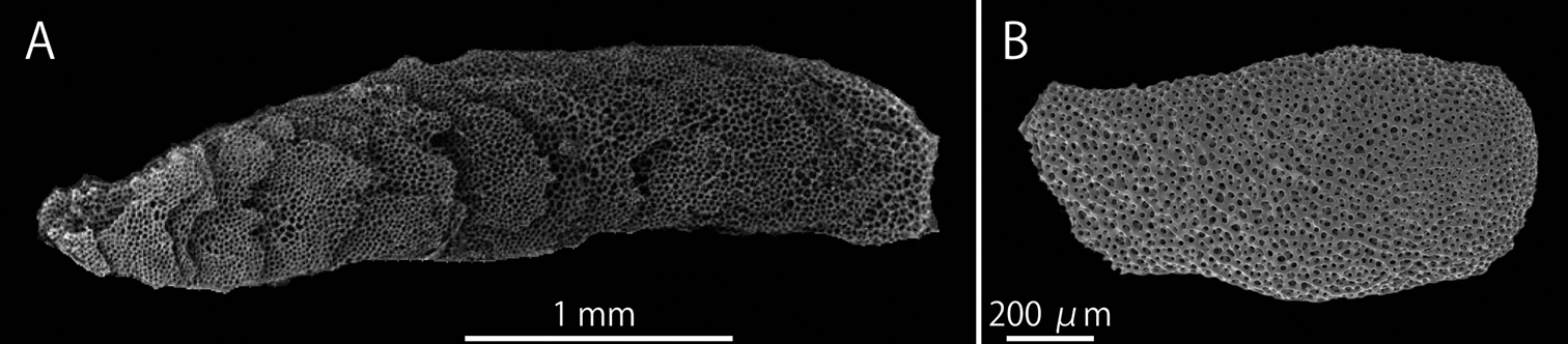
SEM photographs of radial shields of Asteroschema tubiferum (NSMT E-2110) (A) and Squamophis albozosteres sp. n., paratype(MV F-162658) (B). A multi-layered radial shield B single-layered radial shields. The left side of the images are towards the center of the disc and the right side towards the disc margin.
The terms used for the superficial asteroschematid body
ossicles have also been inconsistent in previous descriptions.
Traditionally, “granules”, “scales” and “tubercles” have been used for
the various ossicles on asteroschematid bodies (e.g.
Asteroschema Örsted & Lütken, 1856 (in
Asterias oligactes Pallas, 1788
Four genera are currently recognized within the Asteroschematidae: 1) the type genus Asteroschema erected for the Caribbean species Asterias oligactes Pallas, 1788 (=Asteroschema oligactes); 2) Ophiocreas, also erected for the Caribbean species, Ophiocreas lumbricus Lyman, 1869; 3) the genus Astrocharis, erected for Philippines’ species, Astrocharis virgo Koehler, 1904; and 4) Astrobrachion, erected for the New Zealand species, Ophiocreas constrictus Farquhar, 1900 (=Astrobrachion constrictum). The monotypic genus Ophiuropsis was erected by
In his key to the genera of Asteroschematidae,
Our review of the taxonomic literature and
examination of asteroschematid specimens, has indicated to us that
several of these characters are not useful for defining genera. We have
found that degree of separation of jaws varies in response to animal
preservation. The abrupt increase in arm width, supposedly
characteristic of Astrocharis (see
We propose that four characters are useful for distinguishing the existing genera. The genus Astrobrachion
has ventral arm plates separating the lateral arm plates on the oral
midline throughout the arms, while the other genera have no ventral arm
plates at least from the middle to distal portion of the arms. The
genus Astrocharis
has completely naked radial shields, whereas the radial shields of the
other genera are completely covered by thick skin or epidermal
ossicles. Therefore, the absence/presence of the ventral arm plates and
the covering of the radial shields are useful generic diagnostic
characters as
Tabular morphological key to the genera of the family Asteroschematidae.
| Genus | Shape and arrangement of epidermal ossicles on aboral periphery of the disc and aboral basal portion of the arms | Radial shields | Ventral arm plate on middle to distal portion of the arms | Relative length of the longest arm spines to the corresponding arm segment |
|---|---|---|---|---|
| Asteroschema Örsted & Lütken, 1856* | Cone-shaped and completely in contact, or granule-shaped and slightly in contact | Multi-layered, covered by epidermal ossicles | Absent | Two times longer |
| Ophiocreas Lyman, 1869 | Granule-shaped, slightly in contact or separated, or no epidermal ossicles | Multi-layered, covered by epidermal ossicles or skin | Absent | Two times longer |
| Astrobrachion Döderlein, 1927 | No epidermal ossicles | Multi-layered, covered by skin | Present | The same length |
| Astrocharis Koehler, 1904 | Plate-shaped and completely in contact | Single-layered, naked | Absent | Two times longer |
| Squamophis gen. n. | Plate-shaped and completely in contact | Single-layered, covered by epidermal ossicles | Absent | The same length |
* Except Astrocharis capense and Astrocharis igloo which may be related to Squamophis gen. n. (see
The shapes and arrangement of epidermal ossicles
on aboral surfaces of the discs and arms have been used to distinguish
the four genera (
Astrocharis has been distinguished by its short arm spines (
Asteroschema amamiense Okanishi & Fujita, 2009
Squamophis albozosteres sp. n.
Aboral periphery of the disc and aboral base of the arms covered completely by contiguous plate-shaped epidermal ossicles. Single-layered radial shields completely covered by epidermal ossicles. No ventral arm plates on middle to distal sections of the arms. Relative length of the longest arm spines the same length as the corresponding arm segment throughout the arms.
The generic name is a masculine noun in the subjective case, a compound of Latin, squama (prefix, meaning “scale”) referring to the plate-shaped epidermal ossicles on their body and the Greek ophis (masculine noun, meaning “snake”), referring to their snake-like arms.
(
The genus Squamophis currently comprises two species: Squamophis amamiensis (Okanishi & Fujita, 2009) from south-western Japan, 167–168 m and Squamophis albozosteres sp. n. from north-western Australia, 95–108 m. Asteroschema capense Mortensen, 1925 and Asteroschema igloo Baker, 1980 may also be related to Squamophis amamiensis, based on the similarity of shapes and arrangement of epidermal ossicles (
urn:lsid:zoobank.org:act:9D85F117-BE04-4BF6-A03A-68636966B737
http://species-id.net/wiki/Squamophis_albozosteres
Figs 1B, 3–7MV F 162657, holotype, stn SS05/2007 116, off Broome, northwestern Australia, 16°45.09'S, 121°02.48'E – 16°44.36'S, 121°02.12'E, 100–108 m, rocky bottom, 23.3 °C, 30 Jun 2007, epibenthic sled. MV F162658, two paratypes, stn SS05/2007 188, off Ashmore Reef, northwestern Australia, 12°26.42'S, 123°36.03'E – 12°26.58'S, 123°36.35'E, 95–96 m, rocky bottom, 24, 8 °C, 7 Jul 2007, benthic dredge (Fig. 2).
Epidermal ossicles conspicuous white, slightly domed and round plate-shaped, irregularly placed on aboral side of disc and forming two transverse bands on aboral and lateral sides of each arm segment. Rest of the aboral surface uniformly brown except light purple near the tips of arms and finally without color at the tip.
MV F162657: disc diameter 3.4 mm, arm length approximately 50 mm (Fig. 3).
Disc. Disc five-lobed with slightly notched interradial edges: lacking evidence of fission (Figs 3, 4A). Aboral surface almost flat, but radial shields and their surrounds slightly tumid, covered by white, slightly domed and round plate-shaped epidermal ossicles and brown, flat and polygonal plate-shaped epidermal ossicles (Fig. 4A–C). Epidermal ossicles covered by a thin skin. White epidermal ossicles forming transverse rows at the aboral disc (Fig. 4A), almost uniform in size on aboral disc, 70–120 µm long, approximately 100 µm thick. Brown epidermal ossicles obscured by skin and cannot observed externally (Fig. 4A–C); relatively large near the periphery, 150–250 µm long, approximately 50 µm thick, and relatively small at the disc center, 100–150 µm long, approximately 50 µm thick. Radial shields completely covered by epidermal ossicles, oblong, approximately 1.2 mm long and 0.6 mm wide, not reaching the center of the disc.
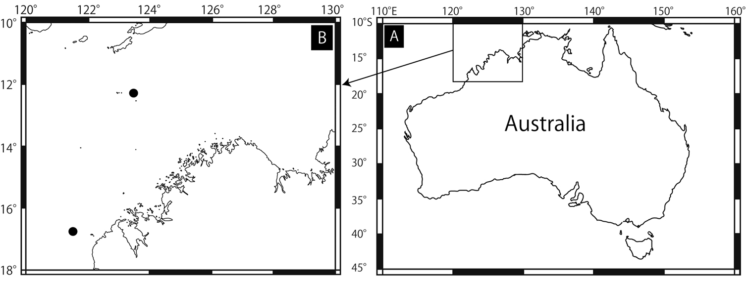
Collected sites of Squamophis albozosteres sp. n. Northern solid circle is for Ashmore Reef and southern one is off Broome.

Squamophis albozosteres sp. n., holotype (MV F162657). A aboral view B oral view. Arrows indicate the arm of another ophiuroid gripped by Squamophis albozosteres.
Oral surface of disc entirely covered by only white, flat and polygonal plate-shaped epidermal ossicles, 50–100 µm long and approximately 50 µm thick (Fig. 4D–G). Four to five triangular teeth forming a vertical row on dental plate. Domed granule-shaped oral papillae lying on either side of jaw (Fig. 4F).
Lateral interradial surface of disc nearly vertical, covered by epidermal ossicles similar to those on oral surface (Fig. 4H). Two genital slits (0.6 mm long and 0.3 mm wide) present in each interradius. No distinct ossicles suggesting existence of madreporites or oral plates observed on any oral interradius, and only epidermal ossicles covered these surfaces (Fig. 4H).
Arms. Arms simple, five in number, no abrupt change in width near the arm base (Fig. 3). The basal portion of the arm 1.4 mm wide and 1.5 mm high, with an arched aboral surface and flattened oral surface. Arms tapering gradually toward the arm tip (Figs 3, 5A, D, G).
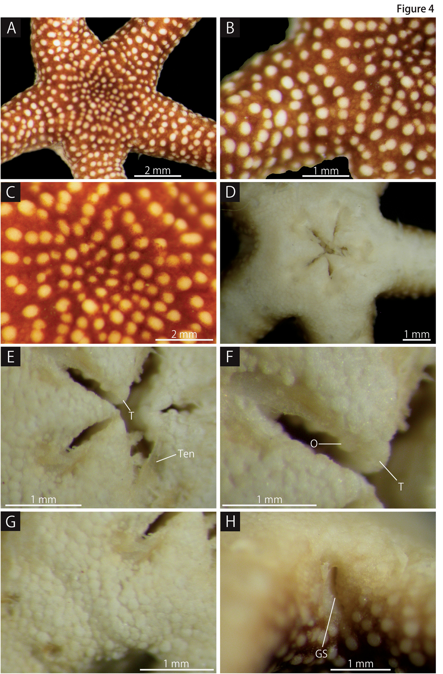
Squamophis albozosteres sp. n., holotype (MV F162657). A aboral disc and basal portion of the arms B periphery of the disc and basal portion of the arm C aboral central disc D oral disc E oral central disc F jaws G oral periphery of the disc H lateral interradius of the disc. Abbreviations: GS - genital slit; O - oral papillae; T - teeth; Ten - Tentacles.
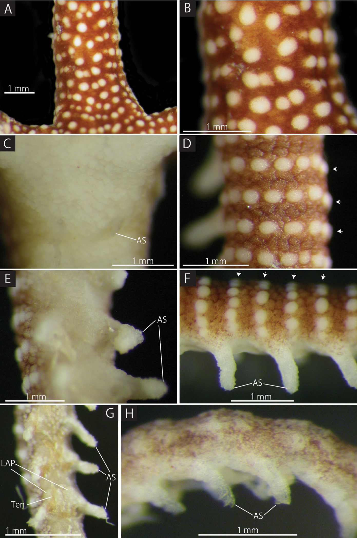
Squamophis albozosteres sp. n., holotype (MV F162657). A, B aboral basal portion of the arm C oral basal portion of the arm D aboral middle portion of the arm, each arrow head indicates a row of white ossicles E oral middle portion of the arm F lateral middle portion of the arm, each arrow head indicates a row of white ossicles G oral distal portion of the arm H lateral distal portion of the arm. Abbreviations: - AS arm spine; LAP - lateral arm plate; Ten - tentacles.
The aboral and lateral surface of the base of arms completely covered by white epidermal ossicles, 150–300 µm long, approximately 60 µm thick, and brown epidermal ossicles, 100–300 µm long, approximately 50 µm thick (Fig. 5A, B), similar to those on aboral periphery of disc. Epidermal ossicles on basal portion of arms covered by thin skin. Brown epidermal ossicles obscured by skin, similar to those on aboral surface of disc. Oral surface of the base of arm covered by white epidermal ossicles, 50–100 µm long, approximately 50 µm thick, similar to those on oral surface of disc (Fig. 5C). From basal to middle portion of the arms, the size of plate-shaped epidermal ossicles decreasing on both the aboral and lateral surfaces (Fig. 5D, F), the white domed ones to 100–200 µm and the brown polygonal ones to 100–150 µm. Brown polygonal ones on oral surface decreasing to 50 µm (Fig. 5E). The distal sections of arms covered by scattered granule-shaped epidermal ossicles of 30 µm, finally disappearing near arm tip (Fig. 5G, H).
First to third tentacle pores lacking arm spines; 4th and more distal pores with one arm spine. Arm spines on basal one-third of arm ovoid, minute, approximately one-third to one-half the length of corresponding arm segment (Fig. 5C). Arm spines in middle one-third of arm the same length as corresponding arm segment, bearing fine thorns at their apex (Fig. 5E, F). Arm spines on distal one-third of arm hook-shaped with conspicuous lateral secondary teeth along inner edge (Fig. 5G, H). Length of hook-shaped arm spines gradually decreasing to two-thirds of the corresponding arm segment on distal third of arm, and number of secondary teeth decreasing from two to one. All tentacles pores lacking a sheath around the cylindrical, narrow tube feet (Figs 4E, 5G).
Lateral and ventral arm plates completely concealed by epidermal ossicles over basal to middle portion of arms, but lateral arm plates visible in distal portion of arms (Fig. 5G).
Color.Aboral surface of disc brown, with white spots highlighting the domed epidermal ossicles. Pigmentation on aboral distal portion of arms lighter and appearing purple, finally disappearing at the tip (Fig. 5H). Oral side entirely white (Fig. 3).
MVF162658: Disc diameter 5.3 mm, arm length at least 200 mm.
Flat and polygonal plate-shaped epidermal ossicles at aboral periphery of disc, approximately 236 µm long and 43 µm thick (Fig. 6A, B), the white, round and domed plate-shaped epidermal ossicles approximately 136 µm long and 40 µm thick (Fig. 6C, D). On aboral surface at base of arm, domed ossicles slightly oblong, approximately 226 µm long and 34 µm thick (Fig. 6E, F), whereas the other ossicles flat and round, granule-shaped, 64 µm long and 20 µm thick (Fig. 6G, H).
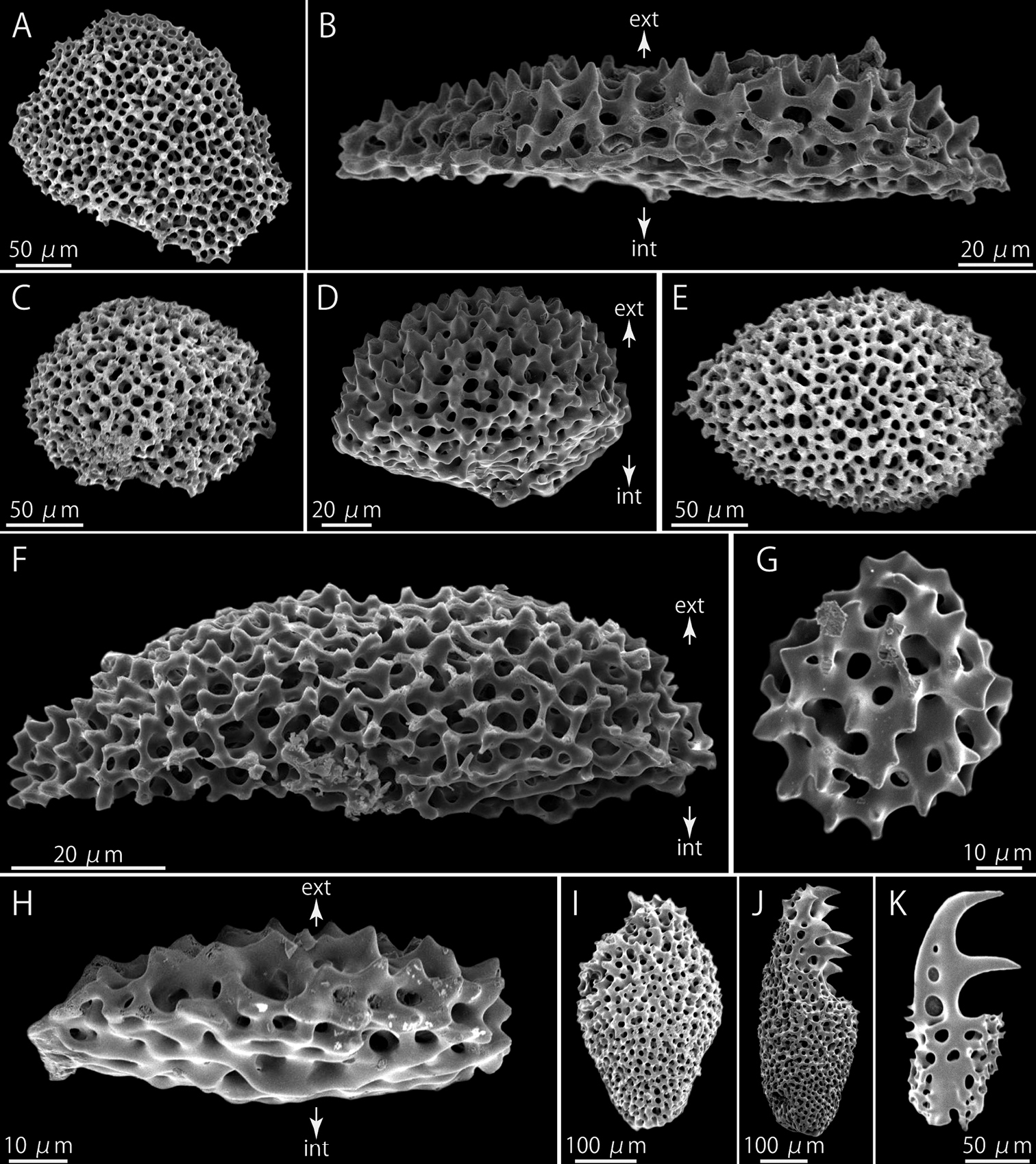
Squamophis albozosteres sp. n., paratype(MV F162658). SEM photographs of internal ossicles. A, B polygonal plate-shaped epidermal ossicles at the aboral periphery of the disc, external (A) and lateral (B) views C, D domed plate-shaped epidermal ossicles at the periphery of the disc, external (C) and lateral (D) views E, F domed plate-shaped epidermal ossicles on the aboral middle portion of the arm, external (E) and lateral (F) views G, H granule-shaped epidermal ossicles on the oral middle portion of the arm, external (G) and lateral (H) views I–K arm spines from basal (I), middle (J) and distal (K) portion of the arm. Arrows indicate the orientation (B, D, F, H): ext - external side; int - internal side.
The radial shields flat and oblong, single-layered, approximately 1.15 mm in length and 0.57 mm in width (Fig. 1B).
Arm spines on basal one-third of arm ovoid (Fig. 6I), in middle cylindrical, bearing fine thorns at tip (Fig. 6J), and distally, they hook shaped with conspicuous secondary teeth along inner edge (Fig. 6K). Number of secondary teeth decreasing gradually to one along distal quarter of arm.
Each lateral arm plate associated with one arm spine and has separate muscle and nerve openings (
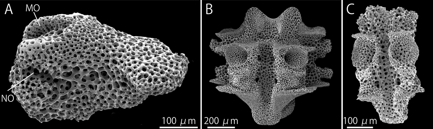
Squamophis albozosteres sp. n., paratype(MV F162658). SEM photographs of internal ossicles. A lateral arm plate from middle portion of the arm B, C vertebrae from middle (B) and distal (C) portion of the arm, oral views. Abbreviations: MO - muscle opening; NO - nerve opening.
Although only three specimens have been
collected, some morphological variation was observed. The smaller
holotype (3.4 mm in disc diameter) has no abrupt reduction in arm
thickness, but the basal portion of the arm on the two larger paratypes
(5.3 mm and 5.6 mm in disc diameter) are slightly widened. The
difference between the three specimens may be due to a difference in
their sexual maturity or reproductive state similar to the congener, Squamophis amamiensis (
North-western Australia; 95–108 m. Type locality: off Broome, 100–108 m (Fig. 2).
The specific name is a masculine noun in apposition formed as a compound of Latin words, albus (adjective, meaning “whitish”) and a plural form of zoster (masculine noun, meaning “ring”), referring to the rings of white plate-shaped dermal ossicles of arms.
Squamophis albozosteres sp. n. and its congener, Squamophis amamiensis,
are similar to each other, however, they can be distinguished by the
morphology of the epidermal ossicles on the aboral body and by
pigmentation. Squamophis albozosteres
has conspicuously white, domed and plate-shaped epidermal ossicles on
the aboral side of the disc and basal to middle portion of the arms,
forming two transverse rows on the lateral and aboral surfaces of each
arm segments (Fig. 5F). Whereas Squamophis amamiensis
has only uniform coloured, flattened and plate-shaped epidermal
ossicles on the corresponding surfaces. The aboral body surface of Squamophis albozosteres is basically brown with white spots and the tips of the arms are light purple, finally with no color, but that of Squamophis amamiensis is uniformly orange or pinkish brown (
We wish to express our sincere gratitude to two anonymous reviewers for their critical reading of the manuscript and constructive comments, and Mark Norman and Chris Rowley (MV) for arranging loans of specimens. Thanks are extended to the captain, crew and scientific staff of the R/V Southern Surveyor SS05/2007. This work was supported by grants from the Research Institute of Marine Invertebrates (Tokyo, Japan), the Showa Seitoku Memorial Foundation, and the Japanese Society for the Promotion of Science (Scientific Research [C] No. 22570104, Research fellowships for Young Scientists No. 22506).