


(C) 2011 Claus Rasmussen. This is an open access article distributed under the terms of the Creative Commons Attribution License, which permits unrestricted use, distribution, and reproduction in any medium, provided the original author and source are credited.
For reference, use of the paginated PDF or printed version of this article is recommended.
Callosphecodes Friese, 1909, a synonym or perhaps subgenus of Sphecodes Latreille, 1804, is known on the basis of one female of Sphecodes ralunensis (Friese, 1909)from New Britain and one female and one male of a similar species, Sphecodes manskii (Rayment, 1935) from northeastern Australia. The male is here described for the first time and the females of the two species are compared for the first time. In spite of considerable collecting, only these three specimens have appeared in over a century. Descriptions and illustrations are provided.
Apoidea, Anthophila, Halictinae, Halictini, Sphecodina, Sphecodes, New Britain, Bismarck Archipelago, Australia, taxonomy, cleptoparasite
Even in parts of the world where there has been little investigation of the bee fauna, taxa of bees so distinctive as to have received genus-group names a century or more ago have usually been collected several times so that multiple specimens are now known. Callosphecodes Friese, 1909, however, until now has been known from only two female specimens of different species from localities over 1500 km apart. A third specimen, a male, is herein reported for the first time. To judge by the lack of pollen manipulating and carrying structures in females, this is a cleptoparasitic group. Many cleptoparasites are uncommon, and it seems possible that Callosphecodes is a rare insect, not only in collections but also in the field.
We follow various earlier authors in considering Callosphecodes to be a synonym or possibly a subgenus of Sphecodes
Latreille, 1804, which is the most common and widespread genus of
cleptoparasitic Halictinae. This cleptoparasitic group was given
subtribal status as the Sphecodina in the tribe Halictini (subfamily
Halictinae) in the phylogenetic study by
Callosphecodes was proposed as a subgenus of Sphecodes by
Subsequent views on the position of Callosphecodes have varied from a distinct genus (
A second specimen of Callosphecodes was described as Mellitidia manskii (
Also, in the Australian National Insect Collection, was found a male, judged on the basis of similarity to the female and on geography, to be Sphecodes manskii. It was collected in 1980 by Josephine C. Cardale in Mount Webb National Park (15.045S, 145.07E), Queensland, Australia, about 100 km from Cairns, the type locality for Sphecodes manskii.
When reviewing the cleptoparasitic groups of Halictidae,
The following descriptive comments, largely following the pattern of Michener’s (1978) account of Sphecodes, are based on the three known specimens of the Callosphecodes group, that is Sphecodes ralunensis and manskii (Figs 1, 6 and 13). The description of Sphecodes by
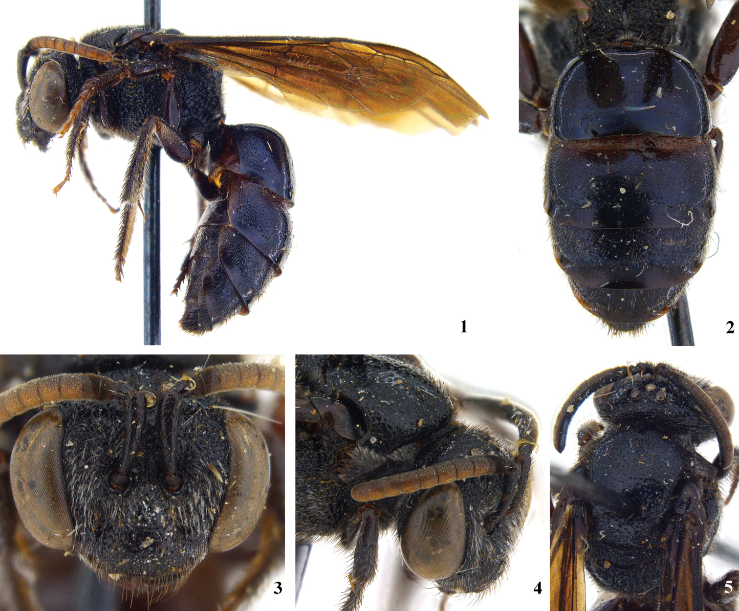
Holotype female of Sphecodes ralunensis: 1 lateral habitus 2 metasoma 3 facial aspect 4 dorsolateral aspect of head and pronotum 5 dorsal aspect of mesosoma and head.
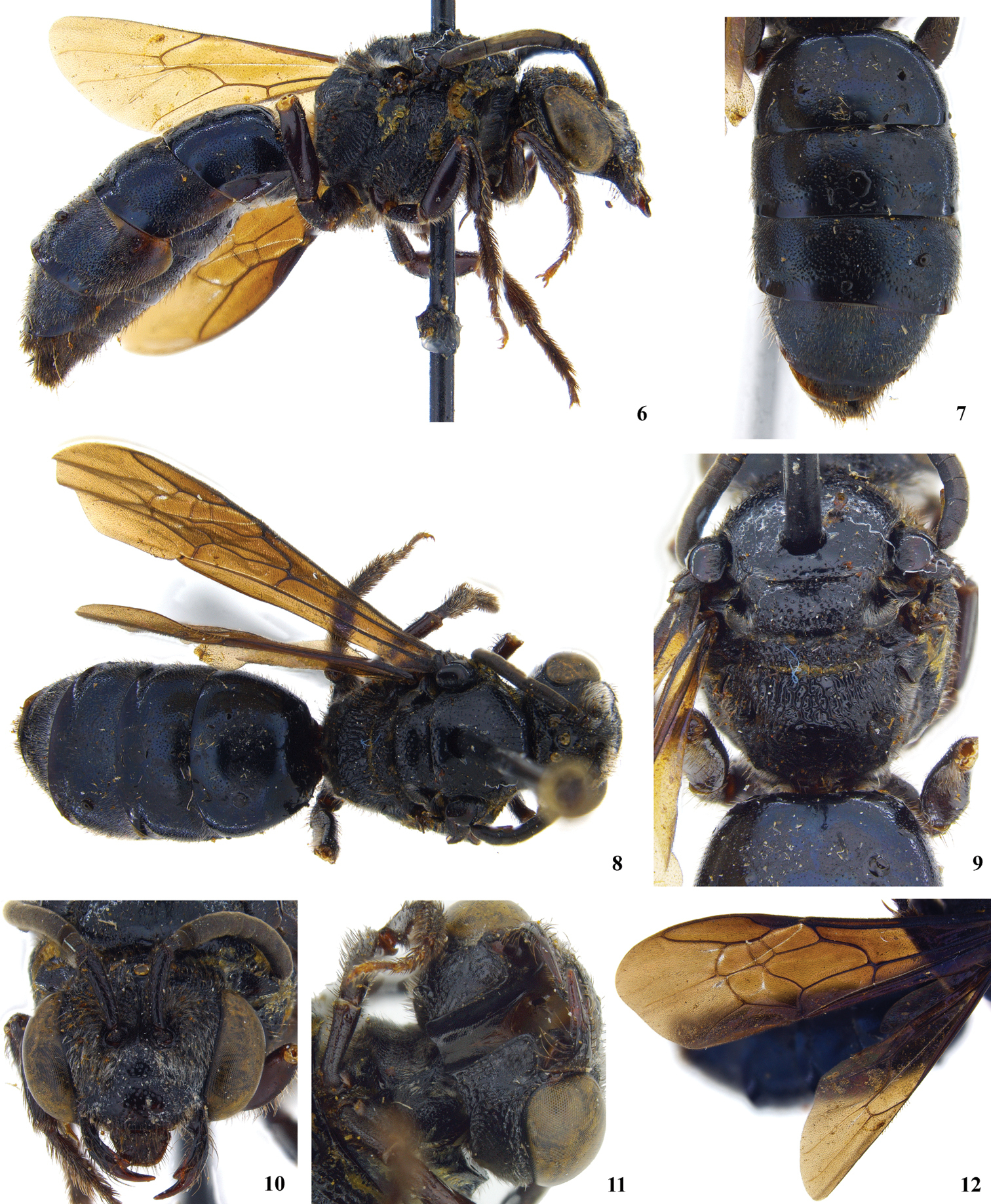
Holotype female of Sphecodes manskii: 6 lateral habitus 7 metasoma 8 dorsal habitus 9 mesosoma including propodeum 10 facial aspect 11 hypostomal carina with tooth at posterior end 12 forewing pattern.
Both sexes: Black, metasomal terga with feeble bluish, purplish, or blue green metallic tints (Figs 2 and 7); wings strongly infuscated (fig. 12). Punctation of head and thorax coarse (fig. 4; moderately fine in Eupetersia); punctures of mesoscutum, especially posteriorly, widely separated (by much more than puncture diameter) by shining surface (fig. 5). Head in facial view much wider than long, clypeus more than twice as wide as long (Figs 3 and 10). Eyes hairless. Hairs of antennal flagellum all very short. Preoccipital carina strong and distinct. Posterior end of hypostomal carina with tooth (fig. 11). Pronotum with horizontal surface of collar almost absent medially, forming lateral angle below which a vertical ridge extends downward; vertical ridge approaching or merging with a more laterally directed ridge that extends toward coxal base; another carina from lateral angle extends across posterior lobe of pronotum. Anterior extremity of mesoscutum convex. Scutellum gently biconvex because of feeble longitudinal median depression. Propodeum with dorsal area strongly areolate, about as long as scutellum, area broadly rounded posteriorly (fig. 9); posterior and lateral surfaces of propodeum with few short plumose hairs in addition to longer hairs. Wings with hairs rather long and dense throughout (as in Eupetersia); stigma moderate; marginal cell pointed at apex; free part of marginal cell beyond submarginal cells longer than part subtended by submarginal cells, which part extends well beyond apex of stigma. Second and third submarginal cells each receiving a recurrent vein (fig. 12). First metasomal tergum broader than long. Second tergum in lateral view with base somewhat depressed forming weak constriction between first and second terga. Posterior margins of terga 2 – 4 broadly depressed, hairless, impunctate.
Female: Mandible with large subapical tooth (fig. 10; unlike Eupetersia). Labrum with broad, flat apical process about two thirds as broad as long. Legs robust, hind femur about three times as long as broad; basitibial plate elevated; long hairs on outer side of hind tibia plumose; hind tibial spine finely serrate. Fifth metasomal tergum, unlike preceding terga, with apical margin fringed except middle part of margin which has smooth, hairless area in front of fringeless part of margin. Pygidial plate broader than in Eupetersia.
Male: Antennae longer than those of female, flagellum thickened (fig. 13; unlike Eupetersia), somewhat crenulate, first flagellar segment broader than long, second longer than first, both first and second shorter than subsequent segments but not very short as in Eupetersia. Labrum not visible on specimen. Second hind tarsal segment longer than third, base broader than base of third. Gonocoxite finely striate, without margined depression as in Eupetersia. Gonocoxite with basal setose lobe (Figs 15 and 16).
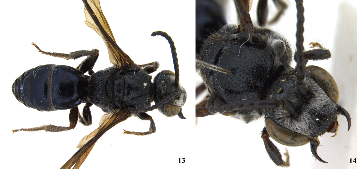
Male of Sphecodes manskii: 13 dorsal habitus; 14 dorsolateral aspect of head and pronotum.
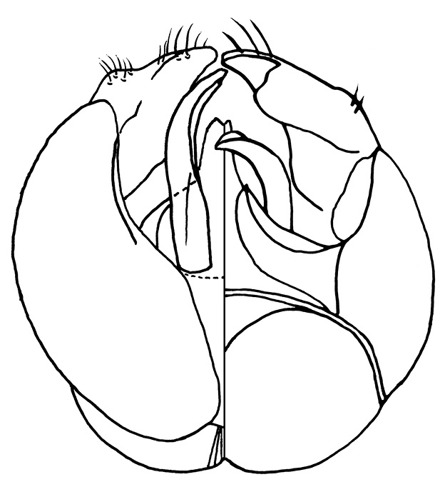
Male genitalia of Sphecodes manskii: 15 Dorsalventral view of genitalia; 16 7th metasomal sternum.
Specific differences: The holotypes (both females) are very similar and we have no way of knowing whether the differences between them are specific differences or indicate variation within a species. The differences (observed by CR) are as follows: Lateral margin of propodeum (immediately below metanotum) in Sphecodes ralunensis largely areolate, in Sphecodes manskii widely strigulate and less areolate. Gena of Sphecodes ralunensis sparsely covered with plumose, light colored setae, in Sphecodes manskii densely covered with white setae. Flagellum in Sphecodes ralunensis ferruginous (fig. 3), in Sphecodes manskii dark brown (fig. 10). Measurements are as follows for the Sphecodes ralunensis holotype female: Total body length about 10 mm; forewing length (including tegula) 8.8 mm; head width 3.1 mm; head length (anterior margin of clypeus to summit of vertex) 2.5 mm; mesoscutum width 2.1; mesoscutum length 2.0 mm. The Sphecodes manskii holotype female: Total body length about 12 mm; forewing length (including tegula) 9.5 mm; head width 3.2 mm; head length (anterior margin of clypeus to summit of vertex) 2.5 mm; mesoscutum width 2.3; mesoscutum length 2.0 mm. Male Sphecodes manskii: Total body length about 11 mm; forewing length (including tegula) 8.1 mm; head width 2.9 mm; head length (anterior margin of clypeus to summit of vertex) 2.5 mm; mesoscutum width 2.3; mesoscutum length 2.2 mm.
We are very thankful to the curators for loan of the specimens: Frank Koch of the Museum für Naturkunde der Humboldt-Universität, Berlin, and Nicole Fisher of the Australian National Insect Collection, Canberra. Also to Carlsberg Foundation for support (C.R.) and Lars Vilhelmsen and Verner Michelsen of the Natural History Museum of Denmark for providing access to the imaging system.