


(C) 2012 Xia Gao. This is an open access article distributed under the terms of the Creative Commons Attribution License 3.0 (CC-BY), which permits unrestricted use, distribution, and reproduction in any medium, provided the original author and source are credited.
For reference, use of the paginated PDF or printed version of this article is recommended.
The two leafhopper species in the genus Parazyginella Chou & Zhang from China are reviewed and illustrated including one new species, Parazyginella tianisp. n. whichis described. A key to separate males of the two species is provided.
Homoptera, Auchenorrhyncha, taxonomy
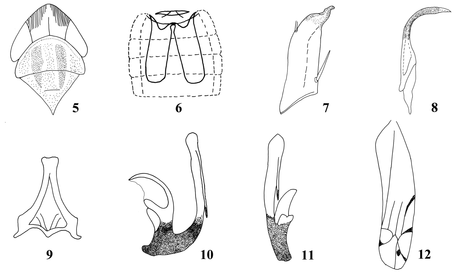
Zyginellini is one of the smaller tribes in the large leafhopper subfamily Typhlocybinae. Members of the subfamily feed on trees, shrubs and herbs and some occur on economic crops including two species of Zyginellini, Zyginella mali (Yang) and Zyginella minuta (Yang), which damage apple trees in China. Members of this tribe can be distinguished by their usual bright coloration with distinct patterns and by the hindwing venation with the vannal veins separate apically, with only one transverse vein visible and submarginal vein extended directly to vein CuA, forming one open cell (Fig. 13).
The Oriental Zyginellini genus Parazyginella was erected by
http://species-id.net/wiki/Parazyginella
Parazyginella lingtianensis Chou & Zhang, 1985
Body flattened. Vertex conically produced, middle length nearly equal to width and length of pronotum; coronal suture distinct. Head and pronotum whitish yellow. Scutellum and venter yellow. Forewing with base of 1st and 4th apical cells at same level; 3rd apical cell triangular and with a dark spot; hind margin of brochosome field and veins in apical area of wing touched with dark brown.
Abdominal apodemes elongate, reaching beyond 5th abdominal sternite.
Male pygofer strongly sclerotized, with short, finger-like process caudo-dorsally and few scattered microsetae. Subgenital plates broad at base, distally abruptly tapered to short narrow apex, with few macrosetae basally. Paramere simple, with central part expanded, apical part tapering to acute apex and strongly bent. Connective with short arms and medial lobe present. Aedeagus asymmetrical with basal part strongly sclerotized with short preatrium and large dorsal apodeme, the latter laterally compressed with distal anterior region membranous and distal posterior region strongly curved anteriorly; shaft elongate, with a single elongate apical process on one side; gonopore obscure.
Parazyginella resembles Zyginella Löw, but differs in its more greatly developed dorsal apodeme of the aedeagus and male pygofer with a dorsal finger-like process and without long macrosetae (Figs 16, 17).
China(Guangxi, Yunnan).
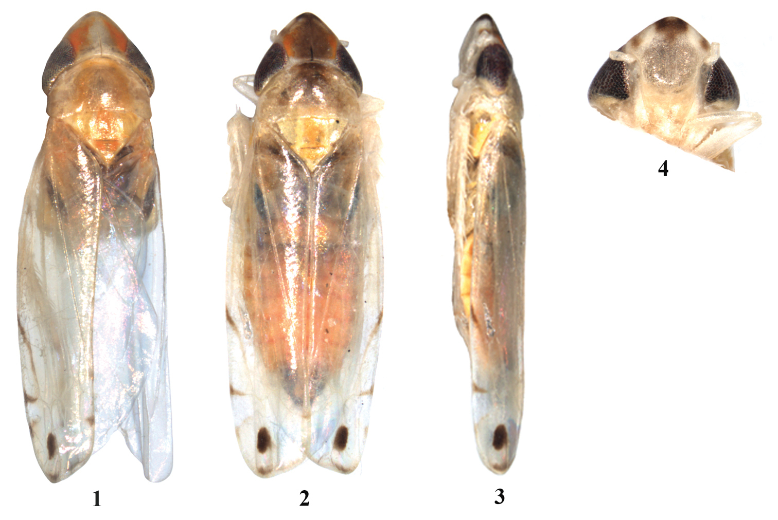
1 Parazyginella lingtianensis, dorsal habitus 2–4 Parazyginella tiani sp. n. 2 dorsal habitus 3 lateral habitus 4 face.
| 1 | Head without brown markings. Aedeagal shaft in lateral view similar in width throughout length, process moderately long and slim, lying parallel to shaft (Figs 10, 11) | Parazyginella lingtianensis |
| – | Head with brown markings (Figs 2–4). Aedeagal shaft in lateral view slightly expanded from midlength to near apex, process long and stout, directed slightly towards basal apodeme (Figs 21, 22) | Parazyginella tiani sp. n. |
http://species-id.net/wiki/Parazyginella_lingtianensis
Figs 1, 5–12Head and pronotum whitish yellow. Scutellum and venter yellow; vertex and pronotum either side of midline marked with orange (Figs 1, 5); 3rd apical cell of forewing with dark elliptical spot (Figs 1, 12).
Abdominal apodemes reaching middle part of 6th abdominal sternite (Fig. 6).
Subgenital plates with one macroseta near base, apex beak-like (Fig. 7). Paramere slightly expanded subapically (Fig. 8). Aedeagal shaft in lateral view similar in width throughout length, process moderately long and slim, lying parallel to shaft (Figs 10, 11).
Male 3.00 mm (including wing).
Holotype, male, China: Guangxi Prov., Lingchuan, Lingtian, 5 June 1984, coll. Lu Xiaolin, lamp (NWAFU).
China (Guangxi).
The male genitalia of the unique type could not be found. Therefore the pygofer (originally not figured) could not be examined and compared to the new species. Also, the original figure of the aedeagus (shown here, Fig. 10) did not show the membranous area of the basal apodeme shown in our new species. We conclude that this area was probably overlooked and add a line to the figure to show its approximate position.
Parazyginella lingtianensis (after
urn:lsid:zoobank.org:act:A19A9268-1164-43C8-8B94-0C0CE69CBA7F
http://species-id.net/wiki/Parazyginella_tiani
Figs 2–4, 13–22Head and pronotum whitish yellow; scutellum and venter yellow; vertex with disc and apex dark brown, orange laterally; face sordid white, with a dark brown patch above antennae; dorsum of abdomen stramineus. Forewing with dark spot in 3rd apical cell (Figs 2, 3).
Abdominal apodemes nearly reaching end of 6th abdominal sternite (Fig. 14).
Male pygofer with short, sclerotized, sickle-like process caudo-dorsally (Figs 16, 17). Subgenital plates with two macrosetae near base, apex digitate with few microsetae (Figs 18, 20). Paramere subapically with row of fine setae on outer margin and row of sensory pits on inner margin; with curved apical part with sinuate ridge (Figs 18, 19). Aedeagal shaft in lateral view slightly expanded from midlength to near apex, process long and stout, directed slightly towards basal apodeme, apex ornamented (Figs 21, 22).
2.95–2.98 mm (including wing).
Holotype, male, China: Yunnan Province, Sanchahe, 7 June 1991, coll. Tian Rungang (NWAFU). Paratypes, two males, seven females, same data as holotype (NWAFU, BMNH).
China (Yunnan).
The new species resembles Parazyginella lingtianensis but differs in having brown markings on the vertex (compare Figs 1 and 2) and different shaped aedeagus as noted in the key.
The new species is named after the collector’s family name in gratitude.
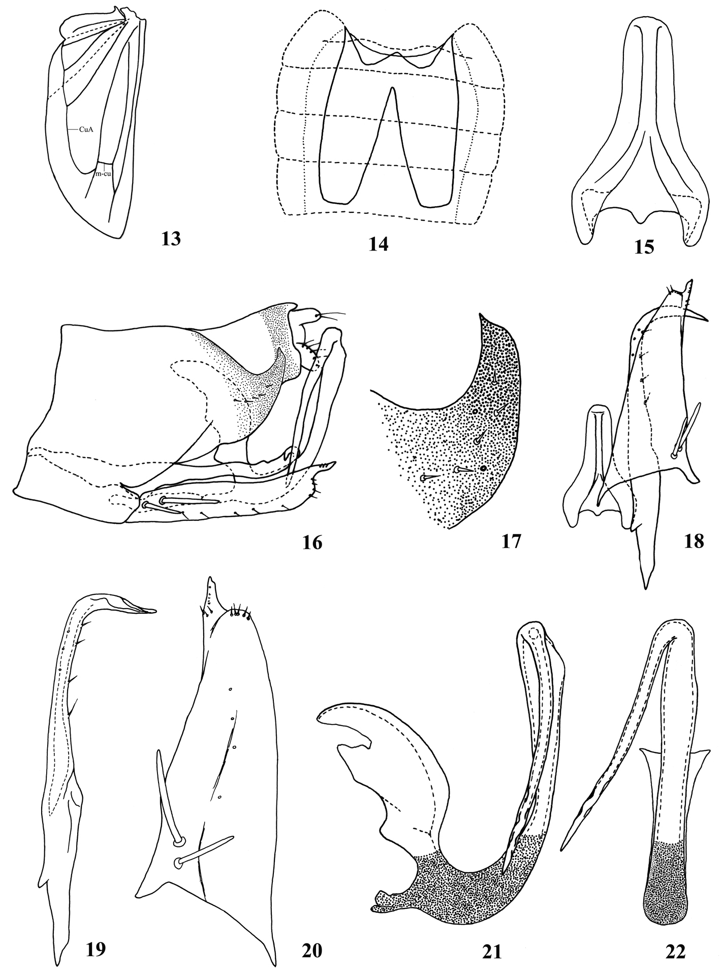
Parazyginella tiani sp. n. 13 Hindwing 14 Abdominal apodeme 15 Connective 16 Genital capsule, lateral view 17 Apical part of male pygofer 18 Paramere, connective, subgenital plate, ventral view 19 Paramere 20 Subgenital plate 21 Aedeagus, lateral view 22 Aedeagus, posterior view.
We express our sincere thanks to Dr. I. Dworakowska (Canada) for her contribution to the knowledge of Chinese Typhlocybinae during her visit to the Entomological Museum, Northwest A & F University, and revising the manuscript. We also appreciate Prof. Dr. J. R. Schrock (Emporia State University, USA), Mr M. Webb (The Natural History Museum, London, UK) for revising the manuscript. The research is supported by “The National Natural Science Foundation of China” (30770262), and “The Ministry of Education of China” (TS2011XBNL061), and also partly by “Basic Science and Technology Project of the Ministry of Science and Technology of China” (2006FY120100) and “Northwest A&F University Grants for Outstanding Young Faculty Members” (Z111020905).