


(C) 2012 Guo-Quan Wang. This is an open access article distributed under the terms of the Creative Commons Attribution License 3.0 (CC-BY), which permits unrestricted use, distribution, and reproduction in any medium, provided the original author and source are credited.
For reference, use of the paginated PDF or printed version of this article is recommended.
A new genus and two new species belonging to subfamily Cecidophyinae, namely Kyllocarus reticulatus gen.n., sp. n. infesting Lithocarpus brevicaudatus (Skan) Hay. (Fagaceae) and Gammaphytoptus schimae sp. n.infesting Schima superba Gardn. et Champ. (Theaceae) are described and illustrated. Both new species are vagrants on their respective host plants. Cecidophyes digephyrusKeifer, 1966 is newly recorded for China.
Eriophyoidea, eriophyoid mites, Cecidophyini, Colomerini, taxonomy, China
The subfamily Cecidophyinae holds two tribes, Cecidophyini and Colomerini, which were differentiated by the former scapular tubercles and setae are absent and the later tubercles and setae are present. So far, sixteen genera and thirty-three species are known from China (
Specimens were located with the aid of a magnifying glass on plant material in the field, and specimens were collected into and preserved in a sucrose-ethanol solution (75%). The mites were cleared in Nesbitt’s solution and mounted in Heinze medium on glass slides at room temperature according to
Type specimens are deposited in the Department of Plant Protection, Guangxi University, Nanning. All measurement units are in micrometers (μm) and rounded off to the nearest full number, and are lengths when not specified. Specimens were examined with an Olympus CX41 (Philippines) microscope with phase contrast. The number of measured specimens is given in parentheses.
Taxonomy Tribe Cecidophyini Keifer, 1966 Genus Cecidophyes Nalepa, 1887Type species. Phytopus galii Karpelles, 1884.
Body fusiform. Prodorsal shield with frontal lobe present; median, admedian lines and submedian lines complete, connected with three transverse lines forming network; scapular tubercles and setae absent. Coxisternal plates sculptured with lines. Legs with normal segments and usual setae, tarsal empodium entire 6-rayed, tarsal solenidion knobbed. Dorsal opisthosoma evenly rounded, dorsal annuli 62–68, with elongated microtubercles; ventral annuli 62–68, with rounded microtubercles, setae h1 absent. Female genital near coxisternal plates, coverflap with two rows of ridges.
10 females, Qingliangfeng National Nature Reserve, Lin’an City (30°10'N, 119°07'E), Zhejiang Province, China, 24. VII. 2007, from Quercus sp. (Fagaceae), collected by Guo-Quan Wang, mounted on individual slide.
USA, China (Zhejiang).
The mites are vagrants on the surfaces of the leaves, no visible damage seen.
Up to date, nine species of Cecidophyes have been known infesting Quercus spp.: Cecidophyes caliquerci (Keifer, 1944) infesting Quercus lobata Nee and Quercus marilandica Muen., Cecidophyes digephyrus Keifer, 1966 infesting Quercus vaccinifolia Kell., Cecidophyes lyrata Keifer, 1959 infesting Quercus lyrata Walt., Cecidophyes pusilla Keifer, 1962 infesting Quercus falcata Michx, Cecidophyes quercialbae Keifer, 1959 infesting Quercus alba L., Cecidophyes querciphagus (Keifer, 1939) infesting Quercus sp., Cecidophyes reticulatus Livshitz, Mitrofanov et Vasil’yeva, 1979 infesting Quercus pubescens Willd., C. tampae Keifer, 1966 infesting Quercus virginiana and Cecidophyes tristernalis (Nalepa, 1898) infesting Quercus cerris L. Among them, only one species, Cecidophyes tampae occurred in China. Cecidophyes digephyrus is second Cecidophyes species from China (
urn:lsid:zoobank.org:act:91681721-9DF7-483B-941C-C882DBF8A4FF
Kyllocarus reticulatus Wang, Wei & Yang, 2012, sp. n.
The new genus with flattened fusiform body, palp genual seta strongly angular prodorsal shield lacking scapular setae (sc) and tubercles, strong wide frontal lobe over gnathosoma, legs normal, except leg II lacking genual seta l’’. Sternal apodeme present; opisthosoma differentiated into broader smooth dorsal annuli and narrower microtuberculate ventral annuli; empodium simple; genitalia very close to coxae, bearing two ranks of numerous ridges.
This cecidophyine mite is very near to Kolacarus in that the genual seta l’’ is absent on Leg II; however, it differs from other cecidophine mites in that palp genual seta d is bent or crooked, possibly minutely bifurcate. It differs from Kolacarus in that the mite has a wide, strong frontal lobe projecting from the prodorsal shield over the gnathosoma; Kolacarus has a normal palp genual seta d, and no frontal lobe on the anterior prodorsal shield.
Kyllo- from Gr. Kyllos, crooked + -carus from Acarus; the name is masculine.
urn:lsid:zoobank.org:act:7358B76E-13F4-4968-82F4-2500479953D0
http://species-id.net/wiki/Kyllocarus_reticulatus
Figs 1–7Body fusiform, white translucency or yellow. Gnathosoma curved obliquely downward, dorsal genual setae (d) bend forming obtuse angle at middle. Prodorsal shield with frontal lobe present; all lines bold and connected with transverse lines forming network; scapular tubercles and setae absent. Coxisternal plates sculptured with lines, prosternal apodeme present, coxigenital annuli 4. Legs segments normal, legs II with genual setae (l’’) absent, tarsal empodium entire, 6-rayed, tarsal solenidion knobbed. Dorsal opisthosoma with shallow median furrow, dorsal annuli smooth; ventral annuli with rounded microtubercles, setae h1 absent. Female genitalia coverflap with two rows of ridges
Female (n = 11). Body fusiform, white translucency or yellow, 172 (150–204), 75 (69–79) wide, 60 (54–63) thick.
Gnathosoma. Curved obliquely downward, 30 (28–31), coxal setae (ep) 6 (6–7), dorsal genual setae (d) bend forming obtuse angle at middle, 11 (10–12); cheliceral stylets 31 (30–33).
Prodorsal shield. 63 (58–70), 69 (65–74) wide, frontal lobe present; all lines bold; median, admedian and submedian lines complete, connected with transverse lines forming network; scapular tubercles and setae absent.
Coxisternal plates. Prosternal apodeme present, coxisternal plates I and II sculptured with lines; anterolateral setae on coxisternum I (1b) 3 (3–4), 13 (12–13) apart; proximal setae on coxisternum I (1a) 5 (5–6), 13 (12–13) apart; proximal setae on coxisternum II (2a) 31 (29–33), 29 (29–30) apart. Coxigenital annuli 4.
Legs. Segments normal. Legs I 34 (30–37), trochanter 2 (2), femur 11 (10–11), femoral setae (bv) 13 (10–15); genu 5 (4–5), genual setae (l’’) 30 (29–32); tibia 7 (6–8), tibial setae (l') located laterally and distally, 15 (13–18); tarsus 8 (7–9), inner fastigial tarsal setae (ft’) 27(25–28), outer fastigial tarsal setae (ft’’) 18 (15–20), unguinal tarsal setae (u’) 5 (4–5); tarsal empodium entire, 12 (11–13), 6-rayed, tarsal solenidion 7 (6–8), knobbed. Legs II 27 (26–30), trochanter 2 (2), femur 10 (10–11), femoral setae (bv) 23 (20–25); genu 4 (4–5), genual setae (l’’) absent; tibia 4 (4–5); tarsus 7 (6–7), inner fastigial tarsal setae (ft’) 24(23–25), outer fastigial tarsal setae (ft’’) 14 (13–15), unguinal tarsal setae (u’) 4 (4–5); tarsal empodium entire, 6 (5–7), 6-rayed, tarsal solenidion 7 (7–8), knobbed.
Opisthosoma. Dorsum with shallow median furrow, dorsal annuli 43, smooth; ventral annuli 63, with rounded microtubercles; setae c2 23 (20–25), on ventral annulus 10th; setae d 71 (63–79), 42 (41–43) apart, on ventral annulus 22th; setae e 11 (8–13), 24 (23–25) apart, on ventral annulus 38th; setae f 24 (22–25), 24 (24–25) apart, on 10th ventral annulus from rear; setae h1 absent, setae h2 31 (26–39).
Female genitalia. Near coxisternal plates, coverflap with two rows of ridges, 24 (23–25), 43 (38–49) wide, proximal setae on coxisternum III (3a) 9 (9–10), 23 (23–24) apart.
Male (n = 2). Body fusiform, 120–140, 58–61 wide.
Prodorsal shield. 53–55, 55–57 wide, frontal lobe present; all lines bold; median, admedian lines and submedian lines complete, connected with transverse lines forming network; scapular tubercles and setae absent.
Coxisternal plates. Prosternal apodeme present, coxisternal plates I and II sculptured with lines; anterolateral setae on coxisternum I (1b) 3, 11 apart; proximal setae on coxisternum I (1a) 5, 11 apart; proximal setae on coxisternum II (2a) 27, 28 apart. Coxigenital annuli 4.
Legs. Segments normal. Legs I 30, trochanter 2, femur 10, femoral setae (bv) 12; genu 4, genual setae (l’’) 27; tibia 6, tibial setae (l') located laterally and distally, 12; tarsus 7, inner fastigial tarsal setae (ft’) 24, outer fastigial tarsal setae (ft’’) 15, unguinal tarsal setae (u’) 4; tarsal empodium entire, 10, 6-rayed, tarsal solenidion 6, knobbed. Legs II 26, trochanter 2, femur 10, femoral setae (bv) 18; genu 4, genual setae (l’’) absent; tibia 4; tarsus 6, iner fastigial tarsal setae (ft’) 21, outer fastigial tarsal setae (ft’’) 12, unguinal tarsal setae (u’) 4; tarsal empodium entire, 5, 6-rayed, tarsal solenidion 7, knobbed.
Opisthosoma. Dorsum with shallow median furrow, dorsal annuli 42, smooth; ventral annuli 62, with rounded microtubercles; setae c2 20, on ventral annulus 10th; setae d 57, 40 apart, on ventral annulus 22th; setae e 7, 21 apart, on ventral annulus 38th; setae f 20, 21 apart, on 10th ventral annulus from rear; setae h1 absent, setae h2 27.
Male genitalia. Near coxisternal plates, 36 wide, proximal setae on coxisternum III (3a) 8, 23 apart.
Type material. Holotype female, China: Zhejiang, Longquan City, Fengyangshan National Nature Reserve (27°53'N, 119°11'E), 27. VII. 2007, collected by Guo-Quan Wang, from Lithocarpus brevicaudatus (Skan) Hayata (Fagaceae). Paratypes, 10 females and 2 males.
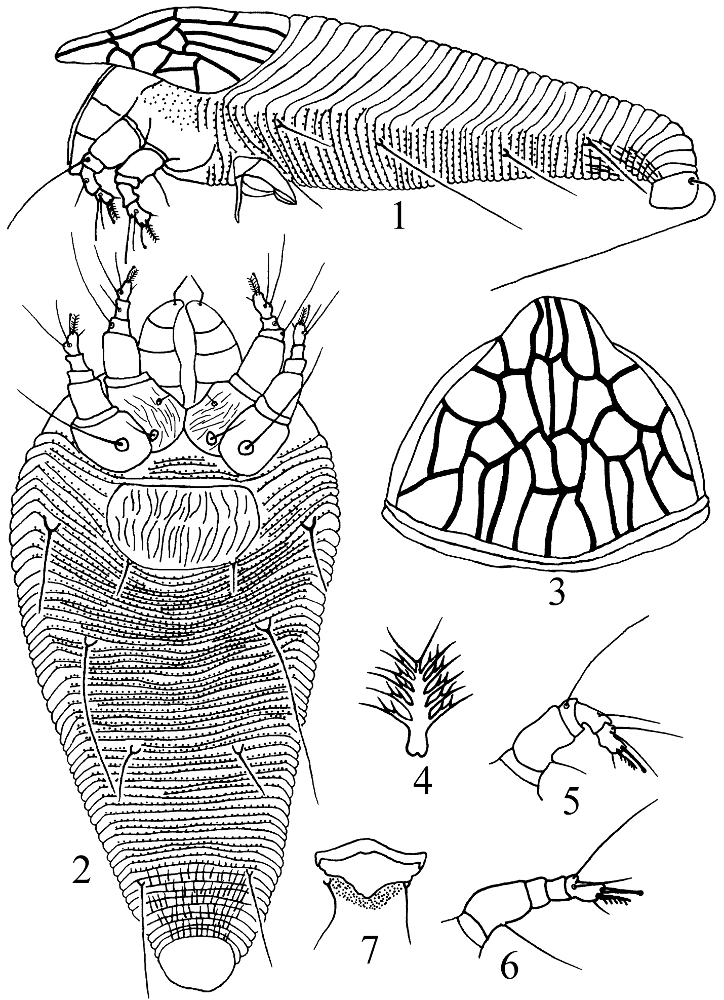
Kolacarus reticulatus sp. n. 1 lateral view of female 2 ventral view of female 3 anterior dorsal view of female 4 tarsal empodium 5 leg I 6 leg II 7 male genitalia
China (Zhejiang).
The species is named after the network-form of the prodorsal shield.
Type species: Gammaphytoptus camphorae Keifer, 1939.
urn:lsid:zoobank.org:act:CEC41DF7-A6BB-4871-9D06-FABC3DDAC219
http://species-id.net/wiki/Gammaphytoptus_schimae
Figs 8–13Body fusiform, yellow. Gnathosoma curved obliquely downward, dorsal genual setae (d) bend forming obtuse angle at middle. Prodorsal shield with frontal lobe present; all lines bold and connected with transverse lines forming network; scapular tubercles and setae absent. Coxisternal plates sculptured with lines, prosternal apodeme present, coxigenital annuli 4. Legs segments normal, legs II with genual setae (l’’) absent, tarsal empodium entire, 6-rayed, tarsal solenidion knobbed. Dorsal opisthosoma with shallow median furrow, dorsal annuli smooth; ventral annuli with rounded microtubercles, setae h1 absent. Female genitalia coverflap with two rows of ridges.
Female (n = 11). Body fusiform, yellow, 183 (169–200), 71 (65–78) wide, 44 (38–52) thick.
Gnathosoma. Curved obliquely downward, 34 (28–35), coxal setae (ep) 2 (2–3), dorsal genual setae (d) 10 (9–11); cheliceral stylets 30 (28–32).
Prodorsal shield. 51 (48–52), 55 (50–63) wide, frontal lobe present; median, admedian and submedian lines complete, connected with three transverse lines forming network; scapular tubercles placed at rear shield margin, 35 (31–39) apart, scapular setae (sc) 8 (8–9), directed backward and divergence.
Coxisternal plates. Prosternal apodeme present, coxisternal plates smooth; anterolateral setae on coxisternum I (1b) 8 (7–9), 13 (12–13) apart; proximal setae on coxisternum I (1a) 25 (19–31), 15 (14–15) apart; proximal setae on coxisternum II (2a) 35 (28–39), 28 (27–30) apart. Coxigenital annuli 4.
Legs. Segments normal. Legs I 36 (34–38), trochanter 2 (2), femur 12 (12–13), femoral setae (bv) 18 (15–22); genu 4 (4–5), genual setae (l’’) 35 (31–40); tibia 10 (9–10), tibial setae (l') located 1/4 from apical, 8 (7–8); tarsus 8 (7–8), iner fastigial tarsal setae (ft’) 20 (18–23), outer fastigial tarsal setae (ft’’) 25 (23–28), unguinal tarsal setae (u’) 5 (5–6); tarsal empodium entire, 7 (7–8), 6-rayed, tarsal solenidion 10 (9–10), knobbed. Legs II 31 (29–34), trochanter 2 (2), femur 11 (11–12), femoral setae (bv) 23 (19–27); genu 3 (3–4), genual setae (l’’) 10 (7–12); tibia 7 (7–8); tarsus 8 (7–8), inner fastigial tarsal setae (ft’) 10 (9–12), outer fastigial tarsal setae (ft’’) 25 (22–29), unguinal tarsal setae (u’) 5 (5–6); tarsal empodium entire, 8 (8–9), 6-rayed, tarsal solenidion 10 (9–11), knobbed.
Opisthosoma. Dorsum evenly rounded, dorsal annuli 59–60, with semi-translucency elongated microtubercles; ventral annuli 81, with filament microtubercles; setae c2 38 (35–40), on ventral annulus 13th; setae d 45 (37–50), 43 (38–45) apart, on ventral annulus 28th; setae e 27 (23–32), 25 (23–26) apart, on ventral annulus 44th; setae f 38 (34–45), 23 (21–26) apart, on 7th ventral annulus from rear; setae h1 absent, setae h2 57 (53–65).
Female genitalia. Near coxisternal plates, coverflap with two rows of ridges, 17 (16–18), 30 (29–22) wide, proximal setae on coxisternum III (3a) 20 (17–25), 13 (13–14) apart.
Male. Unknown.
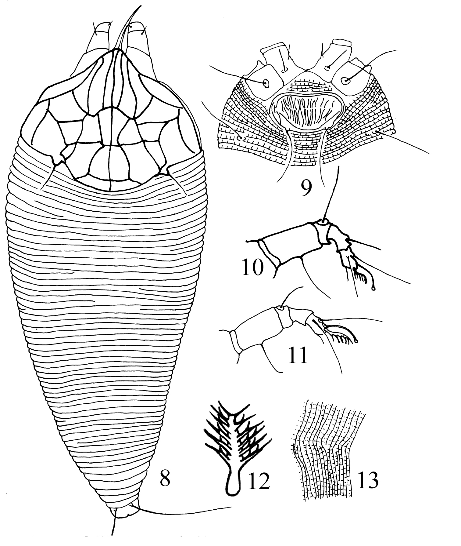
Gammaphytoptus schimae sp. n. 8 dorsal view of female 9 coxigenital area of female 10 leg I 11 leg II 12 tarsal empodium 13 lateral view of annuli.
Holotype female, China: Zhejiang, Longquan City, Fengyangshan National Nature Reserve (27°53'N, 119°11'E), 28. VII. 2007, collected by Guo-Quan Wang, from Schima superba Gardn.&Champ. (Theaceae). Paratypes, 8 females.
China (Zhejiang).
The species is named from the generic name of the type host plant.
This new species is similar to Gammaphytoptus zuihoensus Huang & Wang, 2004, but they can be easily separated as follows: in Gammaphytoptus schimae, median line is complete, setae h1 is absent and infesting Schima superba Gardn.&Champ.; in Gammaphytoptus zuihoensus, median line is incomplete, setae h1 is present and infesting Machilus zuihoensis Hay. var. zuihoensis (
The authors sincerely thank Professor James W. Amrine JR. and the anonymous reviewers for their valuable remarks. Undoubtedly, Prof. Amrine’s very valuable suggestions for the authers to erect the new genus. We would like to thank Prof. Hua Li (College of Agriculture, Guangxi University) for identifying the host plants. This work was supported by the National Natural Science Foundation of China (Grant No. 31160431), the Key Project of Chinese Ministry of Education (Grant No. 211134) and Major Program of Ministry of Science and Technology of the People’s Republic of China (Grant No. 2006FY110500–2).