


(C) 2011 Ricardo Kawada. This is an open access article distributed under the terms of the Creative Commons Attribution License, which permits unrestricted use, distribution, and reproduction in any medium, provided the original author and source are credited.
For reference, use of the paginated PDF or printed version of this article is recommended.
Decevania Huben currently comprises 13 species, the females of which are known for only four. Herein an additional Neotropical Decevania is newly described: Decevania feitosai Kawada, sp. n. from Colombia. The description and identification key were made using the DELTA program. A pictorial key to females of Decevania is provided. Anatomical terminology follows the Hymenoptera Anatomy Ontology project with an atlas for terminologies used for recognition of Decevania species. The distribution maps can be accessed in Google Maps or through of Dryad (repository of data).
Evanioidea, taxonomy, new species
Decevania Huben1 is a small genus of Neotropical Evaniidae with 13 species recognized so far.
Species in this genus are characterized by having 8
flagellomeres, relatively reduced eyes (usually females), wings
frequently large and floppy with reduced venation (C, Sc, M+CU, 1CUa,
1CUb and 2CU only present), fore wing with only one cell enclosed by
tubular veins (costal), and hind tarsomeres 1-3 elongated posteriorly
into spines. According to
Decevania species are sexually dimorphic (antenna, eye, color, facial sculpture, and others) and this complicates association of the sexes and description of new taxa. The head in females is distinctly sculptured and eyes flattened. The antenna is enlarged progressively from the fourth flagellomere apically, antennal pubescence is considerably reduced in flagellomeres IV–X (flattened area) and the posterior region of the metasoma is expanded dorso-ventrally with the ovipositor usually concealed. Males generally have a larger bulging eye, all flagellomeres are equal in diameter, antennal pubescence is evenly distributed with long setae interspersed and the posterior region of the metasoma is constricted dorsoventrally with genitalia protracted, depending on preservation.
The goal of this paper is to disseminate the pictorial key for females of Decevania and describe a new species of this genus from Colombia.
Material and methodsMaterial. The material examined is presented in a list of museums with respective acronyms and countries: CNCI (Canadian National Collection of Insects5) and IAVH (Instituto de Investigación de Recursos Biológicos Alexander von Humboldt, Colombia6). The holotypes are unambiguously identifiable by mean of a red holotype label. The type-material of newly described species are deposited in the IAVH and MZSP (Museu de Zoologia da Universidade de São Paulo, Brazil7).
Images. The best characters for distinguishing species were photographed under a stereomicroscope Leica M205C, magnifying glass attached to video camera Leica DFC 295. The equipment responsible for storing and processing data was a desktop computer with Windows 7 Professional and high-capacity processor Intel (R) Xeon (R) CPU and the software used to combine the images was Leica LAS (Leica Application Suite V3.6.0) Microsystems by Leica8 (Switzerland) Limited. Photos were edited in PhotoShop® using the adjustments (e.g., levels, shadows/highlights), tools (e.g., healing brush, clone stamp) and filters (e.g., unsharp mask).
Distribution map. Google maps9 provides a powerful tool for fast, collaborative research, with some advantages listed below: (1) steady inclusion of data even after publication; (2) use of the same map in other publications, enabling a comparison with previous work; (3) fast inclusion of data through a network of collaboration; (4) accuracy and standardization of data among researchers; (5) the use of the same map, resulting from a publication, elsewhere in the network (blog’s, discussion list, meetings). The locality data reported for all analyzed specimens are a literal transcription of the label. Details on the data associated with these specimens may be accessed at the following link, using Google url shortener10: Decevania Distribution11 in © 2011 Google - Map data © Google or downloaded through the Dryad12, an international repository of data underlying peer-reviewed articles.
Taxonomic procedures. The taxonomic treatment method follows
General terminology. Anatomical terminology follows the Hymenoptera Anatomy Ontology project (HAO14) using the proofing tool available through the
(Unknown female for Decevania brevis Kawada, 2007; Decevania deansi Kawada, 2007; Decevania destituta Kawada, 2007; Decevania elongata Kawada, 2007; Decevania glabra Kawada, 2007; Decevania hemisphaerica Kawada, 2007; Decevania nigra Kawada, 2007; Decevania polita Kawada, 2007; Decevania striatigena (Kieffer, 1910))
Fig. a1–25
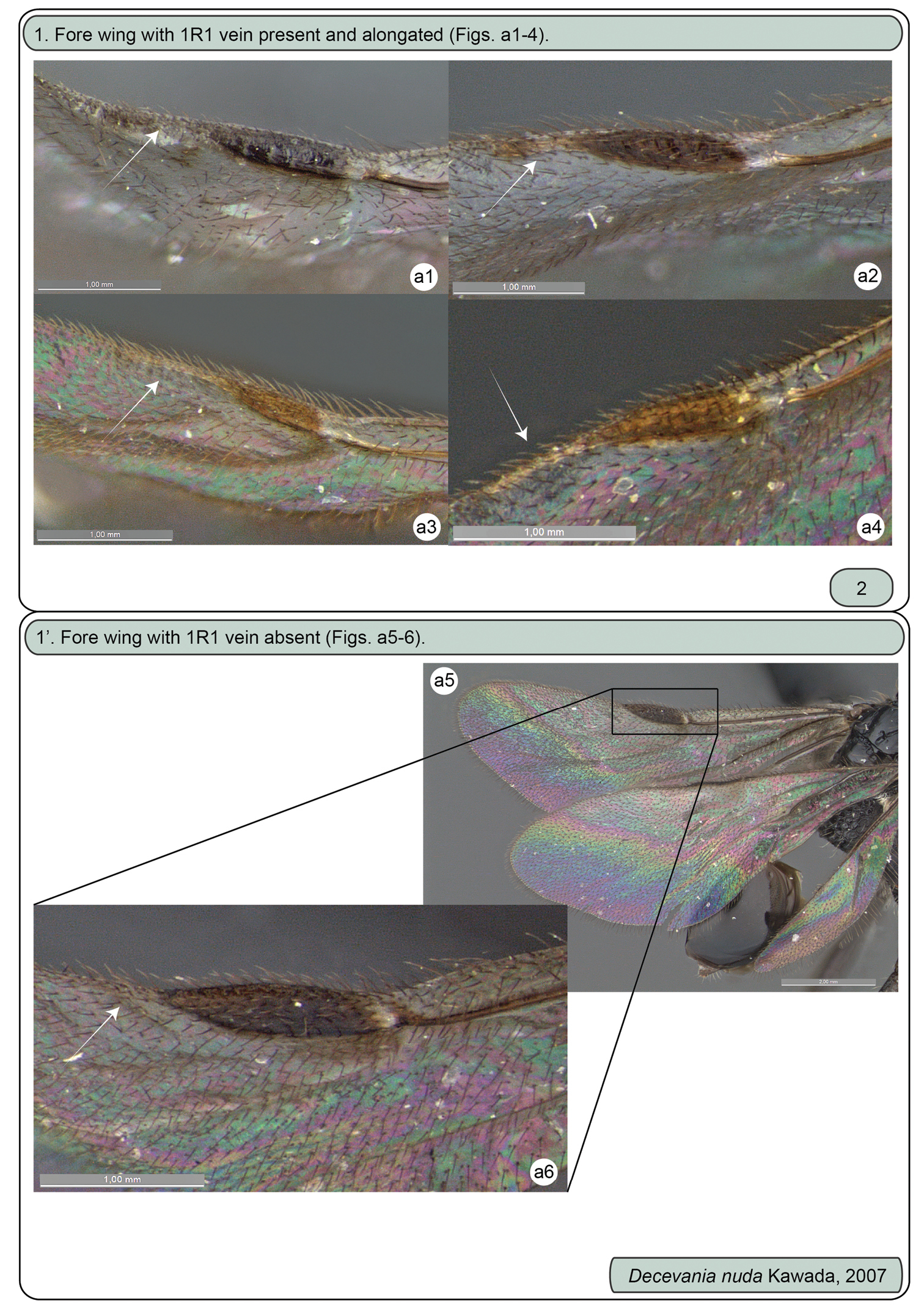
Females. a1 Decevania feitosai sp. n., left fore wing: 1R1 vein a2 Decevania parva (Enderlein, 1901), left fore wing: 1R1 vein a3 Decevania reticulata Kawada, 2007, left fore wing: 1R1 vein a4 Decevania unidentata Kawada, 2007, holotype, left fore wing: 1R1 vein a5-6 Decevania nuda Kawada, 2007 a5 wings and a6 left fore wing: 1R1 vein.
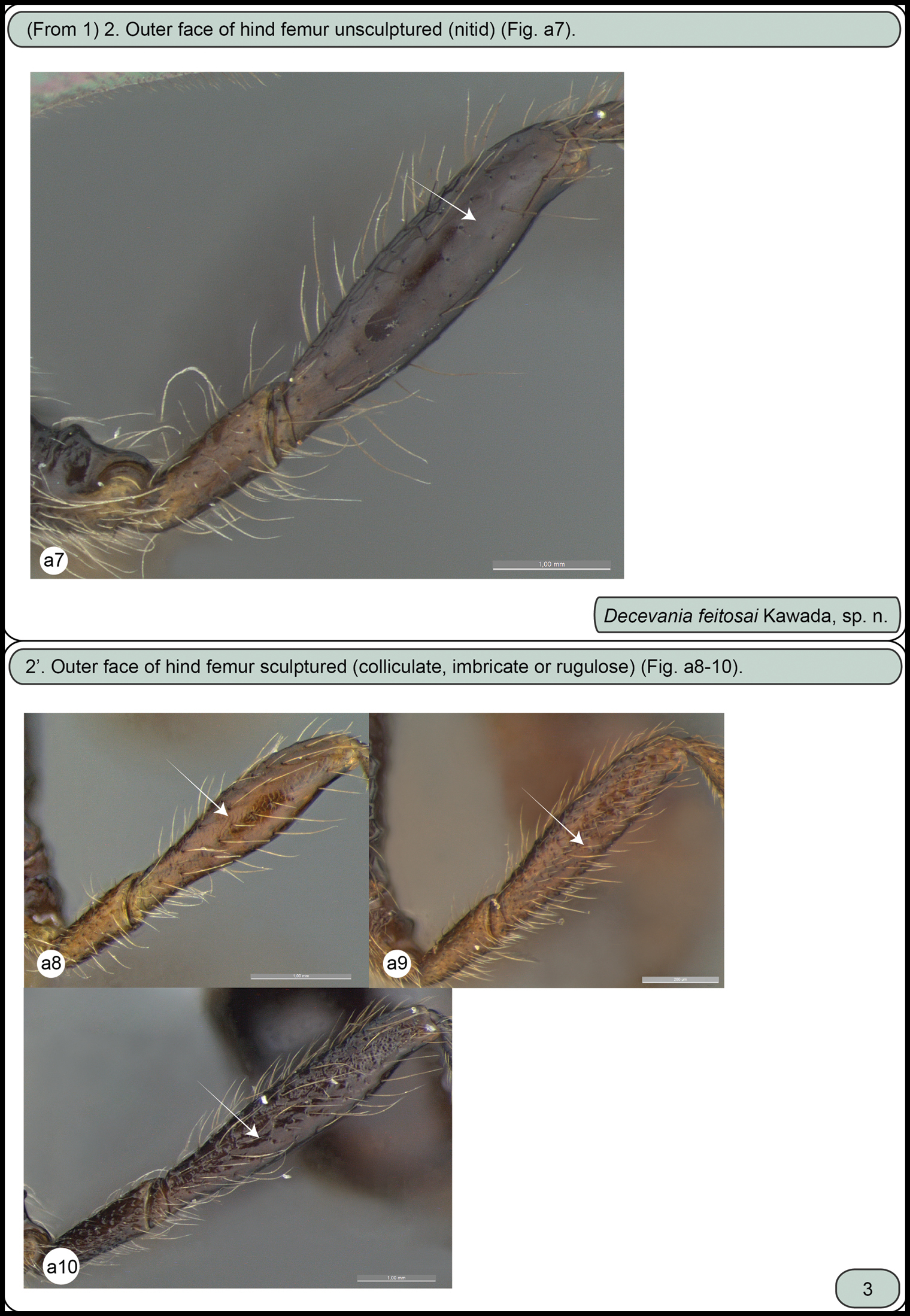
Females. a7 Decevania feitosai sp. n., left hind femur in lateral view a8 Decevania unidentata Kawada, 2007, holotype, left hind femur in lateral view a9 Decevania reticulata Kawada, 2007, left hind femur in lateral view a10 Decevania parva Kawada, 2007, left hind femur in lateral view.
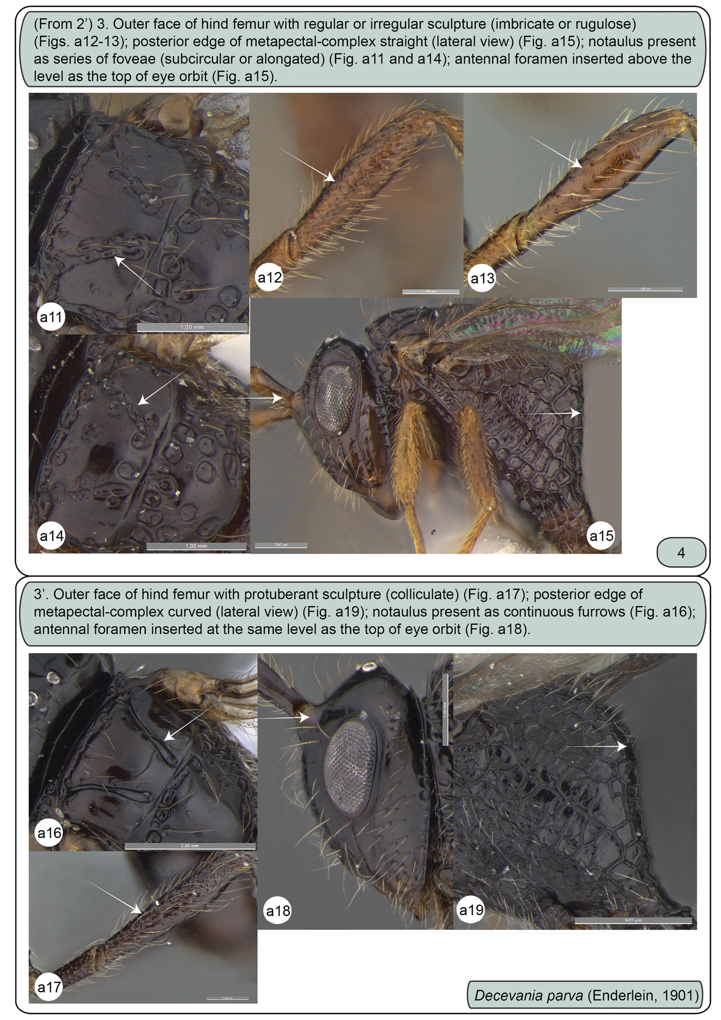
Females. a11 Decevania reticulata Kawada, 2007, mesoscutum in dorsal view a12 Decevania reticulata Kawada, 2007, left hind femur in lateral view a13 Decevania unidentata Kawada, 2007, holotype, left hind femur in lateral view a14 Decevania unidentata Kawada, 2007, holotype, mesoscutum in dorsal view a15 Decevania reticulata Kawada, 2007, head and mesosoma in lateral view a16–19 Decevania parva (Enderlein, 1901) a16 mesoscutum in dorsal view a17 left hind femur in lateral view a18 head in lateral view and a19 metapectal-propodeal complex in lateral view.
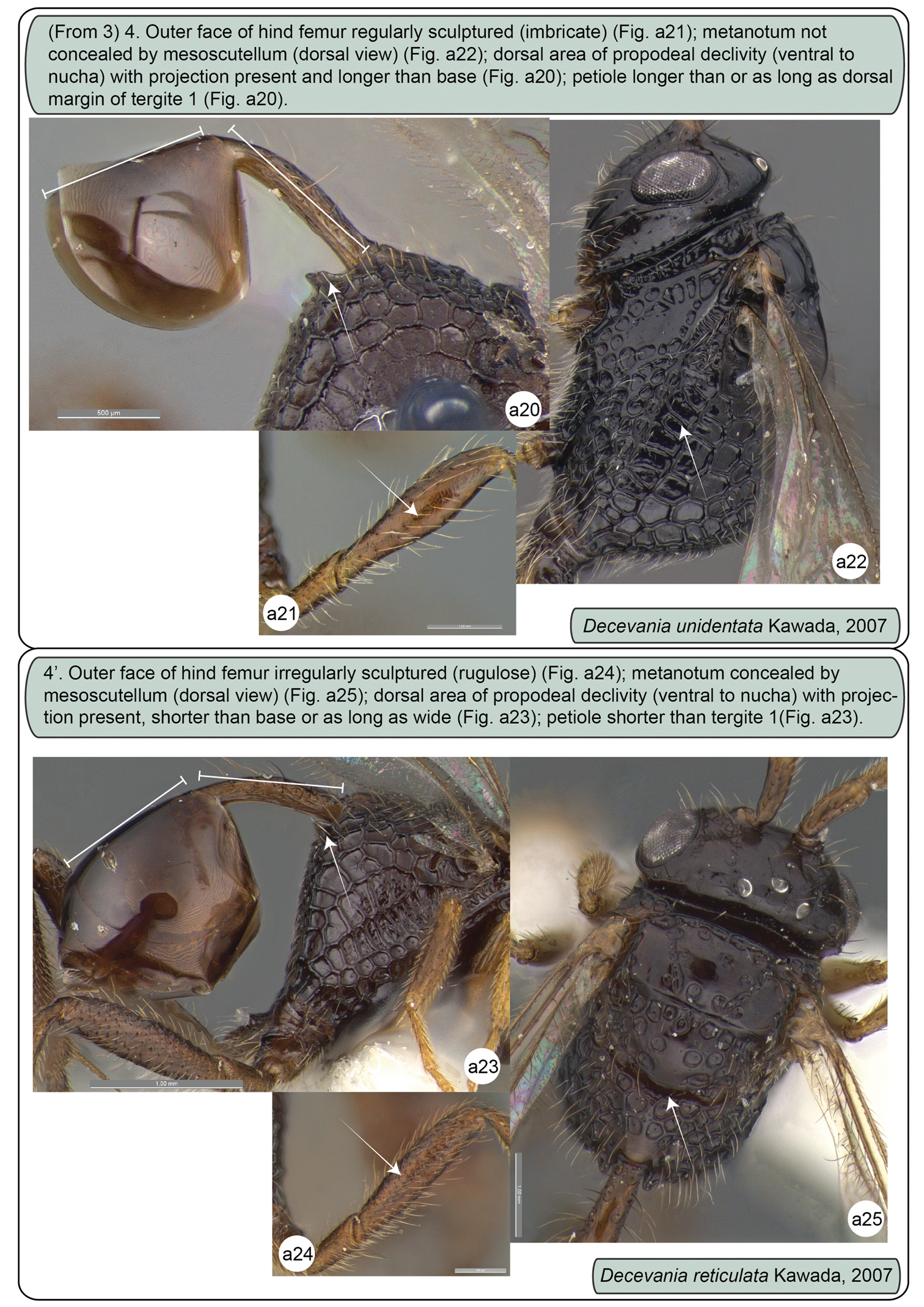
Females. a20-22 Decevania unidentata Kawada, 2007, holotype a20 propodeum and metasoma in lateral view a21 left hind femur in lateral view a22 head and mesosoma in dorsal view a23–25 Decevania reticulata Kawada, 2007 a23 propodeum and metasoma in lateral view a24 left hind femur in lateral view and a25 head and mesosoma in dorsal view.
urn:lsid:zoobank.org:act:C11F980C-6446-4311-B71B-6812939BEC49
http://species-id.net/wiki/Decevania_feitosai
Fig. 1–12Female body length: 1.6 mm (head to propodeum). Head color: black. Mesosoma color: black. Legs color: fore leg: trochanter, trochantellus, tibia, tarsus light-castaneous; femur dark-castaneous. Wings: fore and hind wing hyaline. Metasoma: petiole light-castaneous; tergites: dark-castaneous.
Head (Fig. 1-2, 5-7, 9). Head: long and stiff setae present evenly distributed; close to mesosoma. Vertex: slightly convex in lateral view; nitid with some small and sparse punctures. Ocelli: equal in size; arranged in obtuse isosceles triangle; anterior ocellus: separated from posterior ocellus by one ocellar diameter; anterior ocellus: not reaching the imaginary line between the anterior margin of posterior ocelli; posterior ocelli: separated by three ocellar diameters. Upper face: nitid with some sparse punctures. Eye: subovoid (lateral view); detached from dorsal profile of head; height of eye: as high as anterior margin of mesopleuron. Circumocular sulcus: absent. Postorbital carina: present; extending from anterior base of mandible to 3/4 the height of eye; strongly sinuous; narrower than postgenal sulcus. Antennal foramen: positioned at the same level as the top of the eye orbit; separated by one antennal foramen diameter; antennal rim: elevated laterally. Scape: long and stiff setae present evenly distributed; as long as F8. Pedicel + flagellomere 1: longer than wide; pedicel: as long as F1; flagellum: evenly and densely setose with some sparse and long setae. Median process of lower face: very weak in lateral view (difficult to see). Orbital band: strong, narrow and straight striae to the ventral margin of antennal foramen. Malar sulcus: present and conspicuous, differs from orbital band striae. Malar space: 0.64 times the height of eye (greater length). Clypeus: projecting medially; apical margin dilated and convex laterally. Mandible: two visible teeth, apical tooth longer and sharper than basal tooth.
Mesosoma (Fig. 1-2, 9, 11-12). Pronotum: long and stiff setae present evenly distributed. Pronotal neck: obscured. Dorsal pronotal area: concealed medially. Dorsolateral area of pronotum: expanded posteriorly into a lobe. Pronotal suprahumeral sulcus: scrobiculate, with a large fovea anterior to the lobe. Transverse pronotal carina: acuminate and extending along the anterior margin of pronotum. Mesothoracic spiracular incision: strongly curved and almost closed into an orifice. Lateral and dorsolateral pronotal area: not clearly separated by a carina (inconspicuous). Lateral pronotal area: narrow, same width between the upper eye orbit and occipital carina (widest point); vertical and covered by a row of fovea (transverse pronotal sulcus). Mesonotum: slightly raised (lateral view: compared with propodeum). Mesoscutum: 2.0 times wider than long; nitid with a few, sparse and regular foveae. Anterior mesoscutal sulcus: present as continuous furrow. Notaulus: present as continuous furrow, slightly curved towards the middle and not reaching the posterior margin. Median lobe of mesoscutum: slightly curved anteriorly (lateral view: difficult to see). Parascutal carina: present at posterior half; sulcus: following the parascutal carina and opening posteriorly. Parapsidal line: conspicuous suture, same length of parascutal carina and reaching the posterior margin of mesoscutum. Transscutal articulation: open in the middle and closing to lateral, near the parapsidal line. Mesoscutellum: long and stiff setae present, evenly distributed laterally; nitid in the middle with closed fovea laterally; bulging posteromedially; with a delicate median convexity on the posterior margin, but without overlap on metanotum. Scutoscutellar sulcus: not reaching the transscutal articulation, covered by a large and subcircular fovea. Metanotum: dorsolateral area covered by moderate (cuticle visible) layer of setae. Metanotum and metascutellum: form a continuous structure. Metascutellum: as a flat and nitid structure. Epicnemial carina: without median process (continuous shape). Prespecular sulcus: composed of one fovea. Anterior mesopleural area: covered by a row of rectangular impressions to femoral groove. Speculum: slightly dilated just above the middle of femoral groove. Mesepimeral sulcus: present as a row of irregular and subcircular foveae from posterodorsal mesepimeral area to mesocoxal foramen. Posterodorsal mesepimeral area: scrobiculate (narrow and shallow). Posterior mesepimeral area: curve and elongated posteriorly (closer to metacoxal foramen). Femoral groove: weakly concave; unsculptured medially. Mesopleural pit: absent. Ventral mesopleural area: covered by a subcircular and adjacent fovea; long and stiff setae present evenly distributed. Mesosternum: higher compared to metasternum; mesosternum foveate (irregular) with an open area (punctate) laterally. Mesodiscrimen: present as a flat and inconspicuous sulcus. Mesocoxa: distant 2.5 times (width of mesocoxa) from procoxa; adjacent to metacoxa. Meso- and metacoxa: without a pair of processes between coxae. Metapleuron (metapleural arm to metacoxal foramen): at least 3 times longer than wide. Metapleural carina: straight and parallel with concave lower metapleural area. Upper metapleural area: covered by a row of rectangular foveae. Lower metapleural area: lower region covered by an irregular polygonal fovea; long and stiff setae present, evenly distributed. Metapleural pit: present. Anterior area of metapleural pit: acute isosceles triangle shaped and covered by an irregular fovea. Metapleural epicoxal sulcus: present as a row of large and subrectangular foveae. Metanotum and propodeum: form a continuous structure. Propodeum: irregular foveae (dorsal) to regularly areolate (lateral). Dorsal propodeal area: long and stiff setae present, evenly distributed. Lateral propodeal carina: absent. Lateral propodeal area (upper region): long and stiff setae present evenly distributed. Adpetiolar strip: longer than wide. Nucha: slightly elevated (lateral view). Upper region of propodeal declivity (ventral to nucha): projection present and longer than base. Middle area of propodeal declivity: with long and stiff setae present evenly distributed. Posterior edge of metapectal-complex: curved (lateral view).
Legs (Fig. 4, 10). Protibial spur: apex of calcar longer than apex of velum. Hind leg: long and stiff setae present evenly distributed (longer than outer spur); nitid with sparse punctures (trochanter, trochantellus, femur and tibia). Trochanter: 3.6 times longer (longer point) than wide (widest point). Hind femur: dorsal and ventral margin slightly dilated medially. Hind tibia: longer than hind femur; apical incision of hind tibia: sinuous. Tibial spurs: slightly sinuous; inner tibial spur: extending past the mid length of basitarsus; outer tibial spur: 1.8 times the length of hind basitarsus. Tarsus: minute striae (interspace) and more closer punctures; projections: conspicuous in tarsus 1–3; basitarsus: as long as tarsus 2–4 combined; basitarsus projection: longer than apex of basitarsus (widest point). Tarsal claw: hook-shaped, medially with a minute ventral spine.
Wings (Fig. 8). Apex of fore wing: bordered by long setae. Costal cell: the same length as head + mesosoma combined (dorsal view). Stigmal vein: as wide as costal cell. 1R1 vein: as long as stigmal vein, with slightly dilated apex. M+CU, 1CU and 2CU veins combined: extending past the propodeal declivity. 1CUb and 2CU vein: combined to form an angulated angle (45 degrees). 2CU vein: present with a slight dilatation distally. Hind wing: three hook-shaped hamuli of equal size; fusiform and three times longer than wide. Jugal lobe: present, slender and extending past the propodeal spiracle.
Metasoma (Fig. 11). Petiole: shorter than propodeal declivity; 6–7 times longer than wide; slightly curved distally. Transverse carina on petiole: as a narrow and acuminate rim. Dorsal petiolar area: nitid. Lateral petiolar area: some sparse and elongated punctures; long and stiff setae present, evenly distributed. Ventral petiolar area: fine and delicate longitudinal carina. Metasoma: subovoid (lateral view) with ovipositor concealed; without setae except T6–7 on posterior edge. Tergite 1: longer than petiole.
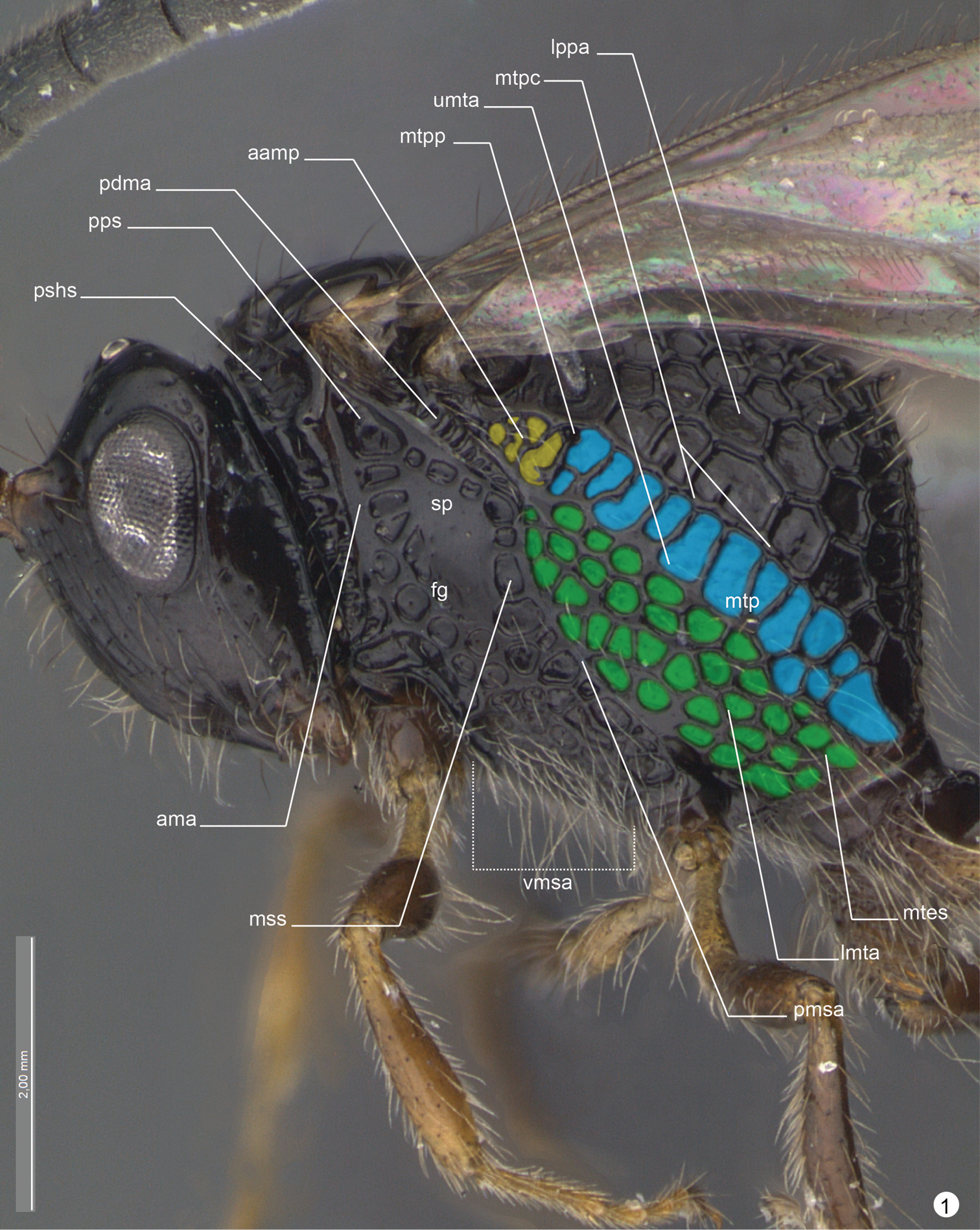
Decevania feitosai sp. n. Holotype, female. Head and mesosoma in lateral view. For terminology see the list in Material and methods. Scale in the figure.
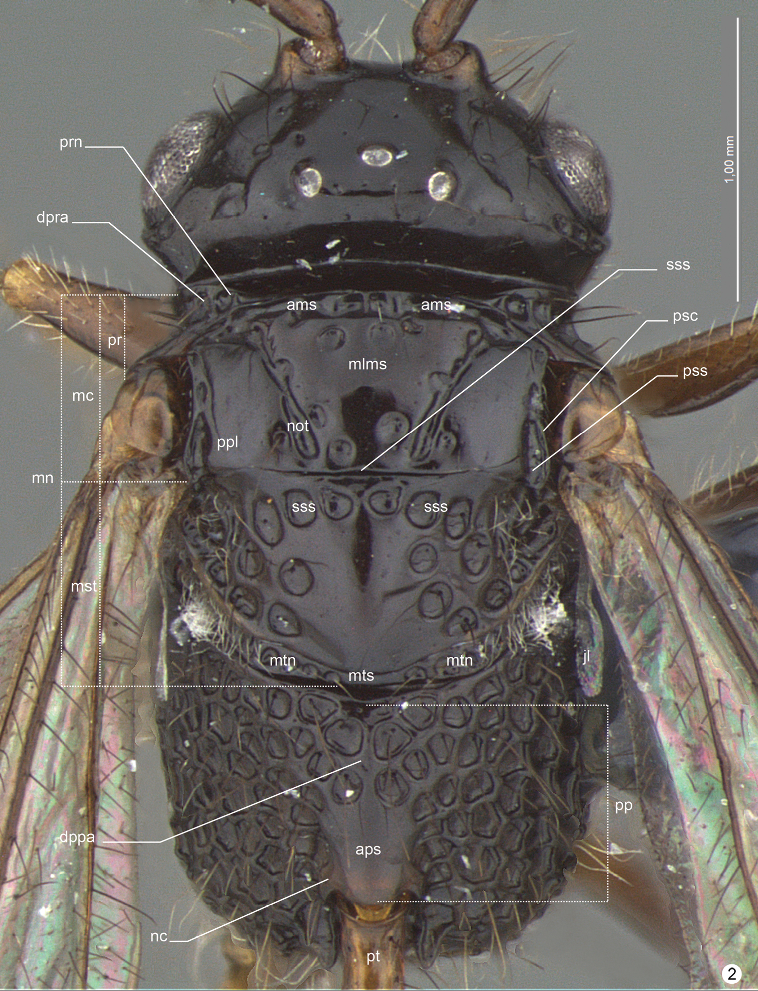
Decevania feitosai sp. n. Holotype, female. Head and mesosoma in dorsal view. For terminology see the list in Material and methods. Scale in the figure.
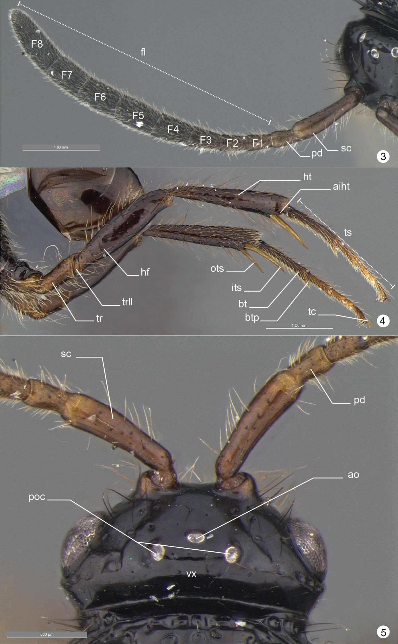
Decevania feitosai sp. n. Holotype, female. 3 right antenna in dorsal view 4 hind legs in lateral view 5 head in dorsal view. For terminology see the list in Material and methods. Scale in the figures.
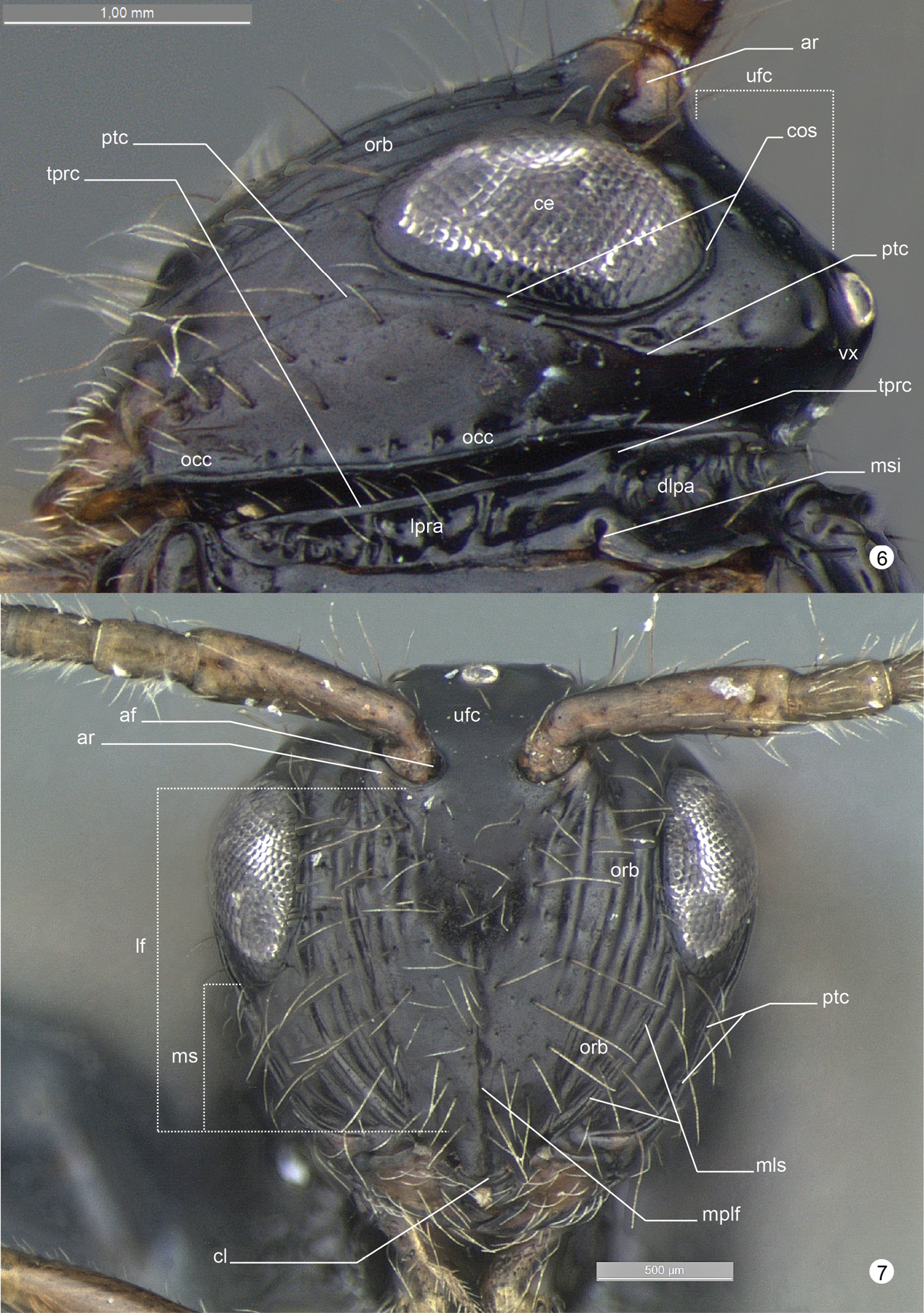
Decevania feitosai sp. n. Holotype, female. 6 head in lateral view 7 head in frontal view. For terminology see the list in Material and methods. Scale in the figures.
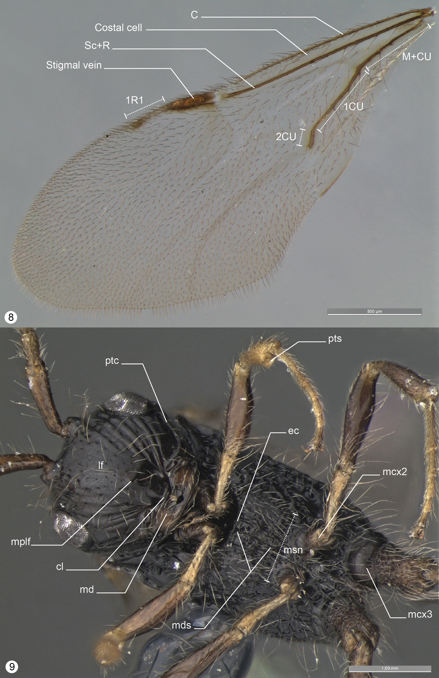
Decevania feitosai sp. n. Holotype, female. 8 left fore wing 9 head and mesosoma in ventral view. For terminology see the list in Material and methods. Scale in the figures.
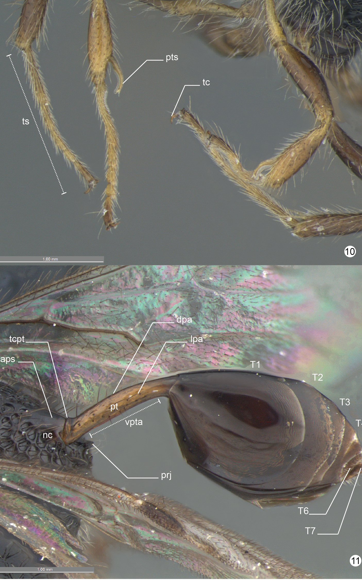
Decevania feitosai sp. n. Holotype, female. 10 fore and mid leg in frontal view 11 metasoma in laterodorsal view. For terminology see the list in Material and methods. Scale in the figures.
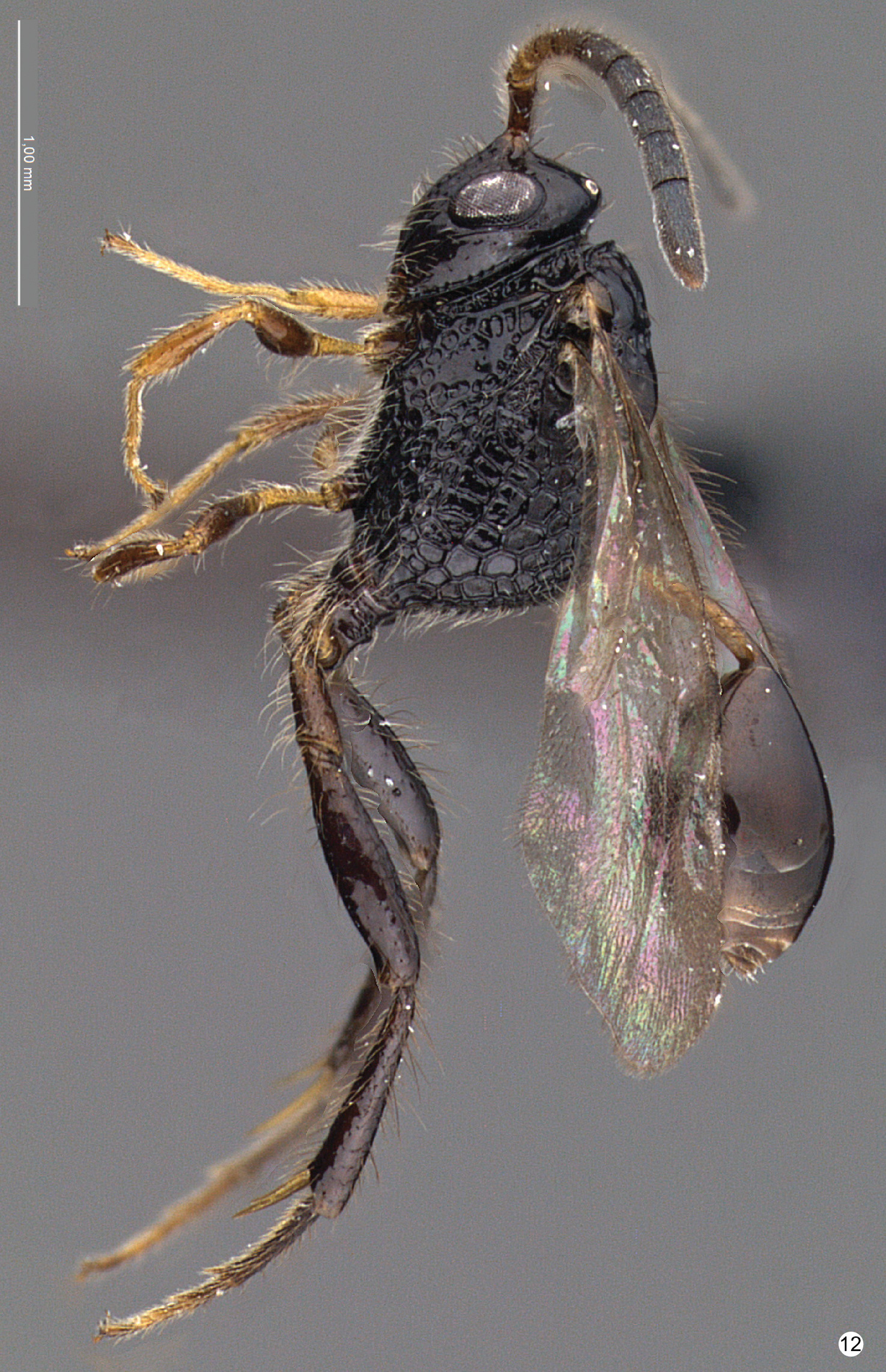
Decevania feitosai sp. n. Holotype, female. 10 habitus in lateral view. Scale in the figure.
Eye: 1.8–2.0 times higher than wide. Postorbital carina: present and complete; conspicuously outlined; detached from the margin of lower eye orbit; sinuous (see malar space); reaching the top of eye orbit (some foveae may also be present and are part of carina). Antennal foramen: inserted at the same level as the top of eye orbit; antennal rim: conspicuously elevated laterally (head lateral view). Median lobe of mesoscutum: slightly curved or flat (lateral view). Notaulus: present as continuous furrow. Metanotum: not concealed by mesoscutellum (dorsal view). Sculpture of hind femur: unsculptured (nitid, autapomorphy for Decevania feitosai sp. n.). Posterior edge of metapectal complex: curved (lateral view). Dorsal area of propodeal declivity (ventral to nucha): projection present and longer than base. Petiole: longer than or as long as dorsal margin of tergite 1. 1R1 vein: present and elongate.
The specific epithet is a patronymic honoring Rodrigo M. Feitosa, colleague and researcher of Formicidae from MZSP.
Decevania Distribution16.
Holotype. Female. COLOMBIA: Risaralda, SFF Otún Quimbaya, El Molinillo, 4°43'N, 75°34'W , 2220 m, Malaise, 17.ii–04.iii.2003, G. López leg., M.3696 (IAVH). Paratypes. 3 females. COLOMBIA: Magdalena, PNN Sierra Nevada de Santa Marta, San Lorenzo, 10°48'N, 73°39'W , 2200 m, Malaise, 09–24.vi.2000, J. Cantillo leg. M. 205 (IAVH 65814); 09–24.vi.2000, J. Cantillo leg. M. 205 (IAVH 65815); 24–30.vi.2000, J. Cantillo leg. M. 211 (IAVH 65816). 4 females. Risaralda, SFF Otún Quimbaya, El Molinillo, 4°43'N, 75°34'W , 2220 m, 03–17.xii.2002, Malaise, G. Walker leg., M. 2972 (IAVH 65827); 4°43'N, 75°34'W , 2220 m, 17.xii.2002–03.i.2003, Malaise, G. Walker leg., M. 2971 (IAVH 65828). Cuchilla Camino, 4°43'N, 75°35'W , 2050 m, 04–17.ii.2003, Malaise, G. López leg., M. 3680 (IAVH). Cuchilla Camino, 4°44'N, 75°35'W , 1960 m, 04–21.iii.2003, Malaise, G. López leg., M. 3669 (IAVH 65826).
Decevania nuda Kawada, 2007. Eye: 1.8–2.0 times higher than wide. Postorbital carina: present and complete; conspicuously outlined; closer to the margin of lower eye orbit; slightly sinuous (see malar space); reaching the top of eye orbit (some foveae may also be present and are part of carina). Antennal foramen: positioned above the level of the top of eye orbit; antennal rim: inconspicuous elevated laterally (head lateral view). Median lobe of mesoscutum: curved (lateral view). Notaulus: present as series of elongate foveae. Metanotum: not concealed by mesoscutellum (dorsal view). Sculpture of hind femur: protuberant sculpture (colliculate). Posterior edge of metapectal complex: angulated (lateral view). Dorsal area of propodeal declivity (ventral to nucha): projection present, shorter than base or as long as wide. Petiole: shorter than tergite 1. 1R1 vein: absent.
Paratype. Female. ECUADOR: Napo, Sierra Azul, 0.67°S, 77.92°W , 2300 m, 21–22.iv.1996, PT, P.J. Hibbs col. (CNCI).
Decevania parva (Enderlein, 1901). Eye: 1.8–2.0 times higher than wide. Postorbital carina: present and complete; inconspicuously outlined; detached from the margin of lower eye orbit; sinuous (see malar space); not reaching the top of eye orbit. Antennal foramen: positioned at the same level as the top of eye orbit; antennal rim: conspicuous elevated laterally (head lateral view). Median lobe of mesoscutum: curved (lateral view). Notaulus: present as continuous furrow. Metanotum: not concealed by mesoscutellum (dorsal view). Sculpture of hind femur: protuberant sculpture (colliculate). Posterior edge of metapectal complex: curved (lateral view). Dorsal area of propodeal declivity (ventral to nucha): projection present, shorter than base or as long as wide. Petiole: longer than or as long as dorsal margin of tergite 1. 1R1 vein: present and elongated.
Female. COLOMBIA: Cundinamarca, PNN Chingaza Bosque, Palacio, 4°31'N, 73°45'W , 2930 m, Malaise, 20.xii.2000–05.i.2001, L. Cifuentes leg., M. 1223 (IAVH 65781).
Decevania reticulata Kawada, 2007. Eye: 1.8–2.0 times higher than wide. Postorbital carina: present and complete; conspicuously outlined; detached from the margin of lower eye orbit; slightly sinuous (see malar space); reaching the top of eye orbit (some foveae may also be present and are part of carina). Antennal foramen: positioned above the level as the top of eye orbit; antennal rim: inconspicuously elevated laterally (head lateral view). Median lobe of mesoscutum: curved (lateral view). Notaulus: present as series of subcircular foveae. Metanotum: concealed by mesoscutellum (dorsal view). Sculpture of hind femur: irregular sculpture (rugulose). Posterior edge of metapectal complex: angulated (lateral view). Dorsal area of propodeal declivity (ventral to nucha): projection present, shorter than base or as long as wide. Petiole: shorter than tergite 1. 1R1 vein: present and elongated.
Paratype. Female. COLOMBIA: Chocó, PNN Utría Cocalito Dosel, 6°1'N, 77°20W , 20 m, Malaise, 04–19.vii.2000, J. Pérez leg., M. 339 (IAVH 65778).
Decevania unidentata Kawada, 2007. Eye: 1.6 times higher than wide. Postorbital carina: present, but some portion not visible; inconspicuously outlined; closer to the margin of lower eye orbit; reaches the top of eye orbit (some foveae may also be present and are part of carina). Antennal foramen: positioned above the level as the top of eye orbit; antennal rim: conspicuous elevated laterally (head lateral view). Median lobe of mesoscutum: slightly curved or flat (lateral view). Notaulus: present as series of subcircular foveae. Metanotum: not concealed by mesoscutellum (dorsal view). Sculpture of hind femur: regular sculpture (imbricate). Posterior edge of metapectal complex: angulated (lateral view). Dorsal area of propodeal declivity (ventral to nucha): projection present and longer than base. Petiole: longer than or as long as dorsal margin of tergite 1. 1R1 vein: present and elongate.
Material examined. Holotype observed. Access through: Evanioidea online17.
Thanks to A. Bennett (CNCI) and C. A. M. Uribe (IAVH) for the loan of material for this study. Julia C. Almeida (MZSP) and Maurício M. da Rocha (MZSP) for discussion and to Antonio C. C. Macedo, Cleide Costa (MZSP), Gabriel Biffi (MZSP), Patricia Mullins (NCSU) and Rodrigo M. Feitosa (MZSP) for constructive comments on the manuscript. This material is based upon work supported by Fundação de Amparo à Pesquisa do Estado de São Paulo (process 2008/04661-3 to Ricardo Kawada).
Abbreviations used on figures
| abbreviation | description | detail | Fig. |
|---|---|---|---|
| aamp | anterior area of metapleural pit | area anterior to metapleural pit, after posterodorsal mesepimeral area and bellow the propodeal spiracle | 1 |
| af | antennal foramen | 7 | |
| ar | antennal rim | 6, 7 | |
| aiht | apical incision of hind tibia | distal margin of hind tibia, site of insertion of inner and outer tibia spurs | 4 |
| ama | anterior mesopleural area | 1 | |
| ams | anterior mesoscutal sulcus | 2 | |
| ao | anterior ocellus | 5 | |
| aps | adpetiolar strip | 5 | |
| bt | basitarsus | 4 | |
| btp | basitarsus projection | projection of the apex of tarsus (at least tarsus 1-3) | 4 |
| ce | compound eye | 6 | |
| cl | clypeus | 7, 9 | |
| cos | circumocular sulcus | furrow that surrounds the compound eye | 6 |
| dlpa | dorsolateral pronotal area | 6 | |
| dpa | dorsal petiolar area | 11 | |
| dppa | dorsal propodeal area | 2 | |
| dpra | dorsal pronotal area | 2 | |
| ec | epicnemial carina | 9 | |
| fg | femoral groove | 1 | |
| fl | flagellum | 3 | |
| F1, F2... | flagellomere | 3 | |
| hf | hind femur | 4 | |
| ht | hind tibia | 4 | |
| its | inner tibial spur | 4 | |
| jl | jugal lobe | 2 | |
| lf | lower face | 7, 9 | |
| lmta | lower metapleural area | 1 | |
| lpa | lateral petiolar area | 11 | |
| lppa | lateral propodeal area | 1 | |
| lpra | lateral pronotal area | 6 | |
| mc | mesoscutum | 2 | |
| mcx2 | mesocoxa | 9 | |
| mcx3 | metacoxa | 9 | |
| md | mandible | 9 | |
| mds | mesodiscrimen | 9 | |
| mlms | median lobe of mesoscutum | 2 | |
| mls | malar sulcus | 7 | |
| mn | mesonotum | 2 | |
| mplf | median process of lower face | 7, 9 | |
| ms | malar space | 7 | |
| msi | mesothoracic spiracular incision | 6 | |
| msn | mesosternum | 9 | |
| mss | mesepimeral sulcus | 1 | |
| mst | mesoscutellum | 2 | |
| mtes | metapleural epicoxal sulcus | 1 | |
| mtn | metanotum | 2 | |
| mtp | metapleuron | divided into two regions by difference of sculpture and flatness: upper metapleural area (usually flat/areolate) and lower metapleural area (usually concave/foveolate) | 1 |
| mtpc | metapleural carina | 1 | |
| mtpp | metapleural pit | 1 | |
| mts | metascutellum | 2 | |
| nc | nucha | 2, 11 | |
| not | notaulus | 2 | |
| orb | orbital band | 6, 7 | |
| ots | outer tibial spur | 4 | |
| pd | pedicel | 3, 5 | |
| pdma | posterodorsal mesepimeral area | 1 | |
| occ | occipital carina | 6 | |
| pmsa | posterior mesepimeral area | 1 | |
| poc | posterior ocelli | 5 | |
| pp | propodeum | 2 | |
| ppl | parapsidal line | 2 | |
| pps | prespecular sulcus | 1 | |
| pr | pronotum | 2 | |
| prj | projection of propodeum | 11 | |
| prn | pronotal neck | 2 | |
| psc | parascutal carina | 2 | |
| pshs | pronotal suprahumeral sulcus | 1 | |
| pss | parascutal sulcus | 2 | |
| pt | petiole | 11 | |
| ptc | postorbital carina | 6, 7 | |
| pts | protibial spur | 10 | |
| sc | scape | 3, 5 | |
| sp | speculum | 1 | |
| sss | scutoscutellar sulcus | 2 | |
| T1, T2... | tergite | 11 | |
| tc | tarsal claw | 4, 10 | |
| tcpt | transverse carina on petiole | 11 | |
| tprc | transverse pronotal carina | 6 | |
| tr | trochanter | 4 | |
| trll | trochantellus | 4 | |
| ts | tarsus | 4, 10 | |
| tsa | transcutal articulation | 2 | |
| ufc | upper face | 6, 7 | |
| umta | upper metapleural area | 1 | |
| vmsa | ventral mesopleural area | 1 | |
| vpta | ventral petiolar area | 11 | |
| vx | vertex | 5, 6 |
Links