


(C) 2011 Ramón A. Dones. This is an open access article distributed under the terms of the Creative Commons Attribution License, which permits unrestricted use, distribution, and reproduction in any medium, provided the original author and source are credited.
For reference, use of the paginated PDF or printed version of this article is recommended.
A new species of armored scale, Mycetaspis ailynaomi Dones and Evans is described and illustrated from specimens collected on mamey (Mammea americana) from Puerto Rico. A key to the species of Mycetaspis is provided.
Sternorrhyncha, Diaspididae, Caribbean, Puerto Rico, Mycetaspis, new species
Mamey (Mammea americana L., Calophyllaceae),
also known as mammee apple, Santo Domingo apricot or South American
apricot, is an evergreen, native to the West Indies and northern South
America, whose fruit is edible. Mamey is confined to tropical or
subtropical climates due to its sensitivity to low temperatures and
seems remarkably resistant to pests and diseases. It has been introduced
successfully into several tropical areas of the Old World (West Africa,
Madagascar, southern Asia, Java, Philippines and Hawaii) but has
not survived well in California and Florida (
Nine species of armored scales have been reported from Puerto Rico on mamey.
The genus Mycetaspis Cockerell, 1897, currently comprises eight species (
We follow the terminology used by
urn:lsid:zoobank.org:act:13FE4406-5E5B-4E18-A31C-8A4E252C795E
http://species-id.net/wiki/Mycetaspis_ailynaomi
Figs 1 – 6Appearance in life was not recorded, but the scale is not pupillarial. Body 1241µm long and 1136µm wide in the holotype; 1347µm long and 998µm wide in the paratype; almost circular. Pygidium slightly produced, almost (1.1 times) as broad as long, 279µm long by 423µm wide and 263µm long by 440µm wide in holotype and paratype, respectively.
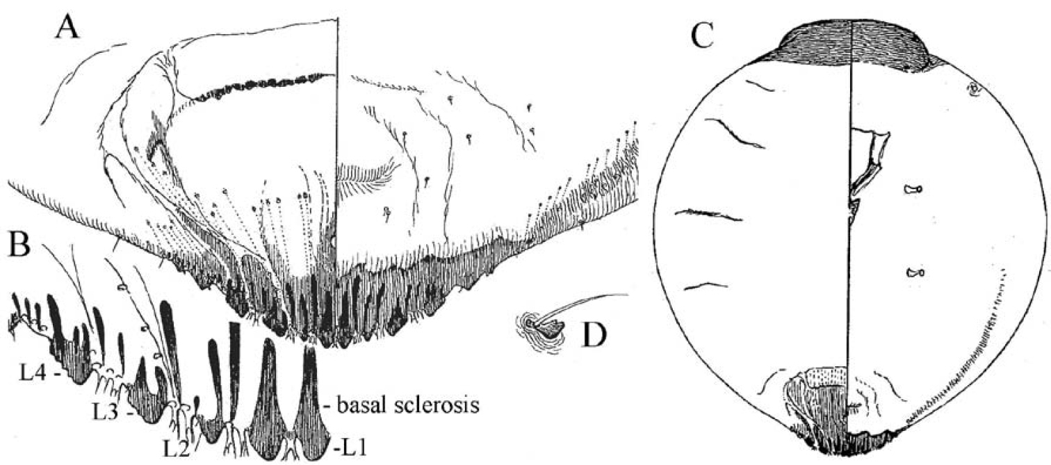
Cephalothorax. Anterior margin of head heavily sclerotized with 10–14 tooth-like, sclerotized lobular processes. Eyes are represented by a sclerotized dot. Antennae each composed of a conspicuous seta and a tubercle. A group of 18–20 microducts in front of each anterior spiracle. A band of microducts between the anterior and posterior spiracles extending outward from the median area to the margin in a slight upward angle without reaching the margin. Pygidium. Lobes. With 4 well-developed lobes (L1-L4); L1 more or less symmetrical, longer than L2-L4, flask-shaped, divergent on the mesal margin, which are shorter than the lateral margin. L2 with mesal margin one third as long as the lateral margin, with 2 or 3 small round teeth. L3 and L4 similar to L2, but more diagonally set with the lateral margin about 4 times as long as the mesal margin. Basal sclerosis. Similar in shape to a paraphysis, arising from the mesal margin of the L1 lobes, about twice as long as the lobe and about one third as wide as the base of the lobe, almost parallel-sided and rounded on the top. Paraphyses. Arranged 2–3-3 on each side of the pygidium. First interlobular space (space between L1 and L2) with a long paraphysis terminating in a club and almost twice as long the basal sclerosis associated with L1; a smaller paraphysis arising from mesal base of L2 and slightly shorter than half the length of the long paraphyses in the space. Second interlobular space with 3 paraphyses: the mesal one arising from the lateral basal corner of L2, similar to the paraphysis arising from the mesal corner of L2 in size and shape; the median paraphysis in the space about twice as long as the mesal paraphysis, approximately the same as the long paraphysis in the first interlobular space in size and shape; the lateral paraphysis from the mesal corner of L3, similar to the mesal paraphysis. The third interlobular space has 2 or 3 paraphyses: a short paraphysis arising at the lateral basal corner of L3, followed by a longer one more than twice as long as the former one. Paraphyses arising from the mesal basal corner of L4 faint or almost obsolete; pygidial margin anterior to L4 also with some short paraphyses. Plates. Plates occurring between lobes, but their numbers are difficult to determine in the available specimens. Plates occurring between L1 and L2 slender and simple, short, not extending beyond the apices of the lobes; plates between L2 and L3 slightly longer and wider with truncate apices; space between L3 and L4 appearing to have 3 short plates, one slender and 2 wider with truncate apices. Anal opening. Small, 14.5 µm in diameter, separated from the bases of L1 by a space about 6.5 times as long as its diameter. Perivulvar pores. Absent.
Unknown.
Two adult females, holo- and paratype, Puerto Rico: 27.vi.2006, M. Resto, on Mammea americana fruit. Specimens are mounted in Canada Balsam. Both specimens are deposited in the U.S. Museum of Natural History (USNM) in Beltsville, Maryland.
Mycetaspis ailynaomi is most similar to Mycetaspis defectopalus Ferris in the shape of the pygidial lobes and the relative lengths and shapes of the paraphyses, but differs from the latter and other species in the genus in having 10–14 sclerotized lobular processes along the anterior margin of the cephalothorax; whereas the anterior margin of the cephalothorax is sclerotized, but smooth and rounded in the other species.
This species in only known to occur on Mammea americana fruit in Puerto Rico. Several embryos were present in both the holotype and paratype specimens.
The species name is the combination of the names of the first author’s daughters, Ailyn and Naomi, as a testimony of his love to them.

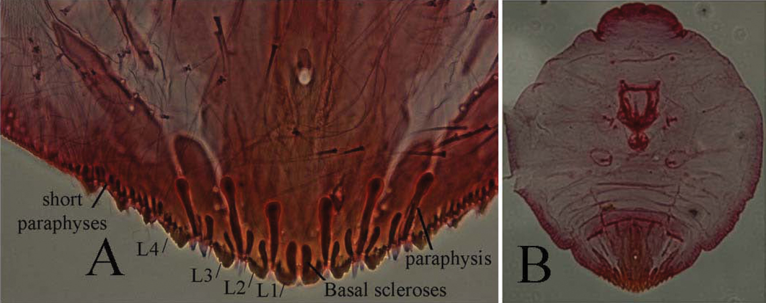
Mycetaspis ailynaomi holotype female 1 habitus 2 detail of lobes on head 3 detail of cluster of thoracic pores 4 detail of pygidial lobes.
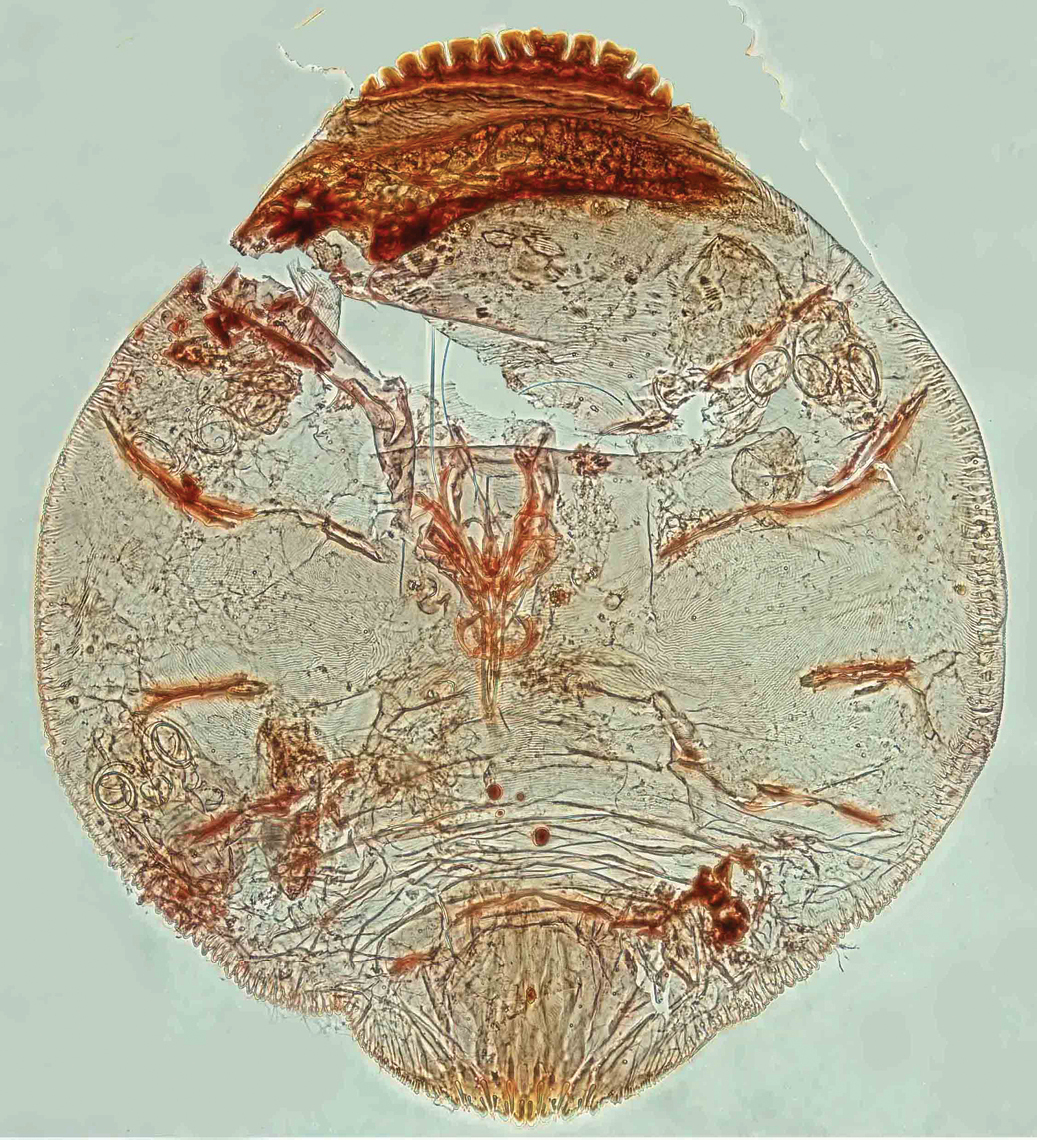
Mycetaspis ailynaomi, habitus of holotype female.
| 1 | Perivulvar pores present; sclerotized area of anterior margin of head flatly rounded with a row of setae; Mexico, Guatemala, Panama, Venezuela | Mycetaspis sphaerioides (Cockerell) |
| 1b | Perivulvar pores absent; sclerotized area of anterior margin of head produced without a row of setae | 2 |
| 2(1b) | L1 each with an elongate, tapering basal sclerosis whose base is about as wide as the base of the L1 lobe | 3 |
| 2b | L1 with the basal sclerosis narrow and arising from the mesal angle, its base is less than half as wide as the L1 base | 4 |
| 3(2) | Perispiracular pores absent. L2 and L3 wider than long; L4 distinct; widespread | Mycetaspis personata (Comstock) |
| 3b | Perispiracular pores present. L2 and L3 longer than wide; L4 merged into sclerotized margin; Brazil | Mycetaspis bezerrai Arruda |
| 4(2b) | Eyes replaced with a thorn-like process; lateral area anterior to L4 with a series of relatively long paraphyses; Argentina, Brazil, Guyana, Mexico, Panama; USA (Texas) | Mycetaspis apicata (Newstead) |
| 4b | Eyes not replaced with a thorn-like process; area anterior to L4 without a series of relatively long paraphyses (short paraphyses present in Mycetaspis ailynaomi) | 5 |
| 5(4b) | Sclerotized area on anterior margin of head with a row of 10–14 large, protruding sclerotized lobular processes; area anterior to L4 with a series of short paraphyses; Puerto Rico | Mycetaspis ailynaomi Dones & Evans, sp. n. |
| 5b | Head smooth, with no processes: area anterior to L4 without a series of short paraphyses | 6 |
| 6(5b) | Anterior margin of head rounded, not incised, smoothly joining the lateral margin of the cephalothorax; Belize, Ecuador, Mexico, Nicaragua, Panama, Peru, USA (Florida, Texas) | Mycetaspis defectopalus Ferris |
| 6b | Anterior margin of head, incised on each side of the apex, giving it a 3-lobed appearance, abruptly joining the lateral margin of the cephalothorax; Brazil | Mycetaspis juventinae Lepage & Giannotti |
Mycetaspis brasiliensis
Hempel was described from Brazil. We do not have an illustration or
specimens of this species to compare with the other species. According
to
Mycetaspis eneideae
Arruda was described from Brazil. No specimens of this species are
available to us and the original illustration lacks sufficient details
helpful for placing it in the key. Based on the original illustration of
the species in
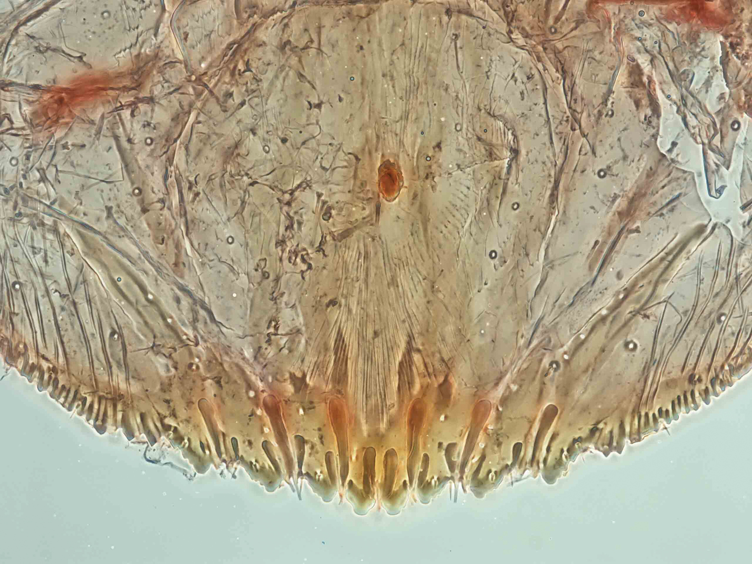
Mycetaspis ailynaomi, detail of pygidial lobes of holotype female.
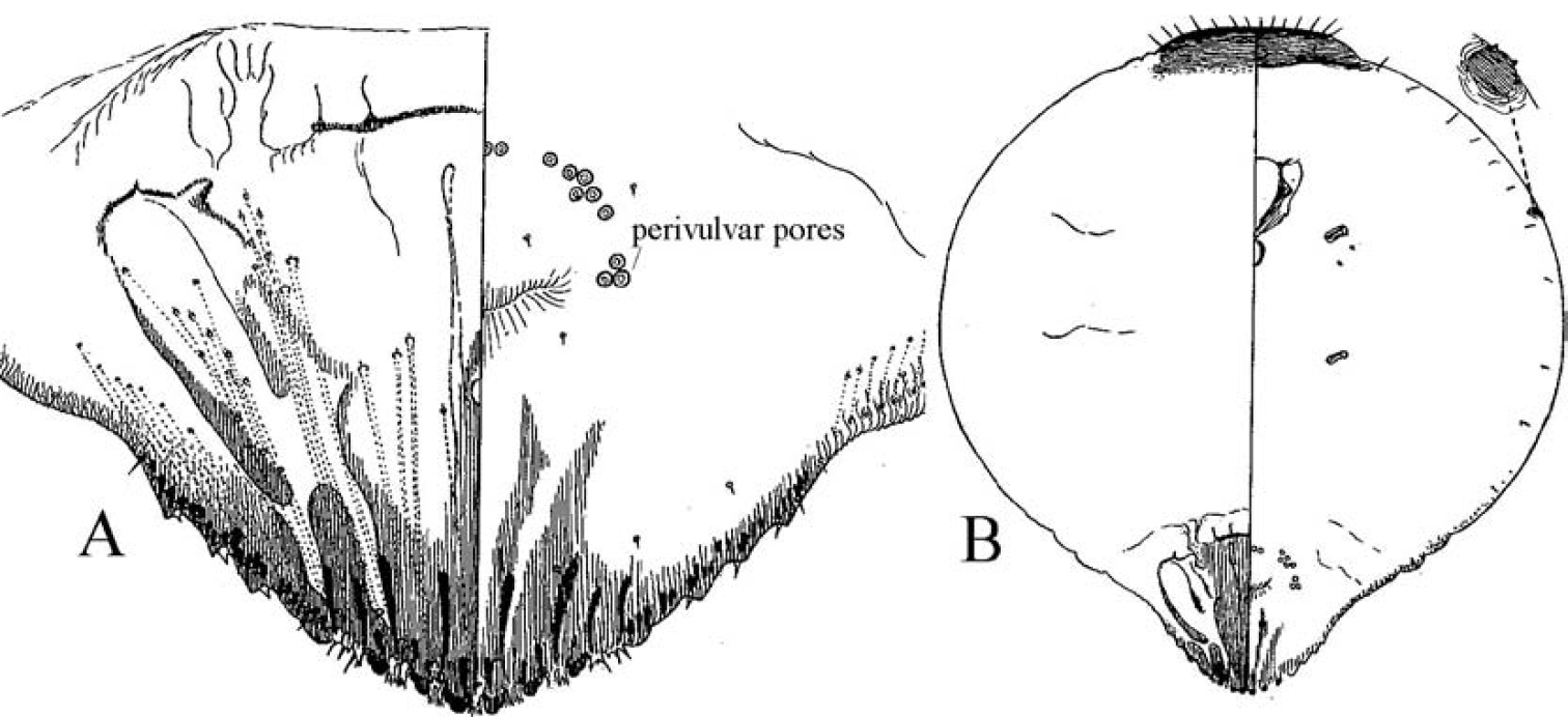
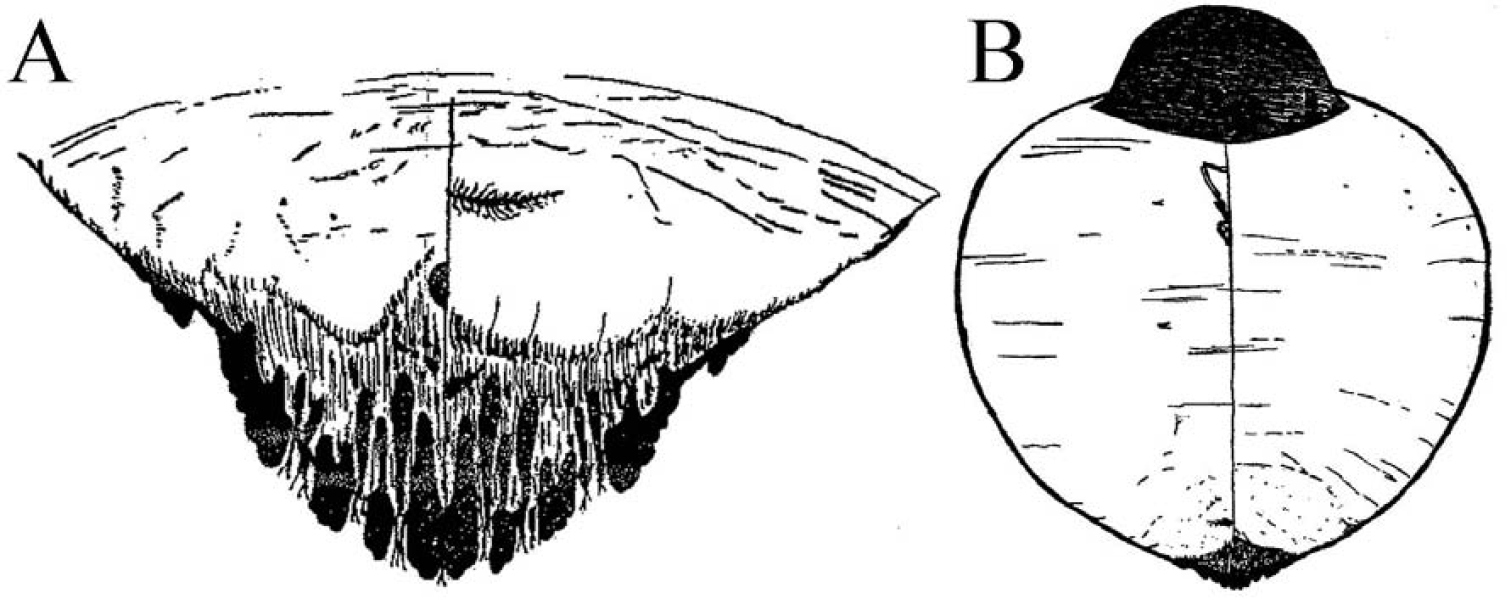
Mycetaspis sphaerioides female a pygidium b habitus (after
Mycetaspis personata female a pygidium b detail of pygidium, c) habitus, d) antenna (after
Mycetaspis bezerrai female a pygidium b habitus (after
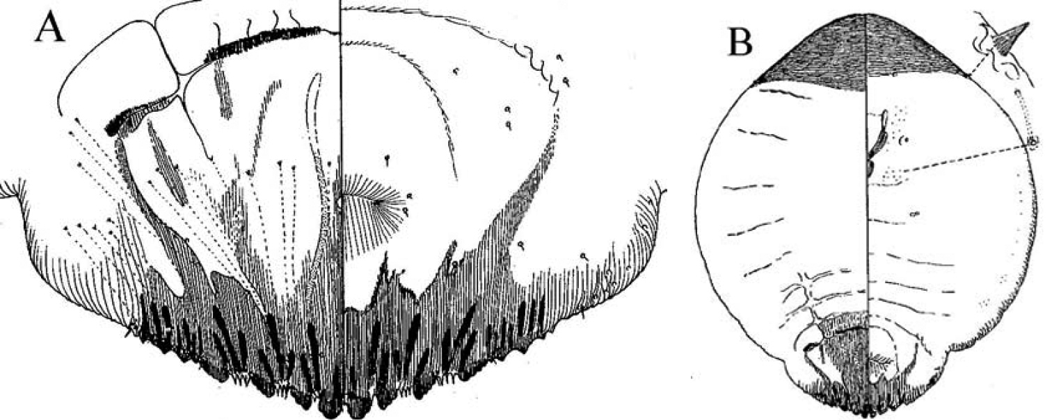
Mycetaspis apicata female a pygidium b habitus (after
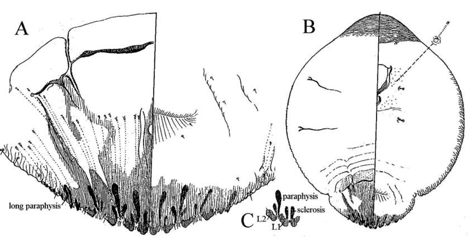
Mycetaspis defectopalus female a pygidium b habitus c detail of L1 and L2 lobes (after
Mycetaspis juventinae female (topotype) a pygidium b habitus.
The first author thanks Mr John W. Dooley, my mentor in the study of the armored scales and, the late Dr Leonce Bonnefil, who introduced me to Entomology. I also thank the San Juan, Puerto Rico Inspection Station where the scale was intercepted and to the Miami Inspection Station for funding this publication. The views and ideas expressed herein are not necessarily those of the USDA.