

(C) 2010 Andreas Schmidt-Rhaesa. This is an open access article distributed under the terms of the Creative Commons Attribution License, which permits unrestricted use, distribution, and reproduction in any medium, provided the original author and source are credited.
For reference, use of the paginated PDF or printed version of this article is recommended.
Chordodes mizoramensis, a new species of freshwater gordiid horsehair worm, is described from Mizoram, NE India on the basis of scanning electron microscopic and morphometric studies. The new species can be distinguished from its congeners in that the apical filaments of the crowned areoles are branched several times, a pattern that has not been observed in other species. An additional distinguishing character is that it has more bulging areoles, which are distributed among simple areoles alone or in groups, do not form clear patterns.
Nematomorpha, Gordiida, Chordodes, new species, hairworm, cuticle
About 350 species of freshwater horsehair worms
(Nematomorpha: Gordiida) are currently known. Of these, only 14 species
(plus an additional undetermined Gordius sp.) have been reported from India (
Chordodes
includes mainly tropical and subtropical species. All horsehair worms
are parasites of arthropods, which leave their host for reproduction
(Hanelt et al. 2005). Praying mantids form the main group of final hosts
for species of Chordodes (see
The specimens investigated were preserved in 70% ethanol, directly after their emergence from the host, an undetermined praying mantis. Pieces about 1 mm long were cut from the mid-body region of each worm. These and the entire posterior ends were prepared for Scanning Electron Microscopy (SEM). The pieces were dehydrated in an increasing ethanol series, critically point dried and coated with gold in a sputter coater. Observation took place using a LEO SEM 1524 under 10 kV. Digital images were taken.
Resultsurn:lsid:zoobank.org:pub:5A212538-A6FC-4C30-BB1F-B56169437AF6
Figs 1–3Mamit Village, Mamit District, Mizoram, India, 23°54'54.94"N, 92°29'16.75"E. Collected July 21, 2010 by Lalramliana and Remsangpuia.
Male specimen from the type locality emerged from Hierodula sp. (type-host). Deposited in the Zoological Museum in the Department of Zoology at Pachhunga University College, Aizawl-Mizoram, India, accession number PUCZM - A/V/1114.
Male specimen from the same host specimen and same locality as the holotype. Deposited in the Zoological Museum in the Department of Zoology at Pachhunga University College, Aizawl-Mizoram, India, accession number PUCZM - A/V/1115.
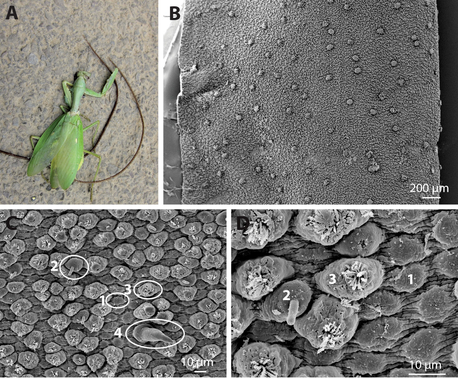
Both specimens emerged from one specimen of Hierodula sp. (Mantodea) (Fig. 1A).
The name refers to the region in which the new species was found, Mizoram in NE India.
The holotype is 200 mm long, with a diameter of 1.3 mm in the mid-body region. Towards the posterior end, the diameter decreases to about 0.7 mm at the level of the cloacal opening. The anterior end is also tapered. The paratype is 265 mm long and has a diameter in the mid-body region of 1.5 mm;, at the level of the cloacal opening the diameter is 0.79 mm. The frontal tip in both specimens is white, whereas the remaining body is medium brown. A pattern of darker patches (the “leopard pattern”) is present in both specimens; in the holotype this is more pronounced than in the paratype.
The cuticle contains six types of areoles (areoles are elevated cuticular structures), for which the terminology of
Chordodes mizoramensis, sp. n. A Hierodula sp., with both specimens of hairworm emerging from it. The darker specimen is the holotype B Overview of a stretched piece of cuticle from an entire section in the mid-body region, showing the distribution of areoles. Elevations are clusters of crowned and circumcluster areoles C Cuticle with simple (1), tubercle (2), bulging (3) and thorn (4) areoles D Magnification of the structure of simple (1), tubercle (2) and bulging (3) areoles. B–D from paratype, SEM.
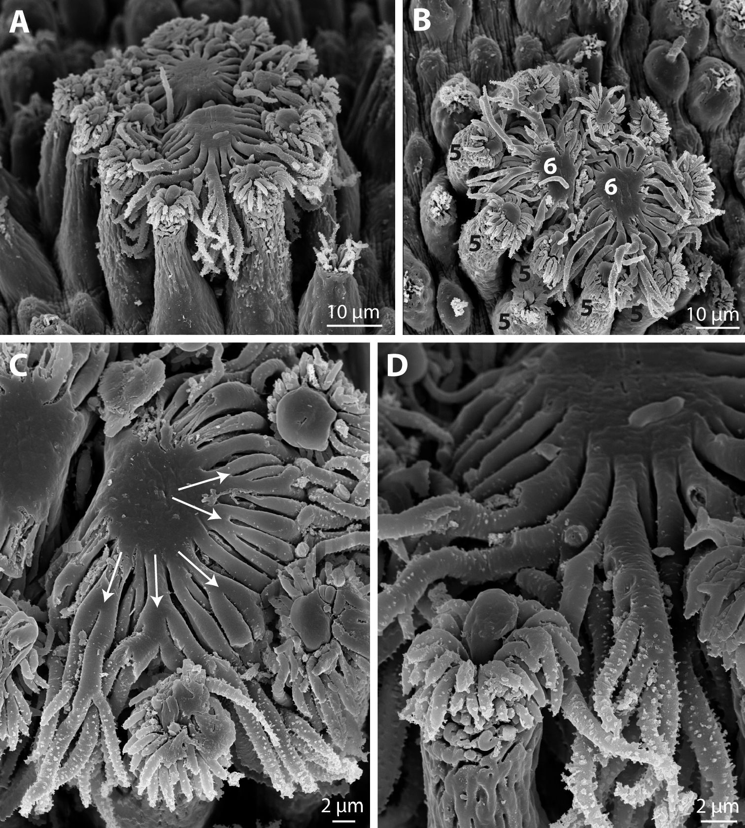
Characteristic for species of Chordodes are crowned areoles, which carry a crown of apical filaments on an elevated “stem”. Crowned areoles occur in pairs and are surrounded by so-called circumcluster areoles (Fig. 2A, B). This last type resembles the bulging areoles, but is longer (as elevated as the crowned areoles) and more slender (Fig. 2A, B). It also carries an apical tuft of short bristles, some of which can be slightly branched. Several circumcluster areoles have a more or less central “plug” among the apical bristles (Fig. 2A–D). This “plug” is variable in shape, in some cases appearing as a drop-like structure emerging from the centre of the areole, but in others it is a broader, more voluminous structure. One pair of crowned areoles occurs in the centre, between the circumcluster areoles. Each crowned areole has a flat, smooth surface, with filaments emerging from the margin, except for the region where both areoles face each other (Fig. 2A–C). The filaments spread flat from the central surface and project between the circumcluster areoles. Their length is about 25 µm. Most filaments divide several times, forming multiple branches (Fig. 2A–D). Only one type of crowned areoles could be found.
Chordodes mizoramensis, sp. n. A–D Crowned (6 in B) and circumcluster areoles (5 in B), C and D at magnifications demonstrating the branching of apical filaments. A–D from paratype, SEM.
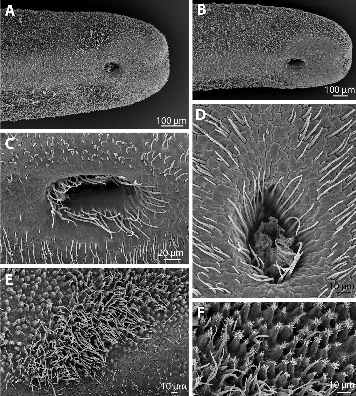
The posterior end of the males is rounded, and a small median incision may be present (Fig. 3A, B). An approximately 150 µm broad ventral strip is free of areoles of the types described above, but forms polygonal or interdigitating compartments with a smooth surface (Fig. 3A, B). This smooth region extends around the ventral cloacal opening, which is about 200 µm anterior of the posterior margin of the worm. The cloacal opening is oval, with a number of long, fine bristles, the circumcloacal bristles, present in a ring emerging approximately 10 µm below its surface (Fig. 3C, D). In the region around the cloacal opening are further bristles; these are abundant and variable in length (Fig. 3C, D). The areoles described above are replaced at the posterior end, at least on the lateral sides, by elevated, conical areoles with an apical tuft of bristles (Fig. 3F). These areoles may represent bulging areoles, but are distinctly pointed apically and more abundant. In a region anterolateral to the cloacal opening is, in the region with areoles, an oval region with more bristles (Fig. 3A, B, E). These are very dense, appear to be all unbranched and have a lengths of up to about 30 µm.
Chordodes mizoramensis, sp. n. A–F Posterior end. A, B Ventral view of posterior end of holotype (A) and paratype (B) showing the distribution of areoles and the ventral cloacal opening C, D Cloacal opening of the holotype (D) and paratype (C), showing circumcloacal bristles and further bristles in the region around the cloacal opening E Field of bristles anterolateral of the cloacal opening (holotype) F Form of areoles posterior to the field of bristles (paratype). A–F SEM.
With about 90 described species, Chordodes is distributed in tropical and subtropical regions worldwide (
The types of areoles present on the cuticle of Chordodes mizoramensis sp. n. represent the “usual” set of areoles present in other Chordodes species, but there are some notable differences. Bulging areoles occur in some, but not all Chordodes species (see
Several Chordodes
species have two types of crowned areoles; those with distinctly longer
apical filaments are present along the ventral and sometimes also the
dorsal mid-line (see
The male posterior end of the new species corresponds, as far as is known, in general with those of the males of other Chordodes species. However, the shape of the areoles on the posterior end (conical, with tuft of bristles on top) may be peculiar to Chordodes mizoramensis.
In summary, Chordodes mizoramensis exhibits some unique features, which justify its description as a new species.
In the key provided by
“crowned areole filaments branched..........Chordodes mizoramensis”
We thank Dr. Tawnenga, Principal, and Dr. K. Lalchhandama, Head, Department of Zoology, Pachhunga University College, Mizoram, India, for providing funding and laboratory facilities. Many thanks also due to Mr. Remsangpuia, who helped with collecting the specimen.


