


(C) 2012 Hong-Thai Pham. This is an open access article distributed under the terms of the Creative Commons Attribution License 3.0 (CC-BY), which permits unrestricted use, distribution, and reproduction in any medium, provided the original author and source are credited.
For reference, use of the paginated PDF or printed version of this article is recommended.
The first record of the genus Semia Matsumura (Cicadidae: Cicadinae, Cicadini) from Vietnam is presented. One new species, Semia spinosa sp. n., is described from southern Vietnam. Photos of the adult, illustrations of the male genitalia, a distribution map and biological data are provided. A key to the species of Semia based on the male adults is also given.
New record genus, Semia spinosa, morphology, Cicadina, Auchenorrhyncha
The cicada fauna of Vietnam has received little attention since the descriptions of
Semia is similar to Leptosemia Matsumura, 1917, Terpnosia Distant, 1892and Euterpnosia Matsumura, 1917, but has a dentate lateral margin to the pronotum (Fig. 2A), transverse male opercula that are nearly contiguous with each other (Fig. 2B) and lacks a tooth-like projection laterally on the male 4th abdominal segment (
Four males of the new species Semia spinosa sp. n., were collected from the Phu Ly, Ma Da-Vinh Cuu Nature reserve (NR), Vinh Cuu district, Dong Nai Province in southern Vietnam. The holotype and two paratypes are deposited in the Institute of Ecology and Biological Resources, Hanoi, Vietnam (IEBR), and one paratype in the Natural History Museum, London (BMNH).
Nomenclature for family, subfamily and tribal classification follows that of
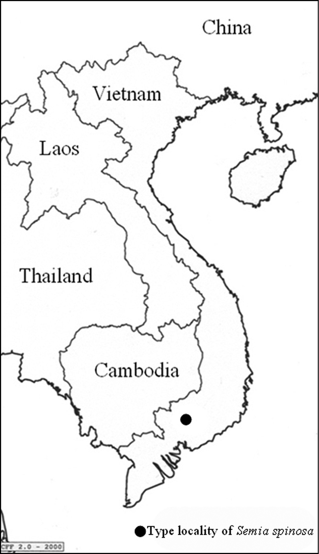
Type locality of Semia spinosa sp. n.; (see text for further details).
Subfamily Cicadinae
Tribe Cicadini
Subtribe Cicadina
http://species-id.net/wiki/Semia
Head nearly as wide as or slightly narrower than base of mesonotum; inner area of pronotum generally concolorous to outer dilatation; male abdomen cylindrical, much longer than distance from head to cruciform elevation and slightly widest across 4th abdominal segment and wider than base of mesonotum; male tymbal cover very small and semicircular, mostly exposing tymbal in dorsal view; male 8th abdominal tergum mostly covered with white powder; ovipositor not protruding beyond abdominal segment 9; male operculum scale-like, roundish, and not extending beyond 2nd abdominal sternum; wings hyaline; 6th apical cell of forewing about as long as or longer than twice of 5th apical cell in median length. Based on
China, Taiwan, Vietnam (Fig. 5).
This genus is similar to Leptosemia, Terpnosia and Euterpnosia (see Introduction).
urn:lsid:zoobank.org:act:B8A6B87E-9A4A-484C-90DE-4C4F7CFB2D8D
http://species-id.net/wiki/Semia_spinosa
Figs 2, 3A–B, 6CThe species name refers to morphological feature such as spinosa for the uncus spines
Holotype ♂: VIETNAM [VC.Ho.0650, Phu Ly, Ma Da-Vinh Cuu NR, Dong Nai Province, 4.viii.2008, light trap, 11°24'42.4"N, 107°06'19.5"E, 100–150m, leg Hoang Vu Tru] (IEBR).
Paratypes (3 ♂♂): 2 ♂♂: VIETNAM [ VC.Ho.0626, 0631, Phu Ly, Ma Da-Vinh Cuu NR, Dong Nai Province, 31.vii.2008, 100–150m, leg Hoang Vu Tru] (IEBR); 1 ♂: [VC.Ho.0765, Phu Ly, Ma Da-Vinh Cuu NR, Dong Nai Province, 2.viii.2008, light trap and netting, 100-150m, leg Ta Huy Thinh] (BMNH).
Head (Figs 2A, 2B, 7, 8): head pale yellowish-brown with following markings: broad median longitudinal band on frons and supra-antennal plate, dark brown; postclypeus dorsally with two oblique oval dark brown patches, in facial view upper half with transverse brown bands, lower half blackish brown; lower half of anteclypeus blackish brown, area between eye and antenna on gena, lorum and apex of rostrum, dark brown. Head including eyes as wide as mesonotum at base; rostrum reaching posterior coxae.
Thorax (Figs 2A, 2B): pale yellowish-green, longitudinal broad band on pronotum narrowed centrally, longitudinal broad band on mesonotum, spot between submedian and lateral sigillae, scutal depression, two spots on lateral margin of mesonotum, central area of cruciform elevation, second anepisternum, anepimeron and katepisternum, dark brown. Pronotal collar with a small dentate projection.
Wings (Fig. 2A): fore and hind wings hyaline, with veins brown or fuscous, and costal margin tawny; fore wings slightly tinged and spotted with infuscations on most veins.
Legs (2B): pale yellow with markings as follows: fore leg with femur, tibia, metatarsus and pretarsus blackish brown, primary spine of femur dark brown, secondary spine dark brown; mid leg with coxa and femur pale yellow, tibia pale brown, apex of femur black, apex and base of tibia black, metatarsus and pretarsus dark brown, mesotarsus pale yellow; hind leg, with femur pale yellow, apex of femur dark brown, tibia pale brown, base of tibia dark brown, tibial spur, tibial comb, and thumb of tibial comb dark brown.
Abdomen (Fig. 2B): pale greenish brown in dorsal view, with a longitudinal broad dark brown streak, tergites 3–7 with their lateral margins edged pale brown (Fig. 2A); pale brown in ventral view, anterior margin of sternites III - VI and sternites VII and VIII dark brown; epipleurites 3–6 lighter than sternites (Fig. 2B).
Operculum (Fig. 2B): pale yellow-green, short, transverse, and not reaching beyond anterior margin of sternite II.
Male genitalia (Figs 3A, B): Pygofer oblong in ventral view, lateral lobe of pygofer narrower than medial lobe, the latter triangular and prominent; dorsal beak acute and blackish brown; uncus brown, darker at apex of uncus lobes, the latter strongly divergent with two apical spines, medial spine shorter and acuter than lateral spine; Anal styles and anal tube dark brown. Aedeagus very slender.
Measurements in mm: (4 ♂♂): body length excluding wings: 27.1–29.0 (28.1); fore wing length: 31.0–32.6 (31.8); head width: 6.7–7.2 (7.0); pronotum width: 6.7–8.1 (7.4).
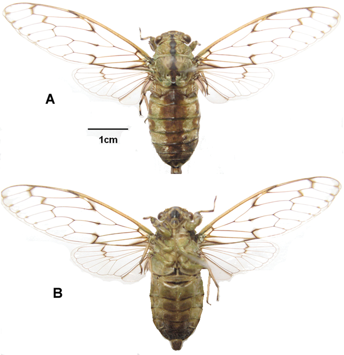
Semia spinosa sp. n. (male): A dorsal view B ventral view.
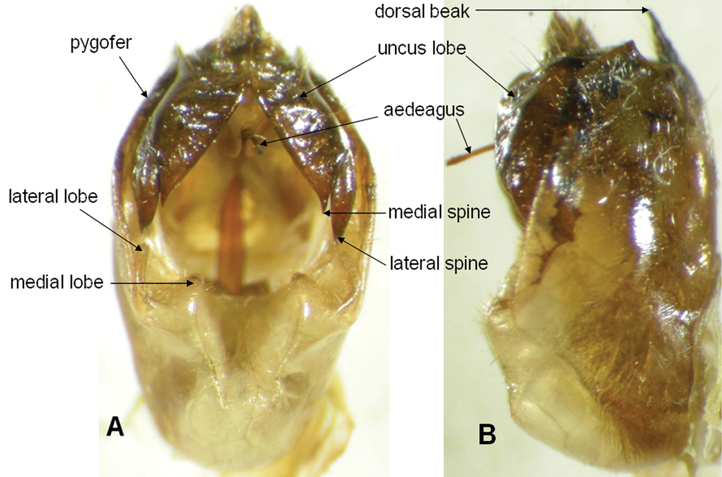
Male genital capsule of Semia spinosa sp. n.: A ventral view B lateral view.
This species was collected by sweeping during the daytime and by light trapping at night in virgin rainforest at an altitude between 100 to150 meters.
Vietnam (Dong Nai Province).
Semia spinosa is distinguishable from Semia watanabei and Semia klapperichi by the body size, which is shorter than 30mm (in male) in Semia spinosa and longer than 35mm (in male) in Semia watanabei and Semia klapperichi, and from Semia klapperichi it differs in the infuscations on the hind wings which lack spots along the ambient veins present in Semia klapperichi (see Figs 4A, C). The new species also differs in the structure of the uncus which has the lobes strongly divergent with acute apical spines (see Figs 6A–C).
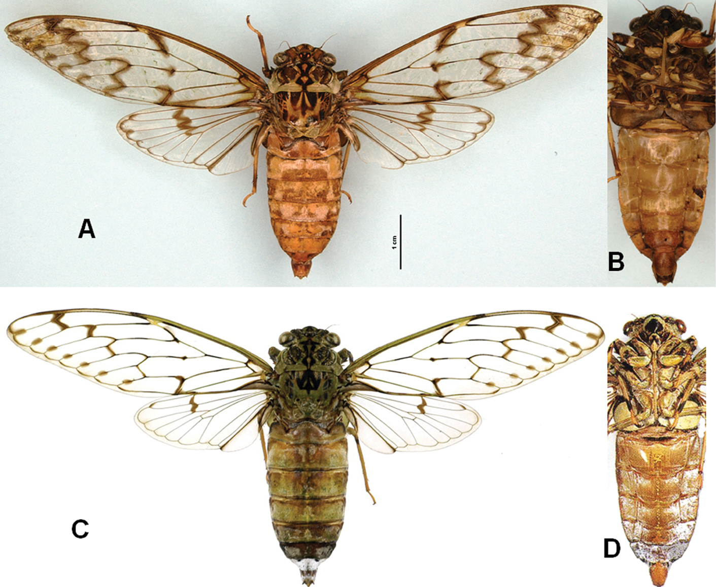
Semia species in dorsal view and ventral view (male): A, BSemia klapperichi(photograph by Dirk Ahrens) C, D Semia wantanabei (after
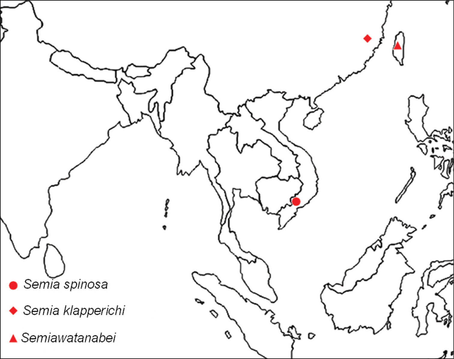
Distribution of the Semina species in the world.
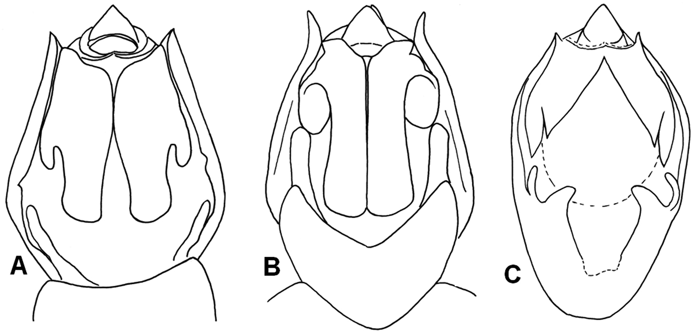
Male genital capsule of Semia species (ventral view): A Semia klapperichi B Semia watanabei (after
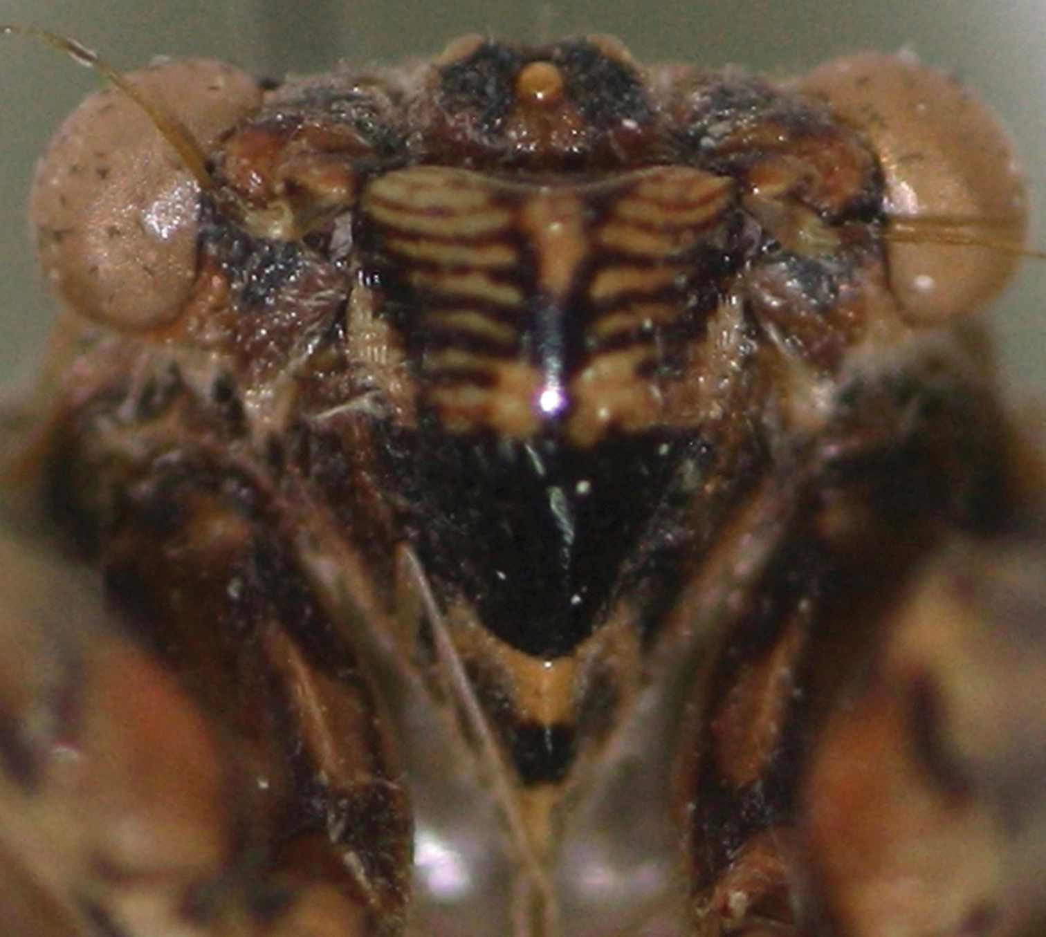
Postclypeus of Semia spinosa sp. n.
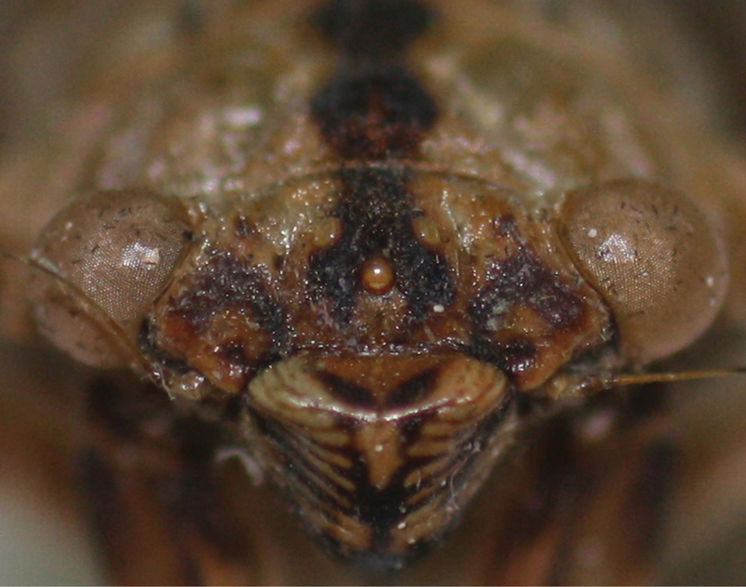
Dorsal part of the postclypeus of Semia spinosa sp. n.
| 1 | Body length <30mm; abodomen with longitudinal broad brown band centrally (Fig. 2A); uncus lobes strongly divergent, each with two apical spines (Fig. 3A) | Semia spinosa sp. n. |
| – | Body length >35mm; abodomen without longitudinal broad brown band centrally; uncus lobes not or weakly divergent without two apical spines | 2 |
| 2 | Body length approximately 38mm; operculum with lateral margin not expanding beyond lateral margin of abdomen; tymbal cover very small, pale brown, with darker margin; tymbal mostly exposed in dorsal view; uncus lobes parallel (Fig. 6B) | Semia watanabei |
| – | Body length 40–45mm; operculum very wide, lateral margin expanding distinctly beyond lateral margin of abdomen; tymbal cover small slightly raised, brown without dark margin; tymbal slightly exposed in dorsal view; uncus lobes weakly divergent (Fig. 6A) | Semia klapperichi |
We thank Prof. Ta Huy Thinh, and Mr. Hoang Vu Tru (Institute of Ecology and Biological Resources, Hanoi) for assistance with field work sharing of specimens and data, Dr. Mick Webb (The Natural History Museum, London), Dr. Max Moulds (The Australian Museum), Dr. Hans Duffels (Netherlands Centre of Biodiversity), and Dr. Jerome Constant (Royal Belgian Institute of Natural Sciences) for their critical reading and valuable comments in improving the contents of the paper. We are grateful to Dr. Dirk Ahrens (Zoologisches Forschungsmuseum A. Koenig) for providing photographs of Semia klapperichi.
The first author is grateful for a scholarship for his PhD dissertation issued by the program of the National Chung Hsing University, Taiwan. We also thank Dr. Yves Samyn and Dr. Patrick Grootaert (Royal Belgian Institute of Natural Sciences) for grant support to the first author; the grant was issued by the capacity building Programme of the Belgian Global Taxonomic Initiative National Focal Point and runs with financial support from the Belgian Directorate-General for Development Cooperation.
The present study was partially supported by the National Foundation for Science and Technology Development (NAFOSTED-106.12.15.09), Vietnam, the International Foundation for Science (IFS-No D/5181-1), Sweden, and the Nagao Natural Environment Foundation, Japan.