


(C) 2011 Matteo Montagna. This is an open access article distributed under the terms of the Creative Commons Attribution License 3.0 (CC-BY), which permits unrestricted use, distribution, and reproduction in any medium, provided the original author and source are credited.
For reference, use of the paginated PDF or printed version of this article is recommended.
Pachybrachis sassii, new species is described from Giglio Island, of the Tuscan Archipelago (Italy). The new species belongs to the nominotypical subgenus and is closely related to Pachybrachis salfii Burlini, 1957, from which it differs in the shape of the median lobe of the aedeagus and in the pattern of the yellow raised spots on the elytra and pronotum. Ecological observations are made. The neotype of Pachybrachis salfii from Colloreto, Monte Pollino (Italy) is designated.
Entomology, taxonomy, Coleoptera, Chrysomelidae, Cryptocephalinae, Pachybrachini, Tuscan Archipelago, neotype
The genus Pachybrachis Dejean, 1836 belongs to the subfamily Cryptocephalinae (Coleoptera, Chrysomelidae) and according to the color of the prothorax and elytra is subdivided into two subgenera: Pachybrachis sensu stricto (hereafter s. str.) and Chloropachys Rey, 1883. Phylogenetic analyses based on mitochondrial (12S rRNA, 16S rRNA) and nuclear markers (18S rRNA and 28S rRNA) (
More than 350 species of Pachybrachis s. str. have been described till now, about 200 of which are distributed in the western hemisphere and 150 in the Palaearctic Region (
In the summer of 2010, during study of a Chrysomelidae fauna of the Mediterranean islands, 11 specimens of a previously unknown Pachybrachis were collected. The specimens appeared closely related to Pachybrachis salfii Burlini, 1957 from which they differed in the structure of the median lobe of the aedeagus and in the pattern of yellow raised spots of the elytra and pronotum. Genital characters together with a pattern of yellow raised spots are commonly used for discrimination of sibling species within this genus (
All the specimens were collected on the host plants by net sweep. Specimen manipulation, dissection, measure, and photographs were completed with the auxiliary use of the stereo microscope Leica MS5 with an ocular micrometer. SEM micrographs of the aedeagus were made using Jeol JSM-5610LV scanning electron microscope. Spermatheca and kotpresse drawings were made using stereomicroscope Leica MS5 with an ocular grid (size 20 × 20 squares 1 cm²). Specimens are deposited at the Civic Museum of Natural History, Milan (MSNM), Matteo Montagna private collection, Anzano del Parco, Como, Italy (MMPC), and Davide Sassi private collection, Castelmarte, Como, Italy (DSPC).
Key to Pachybrachis s. str. with well defined yellow spots or/and stripes on a black background similar to Pachybrachis sassii sp. n. distributed in ItalyDue to a complexity of the genus (
| 1 | Mesoepimeron black, sometimes with blurred indistinct yellow shadow | 2 |
| – | Mesoepimeron yellow | 4 |
| 2 | Last abdominal tergite entirely black, without yellow spots. Mid and hind femora fulvous with lighter spot. Length: 3–3.5 mm. Distribution: Central Europe, Spain, France and Italy | Pachybrachis picus Weise, 1882 |
| – | Last abdominal tergite with yellow spots | 3 |
| 3 | Last abdominal sternite black, without spots. Black spot on pronotum with lateral yellowish dots. Length 3.5–4 mm. Distribution: South Central Europe, from Spain to the Caspian Sea | Pachybrachis tesselatus Olivier, 1791 |
| – | Last abdominal sternite with two yellow spots. Black spot on pronotum without lateral yellowish dots. Length: 3.5–4 mm. Distribution: Spain, France, Italy, Croatia, Serbia and Montenegro | Pachybrachis exclusus Rey, 1883 |
| 4 | Elytron without convex yellow spots or stripes elevated over the background | 5 |
| – | Elytron with convex yellow spots or stripes elevated over the background. Mesoepimeron with yellow sometimes indistinct spot. Last abdominal tergite and sternite completely black. Length 2.8–3.4 mm. Distribution: Giglio island (Italy) | Pachybrachis sassii sp. n. |
| 5 | Last abdominal sternite without two yellow spots. Length: 3.1–4.2 mm. Distribution: Europe and West Siberia | Pachybrachis hieroglyphicus Laicharting, 1781 |
| – | Last abdominal sternite with two yellow spots. Length: 2.8–3 mm. Distribution: Sicily | Pachybrachis siculus Weise, 1891 |
urn:lsid:zoobank.org:act:4AD3247F-94F2-43F3-8F89-FFD6D2F3B4FB
http://species-id.net/wiki/Pachybrachis_sassii
Figs 1, 3, 6Italy, Tuscany: Grosseto, Giglio island, Giglio Campese.
Type-specimens:Holotype male, pinned, with genitalia on a separate card board. Original label: “Italy, Grosseto, Isola del Giglio, Giglio Campese, 42°21.90'N, 10°52.59'E, ca. 30 m a.s.l., 14 Jun 2010, M. Montagna & F. Castiglioni leg. [printed label], “HOLOTYPUS / Pachybrachis sassii / Montagna M. des. [red handwritten label] (MSNM). Paratypes: 5 females and 5 males pinned. Three females and 3 males, original label: “Italy, Grosseto, Isola del Giglio, Via della Cabulgina, 42°22.97'N, 10°53.89'E, ca. 60 m a.s.l., 13 Jun 2010, M. Montagna & F. Castiglioni leg.” [printed label], “PARATYPE / Pachybrachis sassii / Montagna M. des. [red handwritten label]; 2 males and 2 females (MMPC), 1 male and 1 female (DSPC). Two males and 2 females, original label: “Italy, Grosseto, Isola del Giglio, Giglio Campese, 42°21.01'N, 10°52.26'E, ca. 30 m a.s.l., 14 Jun 2010, M. Montagna and F. Castiglioni leg. [printed label], “PARATYPE / Pachybrachis sassii / Montagna M. des. [red handwritten label] (MMPC).
The new species belongs to the subgenus Pachybrachis s.str. The external morphological characters, general habitus, size, black and yellow pattern of elytra and pronotum prompt the ascription of Pachybrachis sassii sp. n. to the fifth artificial group suggested by
Male and female measurements are reported in Table 1.
Body relatively robust, dorsally black with yellow raised spots (Fig. 1), black/yellow ratio variable in male and female (female usually darker than male). Ventral side black, mesoepimera with yellow sometimes evanescent spot. Pygidium black. Vertex, frons, clypeus and upper labrum yellow. Antennal insertions, median longitudinal line on frons (in female wider and triangular in shape) and upper internal margin of eyes black. Vertex with black transverse line. First three antennomeres pale with blackish upper part, from IV to XI blackish to completely black. Pronotum with black/yellow pattern and narrow blackish outline, entirely black in some female. Two large lateral and irregular black spots reach pronotal base at corners, sometimes contact with anterior border in female. Lateral and anterior edges yellow, absent in some female. Evanescent yellow mark present within lateral black spot. Central black “V”-shaped spot connected to lateral spots. Scutellum black, shiny. Elytron with yellow shiny edge extending from humeral callus to suture surrounding scutellum. Humeral callus shiny, proximally yellow, distally black, completely black in some specimens. Suture black. Epipleura yellow with black outline. Apical lunula yellow, covered with evident black punctures in middle, inner and outer yellow branches extending up to half of suture and below humeral calli respectively. Legs brownish, upper part of femora black in male. Mid and hind femora with distinct yellow spots and black knees. Frons flat, interocular furrow fairly impressed and punctured, evanescent in female. Eyes at upper edge with impressed and punctured median line. Head with strong and diffuse punctation, denser in middle and close to internal margins of eyes, evanescent on clypeus. First antennomere swollen, as long as third, second antennomere roundish and shorter than half of third. Eyes prominent, distance between upper lobes smaller than distance between antennal sockets.
Pronotum with maximum width slightly posterior to middle. Longitudinal edge curved in middle, anteriorly not completely visible from above. Posterior edge sinuous in middle, slightly rimmed and bordered by line of punctures. Punctation evanescent on central disc close to median yellow spot, gradually coarser on sides and towards posterior margin, absent on tiny yellow raised ridge surrounding pronotum. Posterior margin of pronotum with single row of punctures. At bottom, two transverse impressions separated from edge by slightly raised area, less evident than in Pachybrachis salfii. Scutellum wide, rectangular, slightly convex, covered with short setae. Elytra slightly wider than pronotum at base, flattened on top. Elytral punctation located mainly on black markings, well impressed and placed in irregular rows anteriorly, starting from median spot becomes almost regular and less impressed. Humeral callus prominent, externally delimited by grooves. Base of elytra, proximally to margin, with evident carina originating at humeral calli and extending along anterior margin up to suture surrounding scutellum. Epipleura narrowly raised. Internal margin of apical lunula evenly cut, puncture clusters separate two inner appendices. Pygidium evenly convex, punctated and pubescent. Urosternite with evident microsculpture and fine whitish pubescence. In male external margin of VII urosternite with row of short and dense setae. Anal sternite with shiny median impression in male, rectangular and not deeply engraved fovea in female. Legs without diagnostic characters, first tarsomere enlarged in male. Median lobe of aedeagus well sclerotized. Phallotreme with sinuous edges, tiny truncated tip surrounded by visible setae, central frenulum with long basal stem expanded anteriorly (Fig. 3a). In lateral view (Fig. 3b) apex sinuous, not curved apically as in Pachybrachis salfii (Figs 4, 5); frenuli narrow with right angle in distal part, not obtuse as in Pachybrachis salfii (Figs 4, 5). Ventral side with evident triangular carina harboring two small cristae at base, apex with robust setae on each side and hairless tip (Fig. 3c). Spermatheca (Fig. 6a) sickle-shaped, wider on curve, slender anteriorly to fine tip, slightly recurved at base; ductus sclerotized and smooth throughout its length, at base robust junction with spermatheca, distal end forming a subtriangular pigmented diverticulum; accessory gland joining spermatheca at base in slightly sclerotized junction opposite to ductus. Ventral sclerites of kotpresse (Fig. 6b) triangular, lateral apices with three spicules each, anterior margin with prominent central carina, posterior margin sinuous and convex in middle; dorsal sclerite strongly transverse (Fig. 6c), two narrow lateral wings and two well sclerotized vertical processes.
The species is dedicated to Dr. Davide Sassi, a friend and well-known specialist in Chrysomelidae.
Pachybrachis sassii inhabits plant geoseries of Cyclamino repandi-Querco ilicis sigmetum (
Male and female measurements of Pachybrachis sassii sp. n.: mean (with 95% confidence interval) in mm, standard deviation (in parentheses), range of variability (in square brackets) in mm. TotL: total length; ProL: length of pronotum; ElyW: width of elytra (measured at the base); ProW: width of pronotum, Ratio L/W (ratio between total body length and width of elytra).
| Specimens | TotL | ProL | ElyW | ProW | Ratio L/W |
| Male | 3.12 ± 0.15 (0.19) | 0.98 ± 0.04 (0.05) | 1.55 ± 0.06 (0.07) | 1.42 ± 0.05 (0.07) | 1.94–2.12 |
| n = 6 | [2.76–3.27] | [0.87–1.02] | [1.42–1.64] | [1.29–1.49] | |
| Female | 3.20 ± 0.10 (0.11) | 0.92 ± 0.03 (0.04) | 1.63 ± 0.05 (0.05) | 1.46 ± 0.03 (0.04) | 1.91–1.98 |
| n = 5 | [3.05–3.38] | [0.87–0.98] | [1.56–1.71] | [1.45–1.53] | [1.45–1.53] |
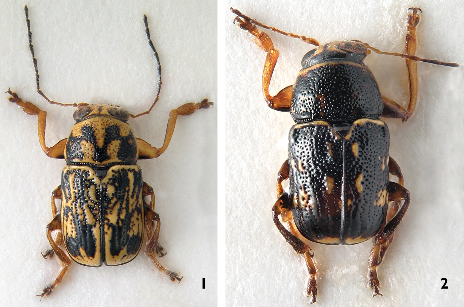
Habitus, dorsal view: 1 Pachybrachis sassii sp. n., holotype 2 Pachybrachis salfii, neotype.
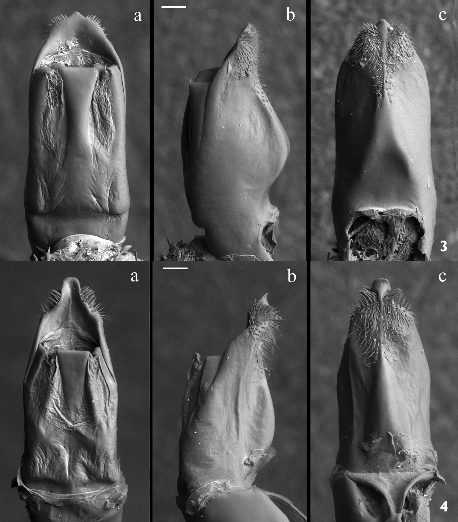
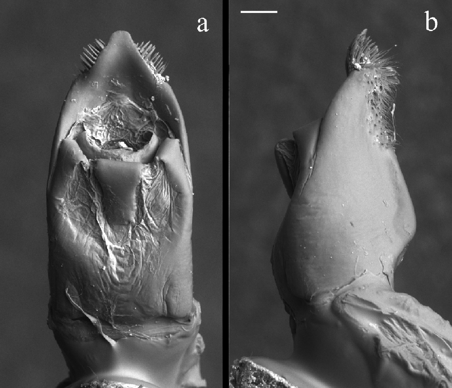
Median lobe of aedeagus; magnification 120×, scale bar = 100 µm 3 Pachybrachis sassii sp. n. holotype; a dorsal b lateral and c ventral view 4 Pachybrachis salfii neotype a dorsal b lateral and c ventral view. Median lobe silhouette is demaged by dehydration, probably due to contact with air for a long time.
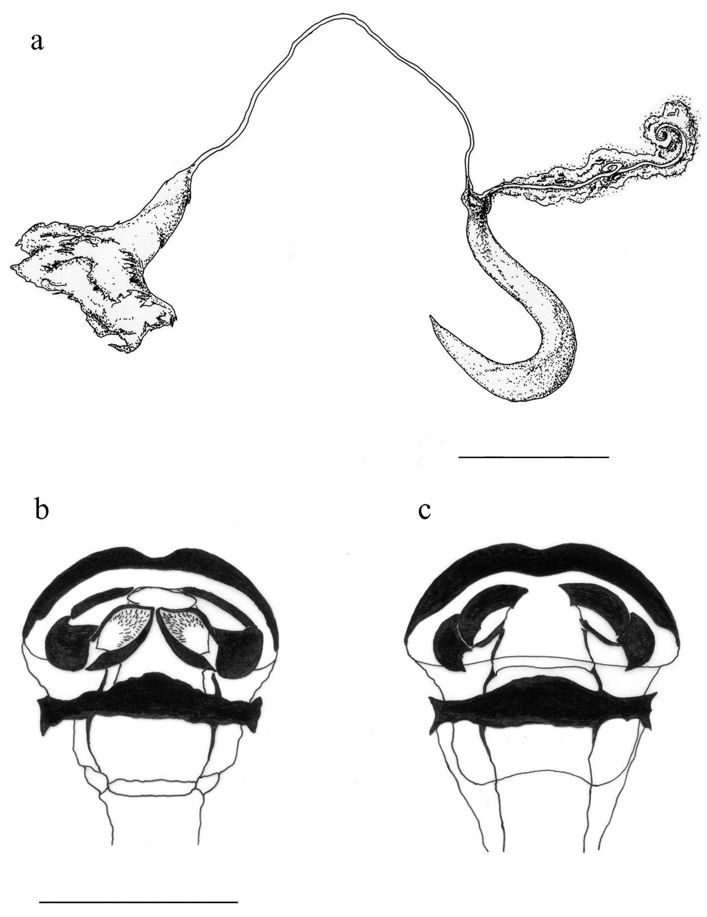
Median lobe of aedeagus: Pachybrachis salfii from Monte Pollino freshly extracted a dorsal and b lateral view. Magnification 120×; scale bar = 100 µm.
Pachybrachis sassii sp. n. a spermatheca b ventral sclerite of kotpresse c dorsal sclerite of kotpresse. Scale bar = 0.3 mm.
Morphological characters of the aedeagus highlight the affinity between Pachybrachis sassii and Pachybrachis salfii, as both taxa show the median lobe with a peculiar triangular carina on the ventral side, nevertheless, they differ in the shape of phallotreme, apical tip and lateral apex profile (Figs 3, 4, 5). The tip of the aedeagus in Pachybrachis sassii is sinuous and not curved anteriorly as in CistusPachybrachis salfii. These two taxa also significantly differ in the black and yellow pattern on pronotum and elytra (Figs 1, 2). Present data does not allow to test different hypotheses for the origin of the new taxon but the pre-Pleistocene transgression that separated Giglio island from Argentario cape could have been the event that lead to the allopatric speciation.
During the present study, the author discovered that the holotype of Pachybrachis salfii Burlini, 1957, deposited in the collection of Museo Zoologico dell’Università Federico II di Napoli, is lost (personal communication of Dr. Nicola Maio, curator, October 2010). According to article 75 of the International Code of Zoological Nomenclature (1999) and in order to clarify the taxonomic status of the nominal taxon, a paratype of Pachybrachis salfii (Burlini, 1957) is designated as neotype. Dr. Carlo Leonardi and Dr. Davide Sassi were previously consulted in order to avoid the proposed designation arousing serious objections. The neotype of Pachybrachis salfii was designated as paratype by
The author aknowledges Dr. Michele Zilioli, Dr. Fabrizio Rigato, and Dr. Carlo Leonardi of Natural History Museum of Milan for the image support and the scientific suggestions. A special thanks to Dr. Federica Castiglioni who participated in sample collection and supported my research. I also would like to thank my friend Dr. Davide Sassi for his critical input and scientific support.
Finally the author would like to thank three referees for their constructive comments, which have resulted in an improved manuscript.