Research Article
Print
Research Article
A taxonomic review of the order Mantodea in Korea based on morphology and DNA barcodes
expand article infoJaeil Shim§, Jeong-Hun Song
‡ National Institute of Agricultural Sciences, Wanju, Republic of Korea
§ Chungnam National University, Daejeon, Republic of Korea
Open Access

Abstract

A taxonomic study of Korean Mantodea using morphological and molecular characters (COI) is presented. Eight species [Amantis nawai (Shiraki, 1908), Acromantis japonica Westwood, 1889, Mantis religiosa sinica Bazyluk, 1960, Statilia maculata (Thunberg, 1784), Tenodera angustipennis Saussure, 1869, T. sinensis Saussure, 1871, Hierodula chinensis Werner, 1929, H. patellifera (Audinet-Serville, 1838)] belonging to six genera in three families are recognized. Interspecific genetic divergence of COI using uncorrected p-distance ranged from 6.7% to 22.4%, while intraspecific divergence ranged from 0% to 2.2% among eight Korean Mantodea species. All eight species were each strongly supported as a single lineage using COI on both neighbor-joining and parsimony trees. An illustrated key, redescriptions, habitus photographs, and illustrations of diagnostic characters of the species of Korean Mantodea are provided to facilitate identification.

Key words

DNA barcodes, Korea, Mantodea, review, taxonomy

Introduction

The order Mantodea comprise approximately 2,400 species in 460 genera, making it a distinctive group of predatory insects (Ehrmann 2002; Otte et al. 2024). They exhibit remarkable diversity in morphology, hunting strategies, and habitat specialization and are widely distributed in tropical and subtropical regions (Otte et al. 2024). Members of this order are generally characterized by a carnivorous diet, mimetic behavior, triangular head, large compound eyes, raptorial foreleg with elongated forecoxa, forefemur and foretibia with characteristic spines, femoral brush for grooming, elongated pronotum, male subgenital plate (coxosternite IX), asymmetrical male genitalia, female subgenital plate (coxosternite VII), and ootheca covered by a secretion (Klass 2001; Klass and Meier 2006; Yager and Svenson 2008; Wieland 2013; Svenson et al. 2015; Hashimoto et al. 2016; Brannoch et al. 2017).

Prior to 1995, only a few entomologists had recorded four species of Mantodea on the Korean peninsula, Mantis religiosa Linnaeus, 1758, Statilia maculata (Thunberg, 1784), Tenodera angustipennis Saussure, 1869, and T. aridifolia (Stoll, 1813) (Okamoto 1924; Bey-Bienko 1930; Doi 1932; Shiraki 1932; Cho 1959; Bazyluk 1960; Ju 1969; ESK and KSAE 1994). Kwon et al. (1996) recorded Amantis nawai (Shiraki, 1908), and Jeon et al. (1999) added three species, Acromantis japonica Westwood, 1889, Hierodula patellifera (Audinet-Serville, 1838), and S. nemoralis (Saussure, 1870), with descriptions and illustrations. Addionally, Shim et al. (2021a) reported the giant Asian mantis Hierodula chinensis Werner, 1929, with a diagnosis and illustrations. To date, nine species of Mantodean insects have been reported in Korea (Kim 2010, 2021; Shim et al. 2021a). While some species have been previously described, accurate identification of species within this group is challenging due to a lack of available morphological information.

In this paper, we present a taxonomic reassessment of Korean Mantodea, recognizing eight species belonging to six genera in three families, including A. nawai (Shiraki, 1908), Ac. Japonica Westwood, 1889, M. religiosa sinica Bazyluk, 1960, S. maculata (Thunberg, 1784), T. angustipennis Saussure, 1869, T. sinensis Saussure, 1871, H. chinensis Werner, 1929, and H. patellifera (Audinet-Serville, 1838). Re-descriptions incorporate salient morphological features critical for accurate identification of these species, including the male genitalia. We also used molecular criteria including genetic divergence and gene tree monophyly using a COI barcode region as a multiple lines of evidence approach for the species identification. Our taxonomic review of Korean Mantodea provides redescriptions, habitus photographs, an interactive key, and diagnoses.

Materials and methods

Studied specimens were mostly collected from inland and the islands of the Korean peninsula. Specimens were collected by direct sweeping, scanning, shifting leaf litter and light trapping. If nymphs or oothecae were found, samples were reared until the adult insect emerged. The collected specimens were killed by freezing to prevent discoloration and were moved to a drying chamber for dehydration at 60 °C for 10 days until completely hardened. The subsequent sample preparation followed methods by Brannoch et al. (2017) and Shim et al. (2021b). Briefly, the male genitalia were incubated overnight in 10% potassium hydroxide (KOH), and washed with distilled water, then 75% ethanol, before storage in glycerol. Depository of specimens examined is as follows: National Institute of Agricultural Sciences Insect Collection (NASIC, Wanju, Korea); Kunsan National University (KsNU, Gunsan, Korea); National Institute of Biological Resources (NIBR, Incheon, Korea).

The specimens were examined with a stereomicroscope (MS5, Leica Microsystem, Wetzlar, Germany). Images were obtained using a Canon DSLR (EOS 5D; Tokyo, Japan) with an attached Canon MP-E 65 mm f/2.8 1–5× lens. Several layers of photographs were combined in Helicon Focus 5.3 software (Helicon Soft Ltd, Kharkov, Ukraine) and edited using Adobe Photoshop CC 2020 (Adobe, San Jose, CA, USA). Measurements were recorded in millimeters using digital Vernier calipers (CD-15APX; Mitutoyo, Sakado, Japan). The terminology of taxonomic characters and measurements of specimens mainly followed Wieland (2013) and Brannoch et al. (2017) for external morphology, and Klass (1997) and Schwarz and Roy (2019) for male genitalia.

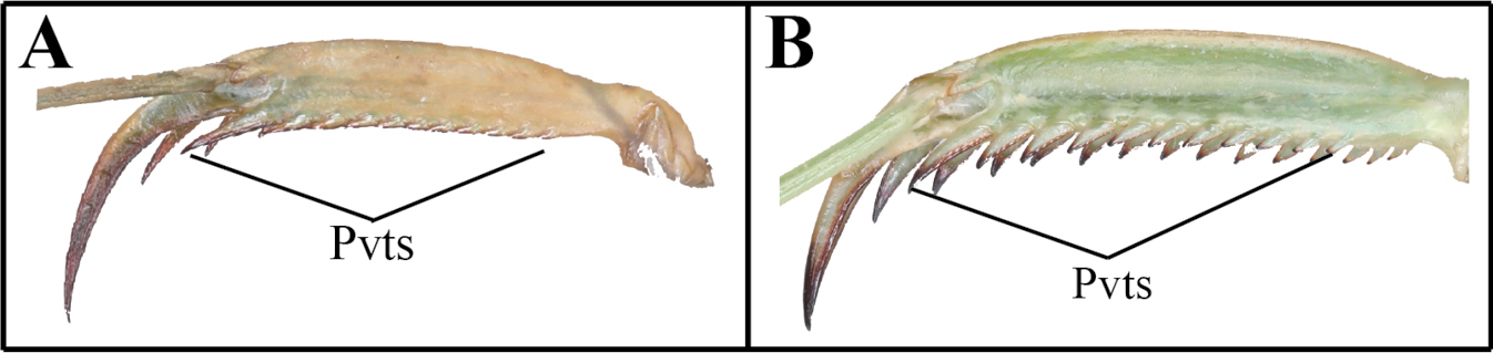
The following abbreviations are used for the foreleg spination formula and male genitalia: spination formula: Avfs = anteroventral femoral spines; Avts = anteroventral tibial spines; Ds = discoidal spines of forefemur; Pvfs = posteroventral femoral spines; Pvts = posteroventral tibial spines. Male genitalia: afa = anterior process of left phallomere (phalloid apophysis); aafa = anterior lobe of phalloid apophysis; fda = posterior lobe of right phallomere; loa = posterior membranous lobe of left phallomere; pafa = posterior lobe of phalloid apophysis; L4B = a sclerite of left phallomere, mostly spoon-shaped; maa = medial arm process of right phallomere; paa = elongated process of left phallomere, titillator; pia = process arising from the midlenth of the ventral wall of right phallomere, located posterolateral area of pva, strongly sclerotized; pva = process arising from midlenth of the ventral wall of right phallomere, located anteromesal area of pia, strongly sclerotized; sdpl = lateral secondary distal process; sdpm = median secondary distal process.

The following abbreviations are used for the provinces of Korean peninsula (Specimens examined): GW: Gangwon-do; GG: Gyeonggi-do; CB: Chungcheongbuk-do; CN: Chungcheongnam-do; GB: Gyeongsangbuk-do; GN: Gyeongsangnam-do; JB: Jeollabuk-do; JN: Jeollanam-do; JJ: Jeju-do (Is.).

For the study of molecular characters, we included a total of 74 specimens for DNA extraction in the dataset and the specimens used are listed in Suppl. material 1. DNA extraction, sequencing and alignments follow the methods described by Shim et al. (2021b). The mitochondrial COI was selected. Primers and amplification strategies are provided in Shim et al. (2021b). Data from GenBank for 50 foreign specimens were incorporated into the study, as indicated in Suppl. material 1. Parsimony (PA) analyses were conducted using MEGA X (Kumar et al. 2018) with 1000 bootstrap replications. A neighbor-joining analysis (NJ) was performed in MEGA X (Kumar et al. 2018) using the Kimura-2-Parameter (K2P) model (Kimura 1980). Bootstrap support values for each node were evaluated via MEGA X with 1000 replicates. Intra- and inter-specific distances in the different taxonomic levels were calculated using an uncorrected pairwise distance method (Srivathsan and Meier 2012).

Taxonomic accounts

Key to species of Mantodea in Korea

1 (A) Pvts fully decumbent Acromantis japonica
(B) Pvts not fully decumbent 2
2 (A) Genicular spurs length as long as Pvfs; second Avfs greatly enlarged Amantis nawai
(B) Genicular spurs length clearly shorter than Pvfs; second Avfs not enlarged 3
3 (A) Forecoxa with a distinct dark or eye spot covering ~ 1/4 to 2/5 of the total length 4
(B) Forecoxa without spot or a faint dark spot covering ~ 1/9 of the total length 5
4 (A) Tibial spur groove only with yellow spot or no spot at all Mantis religiosa sinica
(B) Tibial spur groove with white spot, and dark patch either side Statilia maculata
5 (A) Pronotum long and robust; forewing without stigma pattern; abdominal sternites with longitudinally striped pattern 6
(B) Pronotum clavate in shape; forewing with stigma pattern; abdominal sternites without striped pattern 7
6 (A) Forecoxa base color yellow (in life); hindwing, area of arculus near cells colored dark brown, subcostal to cubitus area brown to dark brown Tenodera sinensis
(B) Forecoxa base color orange (in life); hindwing, area of arculus near cells transparent, subcostal to cubitus area reddish T. angustipennis
7 (A) Furcasternite without pattern Hierodula chinensis
(B) Furcasternite with two-band pattern H. patellifera

Species descriptions

Family Gonypetidae Westwood, 1889

Subfamily Iridopteryginae Giglio-Tos, 1915

Amantis Giglio-Tos, 1915

Cimantis Giglio-Tos, 1915: 154 (synonymized by Beier 1935: 28).

Shirakia Beier, 1935: 47.

Type species

Mantis (Oxypilus) reticulata De Haan, 1842.

Diagnosis

Very small sized mantises. Body with mottled dark spot pattern. Pronotum short, kite- shaped, its dorsal surface with longitudinal striped pattern. Genicular spurs length as long as Pvfs length. Second Avfs is as long as second Ds, much longer than the neighboring spines. Wings brachypterous or macropterous.

Amantis nawai (Shiraki, 1908)

Figs 1–8, 9–14, 15–19

Gonypeta nawai Shiraki, 1908: 47.

Gonypeta maculata Shiraki, 1911: 318.

Amantis nawai (Shiraki, 1908): ESK and KSAE 1994: 44; Kim 2010: 31; Kim 2021: 65. Korean record.

Iridopteryx maculatus (Shiraki, 1911): Kwon et al. 1996: 221. Korean record.

Specimens examined

[NASIC] South Korea: GN: 1♂1♀, Mt. Noja, Dongbu- myeon, Island Geojedo, Geoje-si, 7 VIII 2019, Yeong-Hun Kim; 3♂3♀, Mt. Noja, Dongbu- myeon, Island Geojedo, Geoje-si, 7 VIII 2019, Woojin Jang; JN: 3♂1♀, Island Yeoseodo, Yeoseo-ri, Cheongsan-myeon, Wando-gun, 26 I 2019, Jaeil shim (reared from Ootheca); JJ: 1♀, Andeok Valley, Seogwipo-si, Jeju-do, X 2019, Do-yoon Kim; 2♂1♀, Donnaeko, Seogwipo-si, Jeju-do, 9 IX 2020, Yeong-Hun Kim; 3♂2♀, Seonheul-ri, Jocheon-eup, Jeju-si, Jeju-do, 19 V 2023, Jaeil Shim (reared from ootheca); 2♀, Gamsan-ri, Andeok-myeon, Seogwipo-si, Jeju-do, 23 IX 2023, Jaeil Shim; 1♀, Gamsan-ri, Andeok-myeon, Seogwipo-si, Jeju-do, X 2023, Jisung Kim; Japan: 1♂, Yanbaru, Okinawa, 1–4 I 2020, Wonjun Seong, Forest.

Redescription

Measurements (mm): Body length ♂ 12.1–13.0, ♀ 13.8–15.2; head width ♂ 2.2, ♀ 3.3; head length ♂ 2.9, ♀ 3.9; pronotum width ♂ 2.1, ♀ 2.5; pronotum length ♂ 3.3, ♀ 3.9; forewing (tegmina) length ♂♀ 1.6. Male (Figs 1, 3, 5, 7, 917). Very small sized mantises. Coloration (Figs 1, 3): Body color bright brown to dark brown. Body surface with irregular dark brown spots. Head (Fig. 5): Triangular, slightly broader than pronotum maximum width. Head length 1.3× width. Antenna filiform. Antenna nearly 3× as long as pronotum. Vertex nearly flat, posterior area (Figs 1, 3) with five dark spots. Compound eye slightly protruding, its surface glossy and brown moire pattern (in live specimens). Ocelli small, all of the same shape and size. Epistomal sulcus weakly concave. Prothorax (Fig. 7): Dorsal surface with dark brown longitudinal stripe pattern. Pronotum very short, kite-shaped, lateral margin with minute denticulation. Pronotum length 1.4× as long as maximum width. Metazone nearly 2× as long as prozone. Forelegs (Prothoracic legs) (Figs 917): Coxa and femur slightly longer than pronotum. Coxa dorsal margin (Figs 9, 11) with 9–13 minute forecoxal spines. Dorsal and ventral coxal lobes (Fig. 9) fully divided from each other; dorsal coxal lobes with dark brown spot. Femur, tibia, and first tarsomere darker than body color (Figs 1, 3, 11). Femur interior surface (Fig. 10) with numerous denticles. Genicular spurs (Figs 9, 11) well-developed, as long as Pvts length. Spination formula (Figs 9–11): Avts = 10–12; Pvts = 12–13; Avfs = 11–14; Pvfs = 4; Ds = 4. In 13 Avfs (Figs 9, 10): spines 2 and 13 larger than remaining Avfs; spines 10, 11, and 12 smallest of Avfs. Meso- and metathoracic legs: Meso- and metathoracic legs long and slender, apical area of tibia and first tarsomere dark brown. Tarsi 5-segmented. First tarsomere of mesotarsus subequal to combined length of remaining segments, first tarsomere of metatarsus longer than remaining segments combined. Wings: Brachypterous. Wing venation faded. Abdomen: Cerci setose, not flattened, with nine or ten segments. Male subgenital plate (coxosternite IX) (Fig. 15) nearly rhombus, inter-stylar margin slightly convex; stylus rather short. Male genitalia (Figs 16, 17): Right phallomere with C-shaped pva (Fig. 16); pia sclerotized and weakly wrinkled; fda distal edge margin rounded, surface with few setae. Left phallomere with short and curved paa; afa sclerotized, rectangular (slightly concave at the middle), shape of distal end variable, surface weakly granulated; Korean populations with two types of afa: dorsally curved type (Fig. 16) and posteriorly decumbent type (Fig. 17); loa mostly invisible. Ventral phallomere nearly oblong; sdpm very weakly developed; sdpl slightly expanded (digitiform), right margin of sdpl conspicuously concave. Female (Figs 2, 4, 6, 8). Similar to male, with following differences. Head (Fig. 6): length 1.2× width. Antenna nearly 2× as long as pronotum. Vertex swollen. Prothorax (Fig. 8): Pronotum length 1.5× maximum width. Narrowest width on posterior one-third of pronotum. Forelegs (Prothoracic legs) (Figs 2, 4): Femur color generally same as body color. First tarsomere distal end area dark brown. Abdomen: Post-lateral margin of tergite with dark brown spot. Ootheca (Figs 18, 19). Measurements (mm): Length 3.2–5.3; width 2.8–3.4; height 2.7–3.4; length of emergence area 3.9–6.4; width of emergence area 1.0–1.6. Identification: Prism-shaped, triangular in cross-section. Ootheca attached by ventral surface. External wall colored russet. External coating (Figs 18, 19) beige, consisting of a very thin layer and frothy material, lateral surface and emergence area fully covered. Exhibiting ~ 4–8 egg chambers (Fig. 19). Lateral surface with longitudinal parallel ridges (boundaries). Distal end of ootheca truncate and rough. Distal flap area with long residual process (Fig. 18). Nymph. First instar nymph: Body surface shiny, reddish brown with brightly colored transverse pattern on body segments and legs; postero-medial edge of pro-, meso-, and metathorax tergite slightly protruding.

Figures 1–8. 

Habitus, head and pronotum of Amantis nawai 1 male dorsal aspect (Geojedo island) 2 female dorsal aspect (Geojedo island) 3 male dorsal aspect (Jeju) 4 female dorsal aspect (Jeju) 5 male face 6 female face 7 male pronotum 8 female pronotum.

Figures 9–14. 

Male foreleg of Amantis nawai 9 ventral aspect 10 interior aspect 11 dorsal aspect 12 foreleg dorsal aspect (Jeju) 13 foreleg dorsal aspect (Geojedo island) 14 foreleg dorsal aspect (Yeoseodo island). White arrow = femoral brush.

Biological notes

Amantis nawai is distributed throughout the southern islands of the Korean peninsula, as reported by Kwon et al. (1996). This species is commonly found beneath the litter layer in shaded areas and typically lays its oothecae under dead leaves or between stones on the ground. The species exhibits unique behaviors, including the vibration of its antenna and swinging of its forelegs. The first instar nymphs usually hatch between mid-June and early July, while adults emerge in mid-August.

Distribution

China, Japan, Taiwan, South Korea.

Remarks

Amantis nawai occurs in East Asia (Yamasaki 1981; Chou 2006; Patel and Singh 2016; Oshima 2018). Korean populations of A. nawai exhibit two distinct male genitalia structures, distinguished by the ‘afa’ structure (Figs 16, 17), as well as variable forefemur color patterns (Figs 12–14). The Yeoseodo Island population features a posteriorly decumbent ‘afa’ structure (Fig. 17), and the male forefemur color matches their body color (Fig. 14). In contrast, the Geojedo and Jeju Island populations exhibit a dorsally curved ‘afa’ structure (Fig. 16), and the forefemur color of most male samples is darker than their body color (Figs 1, 3). However, there is no significant intraspecific genetic divergence (COI) between the Yeoseodo and Geojedo Island populations. Additionally, the Jeju population shows only a 0.3% divergence from the other islands.

Figures 15–19. 

Male genitalia and ootheca of Amantis nawai 15 subgenital plate 16 male genitalia (Jeju) 17 male genitalia (Yeoseodo island) 18 ootheca (dorsal aspect) 19 ootheca (lateral aspect). Red arrows = concave area.

Family Hymenopodidae Giglio-Tos, 1915

Subfamily Acromantinae Brunner de Wattenwyl, 1893

Acromantis Saussure, 1870

Type species

Mantis oligoneura De Haan, 1842

Diagnosis

Small-sized mantises. Compound eye with stripe pattern (in live specimens). Anterior area of vertex and lower frons slightly protruding. Pronotum metazone middle area with flat bulge. Genicular spurs clearly shorter than the shortest Pvfs length. Pvts fully decumbent. Meso- and metathoracic femora with weakly expanded postero-ventral femoral lobe. Posterior margins of sternites with a medial lip. Hindwing apex distally truncate.

Acromantis japonica Westwood, 1889

Figs 20–29, 30–33

Acromantis japonica Westwood, 1889: 43.

Acromantis japonica Westwood, 1889: Jeon et al. 1999: 226 (South Korea); Kim 2021: 65. Korean records.

Specimens examined

[NASIC] South Korea: GN: 2♀, Mt. Noja, Dongbu-myeon, Island Geojedo, Geoje-si, 10 IX 2016, Jaeil Shim; 1♂, Mt. Wangjo, Nambu-myeon, Tappo-ri, Island Geojedo, Geoje-si, 23 XII 2019, Jun-Ho Lee; 2♀, Mt. Noja, Dongbu-myeon, Island Geojedo, Geoje-si, 29 IX 2016, Byeongmin Jeong; JN: 3♂, Jeongdo-ri, Wando-gun, 10 I 2019, JaeDong Gim (reared from ootheca); 3♂, Jeongdo-ri, Wando-gun, 26 II 2019, Jaeil Shim (reared from ootheca); 8♂6♀, Is. Bogil-do, Bogil-myoen, Wando-gun, 2 III 2023, Jaeil Shim (reared from ootheca); JJ: 1♂, Mt. Sambang, Namjeju-gun, Jeju-si 28 IX 2000, Miae Kim; 1♂, Hwasun-ri, Andeok-myeon, Seogwipo-si, 12–13 IX 2023, Jaeil Shim; 8♂1♀, Sumang-ri, Namwon-eup, Seogwipo-si, 13–14 IX 2023, Jaeil Shim.

Redescription

Measurements (mm): Total length (vertex to tip of abdomen) ♂ 23.3–26.2, ♀ 28.4–32.2; head width ♂ 4.1–4.2, ♀ 5.0–5.1; head length ♂ 2.4, ♀ 3.4; pronotum width ♂ 2.6–2.7, ♀ 3.4; pronotum length ♂ 6.5–7.0, ♀ 8.2–8.4; forewing (tegmina) length ♂ 17.4–20.0, ♀ 17.0–18.5. Male (Figs 20, 22, 24–28, 30, 31). Small sized mantises. Coloration (Fig. 20): Body greenish brown to brown. Head (Fig. 22): Triangular; width 1.7× length. Antenna > 2× as long as pronotum. Vertex nearly flat, anterior vertex with weakly pronounced spurs. Postocellar tubercle weakly pronounced. Compound eye globular, slightly protruding laterally; compound eye with brightly coloured moire pattern (in live specimens). Ocelli larger than female, middle ocellus nearly globular, lateral two ocelli oblong. Posterior apex of lower frons (Fig. 22) with protruding spur. Prothorax (Figs 24, 25): Pronotum length 2.5–2.6× maximum width. Pronotum (Fig. 24) surface smooth, lateral margin with few denticles; metazone > 2× longer than prozone; middle of metazone with flat bulge (pair); metazone lateral margin slightly concave. Furcasternite (Fig. 25) very slightly convex, middle area with flat protuberance. Forelegs (Prothoracic legs): Foreleg (Figs 20, 26) surface smooth and shiny, pale green. Coxa dorsal margin with 6–10 spines. Dorsal and ventral coxal lobes (Fig. 26) fully divided from each other. Femur dorsal margin (Figs 20, 26) slightly convex at the middle, distal half of margin very slightly concave. Genicular spurs (Fig. 26) well developed, conspicuously shorter than Pvfs, postero-ventral genicular spurs weakly curved outward. Pvts (Fig. 27) fully decumbent. Spination formula: Avts = 12–13; Pvts = 12; Avfs = 10–13; Pvfs = 4; Ds = 4. In 12 Avfs (Fig. 26, 28): spines 2, 4, 6, 8, 10, and 12 size larger than remaining Avfs, dark brown. Ds 2 and 3 dark brown in live specimens. Meso- and metathoracic legs: Meso- and metathoracic legs with transverse dark pattern. Meso- and metathoracic femora (Fig. 20) with weakly expanded postero-ventral femoral lobe. Tarsi 5-segmented. Wing: Wings completely surpassing the end of abdomen (Fig. 20). Forewing costal area bright brown or green, discoidal area brownish and transparent, cubitus brown. Forewing with oblique brown stripes. Hindwing costal area brown, discoidal area brownish and transparent; hindwing apex brown, truncate. Abdomen: Sternites III–VII posterior margin with a medial lip, process slightly expanded. Cerci setose, 13 segments. Male subgenital plate (coxosternite IX) (Fig. 30) irregularly rounded rhomboidal in shape, inter-stylar margin generally concave, but medial area occasionally slightly convex. Ventral surface of subgenital plate with numerous setae. Styli rather short. Male genitalia (Fig. 31): Right phallomere pia sclerotized and surface with few denticles; fda nearly triangular in shape, its surface with few setae. Left phallomere, paa nearly absent (very blunt); afa weakly sclerotized, and distal end bulbous; loa invisible. Ventral phallomere nearly elliptical, without any melanized structures, distal margin truncate or weakly bilobed (sdpm and sdpl may be expanded). Female (Figs 21, 23, 29). Similar to male, with following differences. Head (Fig. 23): width 1.5× length. Antenna as long as pronotum. Prothorax: Pronotum length 2.4× maximum width. Forelegs (Prothoracic legs): Foreleg dorsal surface brown, coxa ventral surface (Fig. 29) red in live specimens. Wings (Fig. 21): Forewing costal area color green. Ootheca (Figs 32, 33). Measurements (mm): Length 10.7–14.8; maximum width 4.3–4.8; maximum height 3.6–4.3; length of emergence area 7.8–10.4; width of emergence area 1.5–2.4. Identification: Nearly rectangular dorsally, hemispherical in cross-section, each edge with slightly expanded attachment area (Fig. 32). Ootheca attached on ventral surface. External wall color brown (Fig. 33). External coating bright brown but surface weakly covered. Exhibiting ~ 10–15 egg chambers (Fig. 33) clearly delimited by prominently visible curved lips. Distal end softly truncate and weakly rough. Nymph. First instar nymph: Body color black and surface shiny (ant mimic); compound eye with white stripes (moiré pattern); posterior margin of pro-, meso-, and metathoracic tergite with white lines; tarsus of meso- and metathoracic legs white. Second to last instar nymph: Body brown to dark brown with brightly colored mottled pattern; vertex spurs well developed; abdomen with expanded lamellar process (lip) in the middle of sternites.

Figures 20–29. 

Habitus, head, pronotum and foreleg of Acromantis japonica 20 male dorsal aspect 21 female dorsal aspect 22 male face 23 female face 24 female pronotum 25 male furcasternite 26 male foreleg ventral aspect 27 foreleg tibia and tarsus (dorsal aspect) 28 foreleg interior aspect 29 female coxa ventral aspect (live specimens). Red arrows = bulge and protuberance.

Figures 30–33. 

Male genitalia and ootheca of Acromantis japonica 30 subgenital plate 31 male genitalia 32 ootheca (dorsal aspect) 33 ootheca (lateral asepct).

Biological notes

Acromantis japonica is found in the southern islands of the Korean Peninsula (Jeon et al. 1999; An 2011). They occur in leaf litter, under broad leaves and on tree trunks. Adults exhibit positive phototaxis, and oothecae are typically laid under stones and the bark of rotten logs. This species overwinters in the egg stage inside their oothecae (Taniguchi 1987; Befu 1992). They exhibit behaviors such as vibrating their antennae and swinging forelegs. Nymphs fold their abdomens back during rest. The first instar nymphs hatch from the end of May to early June, while adults emerge beginning from the end of August.

Distribution

China, Japan, Taiwan, North Korea, South Korea.

Remarks

The genus Acromantis has characteristic stripe patterns on their compound eyes when they are alive (see Nakamine et al. 2017: figs 2, 3), weakly pronounced postocellar tubercles and truncated hindwings (Vyjayandi et al. 2010; Mukherjee et al. 2017; Shcherbakov and Anisyutkin 2018). They occur in East Asia to Australasia (Jeon et al. 1999; Nakamine 2016; Patel et al. 2016).

Family Mantidae Latreille, 1802

Subfamily Mantinae Latreille, 1802

Mantis Linnaeus, 1758

Mantes Geoffroy, 1762: 399.

Type species

Gryllus (Mantis) religiosa Linnaeus, 1758

Diagnosis

Medium to large sized mantises. Body color mostly bright green, yellow, brown. Male antenna reddish, conspicuously thicker than the female’s. Vertex swollen. Forecoxal proximal area with dark spot, ventral surface with numerous yellow spots. Tibial spur groove area with yellow spot. Furcasternite with numerous spots. Metathoracical episternum with a dark lateral spot. Male inter-stylar margin notched.

Mantis religiosa sinica Bazyluk, 1960

Figs 34–46, 47–51

Mantis religiosa sinica Bazyluk, 1960: 255.

Mantis religiosa sinica Bazyluk, 1960: 255; Kim 2010: 31; Kim 2021: 65. Korean record.

Mantis religiosa (Linnaeus, 1758): ESK and KSAE 1994: 44. Korean record.

Specimens examined

[NASIC] South Korea: GW: 3♂, Hotel Ramada, Daegwallyeong Pass, Pyeongchang-gun, 22 VIII 2019, JuHyeong Sohn; GG: 1♀, Jeongok-eup, Yeoncheon-gun, 22 VIII 84, Sunhee Jang; 1♀, Mt. Acha, Gwangjin-gu, Seoul, 8 IX 1977, Sunhee Yoon, Sungshin Univ.; 1 Nymph 1♂4♀, Island Gureopdo, Gureop-ri, Deokjeok-myeon, Incheon, 17 VII 2019, Byeongmin Jeong (reared from nymph); 2♂1♀, Island Gureopdo, Gureop-ri, Deokjeok-myeon, Incheon, 28 VI 2023, Jaeil Shim, Wonjun Sung (reared from nymph); CN: 1♂1♀, Coastal Dune, Sindu-ri, Wonbuk-myeon, Taean-gun, 1 IX 2001, Haechul Park, near the grassland; 4♂6♀, Sonhwang-ri, Woongcheon-eup, Boryeong-si, 12 VII 2023, Jaeil Shim, near the grassland (reared from nymph); 2♂3♀, Coastal Dune, Sindu-ri, Wonbuk-myeon, Taean-gun, 12 VIII 2023, Jaeil Shim, near the grassland; GB: 2♂, Mt. Angi, Songhyeon-dong, Andong-si, VIII 2022, Jaeil Shim; 3 Nymphs, Gyeongjeong Beach, Gyeongjeong-ri, Chuksan-myeon, Yeongdeok-gun, 20 VII 2023, Jaeil Shim; GN: 2♀, Mt. Noja, Dongbu- myeon, Island Geojedo, Geoje-si, 15 IX 2021, Jaeil Shim; JB: 1♂, Byeonsan-myeon, Buan-gun, VIII 1999, Jeonbuk Nat. Univ; 1♂, Mt. Jeoksang, Muju-gun, 8 IX 1999, Taewoo Kim; 1♀, Kunsan Nat. Univ., Gunsan-si, 31 VIII 2019, JuHyeong Sohn; 6♂8♀, Is. Yamido, Okdo-myeon, Gunsan-si, 5 VII 2023, Jaeil Shim (reared from nymph); JJ: 3 Nymphs, Gwangchigi Beach, Goseong-ri, Seongsan-eup, Seogwipo-si, 16 V 2021, Jaeil Shim; Hungary: Mantis religiosa religiosa, 1♂, Mt. Csakyar, Vertes, 23 VIII 2003, J.C. Sohn, Haraszt hegy 250m.

Redescription

Measurements (mm): Total length (vertex to tip of abdomen) ♂ 42.3–55.2, ♀ 50.8–72.4; head width ♂ 5.1–5.4, ♀ 6.2–6.5; head length ♂ 3.8–4.2, ♀ 5.4–5.6; pronotum width ♂ 4.0–4.2, ♀ 5.6–6.0; pronotum length ♂ 13.2–14.0, ♀ 16.6–18.0; forewing (tegmina) length ♂ 38.4–43.2, ♀ 36.1–47.7. Male (Figs 34, 36, 38, 4249) Medium to large sized mantises. Coloration (Figs 34, 35): Body bright green, brown and yellow. Head (Fig. 36): width 1.3× length. Vertex swollen; with pale transverse magenta line along dorsal apex (in live specimens) (Fig. 38). Compound eye globular, anteriorly protruding; dorsal surface with two transverse stripes (in live specimens). Ocelli large, oval, pale yellow. Antenna filiform; pedicel, scape and initial flagellum pale, remaining flagellum orangish to reddish brown (Figs 34, 36). Antenna length > 2× as long as pronotum, conspicuously thicker than in female. Lower frons posterior apex weakly protruding; pale transverse line at posterior one-third of lower frons (in live specimens). Epistomal sulcus transverse. Prothorax (Fig. 38): Pronotum flattened dorso-ventrally, its length 3.3× as long as maximum width. Prozone lateral margin with small denticles. Metazone 3× as long as prozone; lateral margin smooth. Medial keel protruding. Furcasternite (Fig. 41) posterior area with numerous dark spots. Forelegs (Prothoracic legs) (Figs 42–46): Coxa dorsal margin with 6–11 spines (Fig. 43), small denticles located between its spines. Coxa ventral surface (Figs 42, 43) with large oblong black spot or eye spot in proximal area; remaining surface with 15–24 yellow spots, center of each spot with small seta. Coxal lobes fully divided from each other. Tibial spur groove (Figs 42, 44) with yellow spotted (blotch) pattern. Genicular spurs minute. Ventral surface of tibia (Fig. 44) yellow to orange. Spination formula (Figs 44, 45): Avts = 12–13; Pvts = 7; Avfs = 12–13; Pvfs = 4; Ds = 4. In 13 Avfs (Figs 44, 45): spines 2, 4, 6, 8, 10, 12 and 13 larger and black, spines with black spot at the base. Tarsus with ventral area brownish. Meso- and metathorax and legs: Metathorax episternum (Fig. 47) with dark triangular spot. Meso- and metathoracic legs long and slender; tarsi 5-segmented. Wings: Forewing completely surpassing the end of abdomen. Stigma elongate, slightly protruding; color same as forewing venation. Forewing anterior margin brown, discoidal area mostly hyaline. Hindwing transparent but apex brownish. Abdomen: Cerci setose, elongated, and thick, not flattened, brown; 17 segments. Male subgenital plate (coxosternite IX) (Fig. 48) irregularly elliptical in shape, inter-stylar margin notched in V-shape. Styli rather long. Male genitalia (Fig. 49): Right phallomere smooth, forming a C-shaped pva; pia sclerotized and weakly wrinkled; fda triangular. Left phallomere with elongate and curved paa, its distal end sharp and sclerotized, paa anterior margin one-fourth to one-third with one or two short projections; afa strongly sclerotized, rounded, anterior margin obliquely curved dorsally, surface granulated; L4B curved spoon-shaped. Ventral phallomere (Fig. 49) nearly rhomboidal; sdpm elongated, blunt finger-like and slightly curved dorsally, distal end and right margin sclerotized and surface with numerous denticulation; sdpl slightly protruding (blunt projection). Female (Figs 35, 37, 39, 40). Similar to male, with following differences. Head (Fig. 37): Vertex convex. Head width 1.1× as long as length. Antenna as long as pronotum. Prothorax (Figs 39, 40): Pronotum length 2.9–3.0× as long as maximum width. Metazone 2–3× as long as prozone. Ootheca (Figs 50, 51). Measurements (mm): Length 17.2–25.1; maximum width 11.4–14.3; maximum height 9.3–10.5; length of emergence area 17.0–21.6; width of emergence area 3.2–5.3. Identification: Oblong, nearly hemispherical in cross-section. External wall bright brown to brown (Fig. 50). External coating beige on egg chamber surface, pale on emergence area (flap). Exhibiting ~ 20–30 egg chambers (Fig. 51) clearly delimited by visible slightly curved lips. Distal end of ootheca narrowed into residual process; residual process attached to substrate. Nymph. First instar nymph: Body brown, vertex dorsal apex with pair of dark spots. Mid to last instar nymph: In brown morph, dorsal surface of body with a few stripes.

Figures 34–46. 

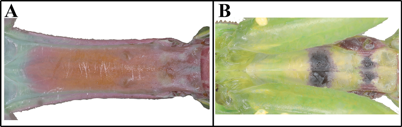
Habitus, head, pronotum and foreleg of Mantis religiosa sinica 34 male dorsal aspect 35 female dorsal aspect 36 male face 37 female face 38 male pronotum 39 female pronotum (inland) 40 female pronotum (Gureopdo island) 41 furcasternite 42 foreleg ventral aspect (live specimens) 43 coxa ventral aspect 44 forleg ventral aspect 45 foreleg interior aspect 46 foreleg dorsal aspect. Abbreviation: Tsg = tibial spur groove. Red arrows = dark spots of furcasternite.

Biological notes

Mantis religiosa sinica is sparsely distributed on the Korean Peninsula. This species prefers broad grasslands and bushy areas, comprised of shrubs and grass in sandy fields as its habitat. It exhibits positive phototaxis, meaning it is attracted to light. When threatened, it makes a hissing sound by rubbing its hindwings against the abdomen. The first instar nymphs hatch from the end of May onwards while the adults emerge in August.

Distribution

China, Japan, South Korea.

Remarks

Mantis religiosa (Linnaeus) is a widely distributed Paleotropical and Holarctic species (Lia 2007; Patel and Singh 2016; Otte et al. 2024). Bazyluk (1960) and Roy (1968) provided illustrations of the sdpm variations and Schwarz and Roy (2019) presented figures of male genitalia. Additional morphological information was provided by Ehrmann and Borer (2015), Shcherbakov and Anisyutkin (2018), and Shcherbakov and Govorov (2020). Bazyluk (1960) classified this species as the subspecies M. religiosa sinica, which is found in East Asia including Korea. The population on Gureopdo Island exhibits a shorter pronotum, smaller body size, and shorter forewing length compared to inland populations. However, there are minimal genetic differences in the partial COI regions (0%–0.2%). Furthermore, all specimens were supported as a single lineage using COI on both NJ and PA trees (Fig. 138).

Figures 47–51. 

Metathorax, male genitalia and ootheca of Mantis religiosa sinica 47 metathorax episterum 48 subgenital plate 49 male genitalia (small box = variation of sdpl) 50 ootheca (dorsal aspect) 51 ootheca (lateral asepct). Red arrow = dark pattern.

Statilia Stål, 1877

Type species

Pseudomantis nemoralis Saussure, 1870

Diagnosis

Medium sized mantises. Vertex nearly flat. Pronotum slender. Ventral surface of foreleg shiny, densely patterned, forcoxa proximal area with dark pattern. Tibial spur groove with whitish yellow to yellow spots. Male inter-stylar margin notched. Male genitalia: sdpl and sdpm well developed, angled at 90°.

Statilia maculata (Thunberg, 1784)

Figs 52–62, 63–70

Mantis maculata Thunberg, 1784: 61.

Pseudomantis haanii Saussure, 1871a: 37, 1871b: 400.

Statilia maculata var. hyalina Giglio-Tos, 1927: 410.

Statilia haani var. major Werner, 1922: 154.

Statilia maculata continentalis Werner, 1935: 495.

Statilia maculata (Thunberg, 1784): ESK and KSAE 1994: 44; Kim 2010: 31; Kim 2021: 65. Korean record.

Statilia nemoralis (Saussure, 1870): Jeon et al. 1999: 227 (misidentification); Kim 2010: 31 (misidentification). Korean record.

Specimens examined

[KsNU] South Korea: CN: 1♂, Mt. Bongsoo, Daechung-myeon, Yesan-gun, 18 V 2017 Hongjoon Choi; JB: 1♀, Eunpa Lake, Jigok-dong, Gunsan-si, 3 IX 2016; Donghwan Na; 1♀, Miryong-dong, Gunsan-si, 27 VI 2017, Eunhye Jeon; [NASIC] South Korea: GW: 1♂, Ssangyong, Yeongwol-gun, 30 IX 1999, Miae Kim; GG: 1♀, Mt. Umyeon, Seocho-gu, Seoul, 1 IX 1991, Jiyung Oh; 1♀, Jamsil 6-dong, Songpa-gu, Seoul, 20 X 1997, Soyeon Kim; 1♀, Mt. Nam, Hada-ri, Heungcheon-myeon, Yeoju-si, 3 IX 2000, Yeongbo Lee; 1♀, Seodun-dong, Suwon-si, 4 IX 2000, Taewoo Kim; 1♂, Seodun-dong, Suwon-si, 20 IX 2000, Taewoo Kim; 1♂, Seodun-dong, Suwon-si, 20 IX 2001, Jeonghun Hwang; ♂1, Haguidong, Uiwang-si, 24 IX 2003, Jeongsun Lee; 1♂, Mt. Gwanggyo, Jangan-gu, Suwon-si, 10 X 2003, Mikyeong Ahn; CB: 1♀, Mt. Nam, Cheongju-si, 8 VIII 2000, Hyea Lee; 1♂, Geumseok-ri, Geumwang-eup, Eumseong-gun, 14 IX 2019, Seong-Gyu Lee; CN: 1♂1♀, Coastal Dune, Sindu-ri, Wonbuk-myeon, Taean-gun, 1 IX 2005, Yeongbo Lee; GB: 1♀, Mt. Sobaek, Punggi-gun, 5 IX 2000, Miae Kim; 1♂3♀, Mt. Angi, Songhyeon-dong, Andong-si, VIII 2022, Jaeil Shim; 2♂6♀, Hotel Interburgo, Manchon-dong, Suseong-gu, Daegu-si 29 X 2023, Jaeil Shim; GN: 1♂, Sinhyeon-ri, Geoje island, Geoje-si, 11 IX 2002, Miae Kim; 2♂1♀, Mundong Waterfall, Geoje-si, 19 IX 2006, Miae Kim; ♀1 (green), Recreational Forest, Dongbu-myeon, Geojedo, Geoje-si, 29 IX 2020, Wonjun Sung; JB: 2♂4♀, Gueok, Yongjin-myeon, Wanju, 13 VIII 2013, Hanjun Bae; 1♂, Iseo-myeon, Wanju-gun, 26 VII 2016, Taeman Han; 1♀, Iseo-myeon, Wanju-gun, 29 VII 2014, Kyusuk Lee; 1♂1♀, Apartment, Iseo-myeon, Wanju-gun, 6 VIII 2015, Kyusuk Lee; 1♀ (green), National Institute of Agricultural Sciences, Iseo-myeon, Wanju-gun, 24 VIII 2016, Hyeop Lee; 1♀, Miryong-dong, Gunsan-si, 27 VI 2017, Eunhye Jeon; 2♂, Iseo-myeon, Wanju-gun, 23 VIII 2019, Jaeil Shim; 1♂2♀, Mt. Moak, Gui-myeon, Wanju-gun, 7 IX 2019, Jaeil Shim; 7♂5♀, Jangsu-eup, Jangsu-gun, 5 IX 2019, Jaeil Shim; JN: 3♀, Geumgok-dong, Buk-gu, Gwangju-si, 20 III 2021, Jaeil Shim; 4♀, Near Yeosu Airport, Sinpung-ri, Yulchon-myeon, Yeosu-si, IX 2021, Byeongmin Jeong; 2♂1♀, Is. Bogil-do, Bogil-myoen, Wando-gun, 2 III 2023, Jaeil Shim (reared from ootheca); 8♂9♀, Island Yeoseodo, Yeoseo-ri, Cheongsan-myeon, Wando-gun, VI 2023, Jaeil shim (reared from Ootheca); JJ: 1♂, Seonheul-ri, Jeju-si, 28 IX 2000, Taewoo Kim; 1♂, Sinae 1-ri, Jeju-si, 18 X 2001, Mikyeong Ahn; 1♂, Bijarim Forest, Jeju-si, 22 IX 2006, Miae Kim; Japan: 1♂, Asakura, Kyushu, 24 VIII 2013, Sangwook Park; [NIBR] South Korea: GG: 1♀, Namhansanseong, Seongnam, 28 IX 1997, Jeong Yun Chang.

Figures 52–62. 

Habitus, head, pronotum and foreleg of Statilia maculata 52 male dorsal aspect 53 female dorsal aspect 54 female dorsal aspect (green morpho-type) 55 male face 56 female face 57 male pronotum 58 female pronotum 59 furcasternite (brown morpho-type) 60 furcasternite (green morpho-type) 61 femur ventral aspect (green morpho-type) 62 foreleg ventral aspect (live specimens). Abbreviation: Tsg = tibial spur groove. Red arrows = dark spot of furcasternite.

Redescription

Measurements (mm): Total length (vertex to tip of abdomen) ♂ 35.2–43.1, ♀ 43.7–57.7; head width ♂ 4.5–5.0, ♀ 5.4–5.8; head length ♂ 2.9–3.2, ♀ 4.1–4.3; pronotum width ♂ 2.9–3.2, ♀ 4.4–4.6; pronotum length ♂ 12.1–14.0, ♀ 14.0–15.3; forewing (tegmina) length ♂ 26.4–31.2, ♀ 33.1–36.1. Male (Figs 52, 55, 57, 63–68) Medium sized, body slender. Coloration (Figs 52, 53): Beige to dark brown. Head (Fig. 55): Triangular. Head width 1.5–1.6× length. Vertex flat; dark transverse stripe on dorsal apex. Vertex and juxtaocular bulge with sparse, pale, transversely striped pattern. Compound eye large, globular, protruding laterally; in live specimens, dorsal and lateral surface with two brightly colored lines. Ocelli large, oblong. Antenna filiform, slightly longer than pronotum. Lower frons posterior apex protruding very slightly. Epistomal sulcus slightly concave. Prothorax (Fig. 57): Pronotum slender, narrow; length 4.1–4.3× as long as maximum width; dorsal surface smooth. Pronotum lateral margin with numerous denticles. Medial keel protruding. Metazone 3× as long as prozone. Membranous area between basis of forecoxa attachment, shiny and pinkish pearl in color (in live specimens). Furcasternite (Fig. 59) anterior area with rectangular dark spot, remaining posterior area reddish brown to magenta. Forelegs (Prothoracic legs) (Figs 6265): Coxa very slender, triangular; dorsal margin (Figs 59, 62) with 6–8 white spines; proximal area of ventral surface (Fig. 59) with large and shiny rectangular black spot. Coxal lobes divided from each other. Femur ventral surface shiny, with two black spots (Figs 62, 63), large rectangular spot preceding the tibial spur groove, linear spot (transverse line) distad to tibial spur grooves; tibial spur groove (Fig. 63) with white or whitish yellow pattern; distal area of ventral surface (Fig. 62) pale pink to magenta (in live specimens). Spination formula: Avts = 11; Pvts = 6–7; Avfs = 14; Pvfs = 4; Ds = 4. Tarsomere ventral surface (euplantula) dark brown. Basal rim of Pvts with dark spot. In 14 Avfs (Figs 63, 64): spines 2, 4, 6, 8, 10, 12, and 14 larger in size than remaining Avfs; spines 1, 2, 4, 6, 8, 10, 12, and 14 black, with small dark spot at the base. Meso- and metathoracic legs: Tarsi 5-segmented. Wings (Fig. 66): Forewing completely surpassing the end of abdomen. Stigma elongate, slightly protruding. Hindwing costal area reddish brown; discoidal area with darkish smoky mottled pattern but cross veins clearly transparent. Abdomen: Cerci setose, not flattened, brown, with 14 segments. Male subgenital plate (coxosternite IX) (Fig. 67) irregular rhomboidal, inter-stylar margin deeply notched in V-shape. Styli rather long. Male genitalia (Fig. 68): Right phallomere forming a V-shape pva; pia sclerotized and weakly wrinkled; fda triangular. Left phallomere with elongate and curved paa, surface smooth, distal end blunt and slightly swollen; afa sclerotized, small and irregularly rough, surface weakly granulated; L4B C-shaped. Ventral phallomere (Fig. 68) irregular rhomboidal; sdpm sclerotized, wide triangular; sdpl anteriorly curved hook-shaped with blunt apex, margin between sdpm and sdpl flat or slightly concave. Female (Figs 53, 56, 58, 59). Similar to male, with following differences. Coloration (Fig. 53): Body and forewing color beige to dark brown. Head (Fig. 56): width 1.3× as long as length. Antenna 2× longer than prozone length. Prothorax (Figs 58): Pronotum length 3.1–3.3× as long as maximum width. Green morphotype female (Figs 54, 60, 61). Prothorax: Rectangular black spot (Fig. 60) of furcasternite absent or very weakly developed, remaining posterior area greenish. Forlegs (Prothoracic legs) (Figs 60, 61): Tibial spur grooves (Fig. 61) with rectangular pale spotted pattern. Distal area of femur ventral surface pale pink or green. Wings (Fig. 54): Hindwing transparent or anal area with few mottled dark spots. Ootheca (Figs 69, 70). Measurements (mm): Length 20.4–28.9; maximum width 6.1–10.2; maximum height 6.4–8.3; length of emergence area 15.3–24.2; width of emergence area 2.2–3.5. Identification: Fusiform in shape, nearly hemispherical in cross-section. Proximal end with medial elevation of emergence area. Ootheca attached by its ventral surface. External wall bright brown. External coating weakly covering lateral zone of emergence area; beige in color. Exhibiting ~ 25–50 egg chambers clearly delimited by visible prominently oblique lips (Fig. 70). Distal end of ootheca narrowed into residual process, greatly elongated, and attached to substrate. Nymph. First instar nymph: Body dark brown, leg with few brightly colored stripes. Mid to last instar nymph: Forefemur distal area (pale pink to magenta) covered by mottled black or brown spots.

Figures 63–70. 

Foreleg, wings, male genitalia and ootheca of Statilia maculata 63 foreleg ventral aspect 64 foreleg interior aspect 65 foreleg dorsal aspect 66 wings 67 subgenital plate 68 male genitalia 69 ootheca (dorsal aspect) 70 ootheca (lateral asepct). Abbreviation: Ts = tibial spur Tsg = tibial spur groove. Red arrow = stigma.

Biological notes

Statilia maculata is found throughout the Korean Peninsula. This species has shown a remarkable adaptability to urban and suburban environments, and is often observed on building walls and streetlights, exhibiting positive phototaxis. They are known to deposit their oothecae under stones and in cracks of tree bark. Notably, S. maculata is capable of producing a hissing sound by rubbing its hindwings and abdomen together. This species typically hatches in early June, with adults emerging in the middle of August.

Distribution

China, Japan, Nepal, South Korea, Taiwan. Invasive in Eastern USA and Russia.

Remarks

Statilia maculata is predominantly found in the eastern Palearctic regions (S. maculata has also been introduced to Eastern USA and Russia), whereas S. nemoralis is described from the Philippines and, erroneously, from various Southeast Asian countries (Ehrmann 2002; Patel and Singh 2016; Schwarz et al. 2018). These two species, with S. nobilis (Brunner de Wattenwyl, 1893), are frequently confused in the taxonomic literature, leading to numerous misidentifications (see details in Schwarz et al. 2018); for example, both S. nobilis and S. maculata have been erroneously reported as S. nemoralis (Wang 1993; Jeon et al. 1999; Oshima 2017; Schwarz et al. 2018; Oshima 2020; Shcherbakov and Govorov 2020). Notably, the green morph is relatively uncommon in female S. maculata (Fig. 54) but prevalent in both sexes of S. nobilis and S. nemoralis (Zhu et al. 2012; Ehrmann and Borer 2015; Schwarz et al. 2018).

Jeon et al. (1999) initially reported S. nemoralis in Korea based on two female specimens lacking black spots on the furcasternite. Differences in the male genitalia, specifically the margins of the sdpl and sdpm, are reliable for distinguishing between S. maculata and S. nobilis (Schwarz et al. 2018). On the other hand, the lack of a black spot on the furcasternite is a common feature of the green morph of S. maculata. The occurrence of the Philippine Statilia nemoralis is continental SE Asia needs confirmation. Subsequent examinations revealed that the specimens initially identified by Jeon et al. (1999) as S. nemoralis were, in fact, misidentifications of S. maculata.

Subfamily Tenoderinae Brunner de Wattenwyl, 1893

Tenodera Burmeister, 1838

Mantis (Tenodera) Burmeister, 1838: 534.

Paratenodera Rehn, 1903: 705.

Type species

Mantis fasciata Manuel, 1797

Diagnosis

Large sized mantises. Male body slender, female robust. Ventral surface of forefemur patterned with minute spots. Area between forecoxae attachment point of yellow to orange in color. Hindwing with dark mottled pattern. Abdominal sternites with yellowish longitudinal stripes at middle. Male genitalia: aafa and pafa well developed, pafa spoon- or blade-shaped; loa elongate (Jensen et al. 2010).

Tenodera angustipennis Saussure, 1869

Figs 71–81, 82–87

Tenodera angustipennis Saussure, 1869: 69.

Tenodera angustipennis Saussure, 1869: ESK and KSAE 1994: 44; Kim 2010: 31; Kim 2021: 65. Korean record.

Specimens examined

[KsNU] South Korea: JB: 1♀, River Geumgang, Gunsan- si, 15 IX 2012, Hyojoong Kim, near the river mouth; 1♂, Kunsan Nat. Univ., Gunsan-si, 15 X 2015, Soyeon Kim; 1♀, Eunpa Lake, Gunsan-si, 10 V 2016, Sihyun Kim; 1♀, Kunsan Nat. Univ., Gunsan-si, 20 VIII 2017, Juyeong Oh; [NASIC] South Korea: GW: 1♂, Songjuk Coastal area, Ganseong-eup, 19 IX 2001, Miae Kim; GG: 1♂, Gaepo-dong, Seoul, 1 IX 1991, Hyeonjeong Jo; 2 Nymphs, Seodun-dong, Suwon-si, 13 VIII 1998, Graduate School of Korea Univ.; 1♂, Gosaek-dong, Suwon-si, 20 VIII 1998, Seongsun Jang, ridge of rice field; 1♂, Yulgeuk 2-ri, Heungcheon-myeon, Yeoju-si, 24 VIII 1998, Yeongbo Lee, ridge of rice field; 1♂, Seodun-dong, Suwon-si, 11 IX 1998, Graduate School of Korea Univ.; 1♂1♀, Yulgeuk 2-ri, Heungcheon- myeon, Yeoju-si, 30 IX 1999, Yeongbo Lee, ridge of rice field; 1 Nmyph, Seodun-dong, Suwon-si, 6 VII 2000, Taewoo Kim; 3 Nmyphs, Seodun-dong, Suwon-si, 20 VII 2000, Taewoo Kim; 2♂1♀, Seodun-dong, Suwon-si, 20 IX 2000, Taewoo Kim; 5 Nymphs, Shihwa, Siheung-si, 30 VII 2003, Jaecheon Son; 1♂, Sinheung College, Howon-dong, Uijeongbu-si, 26 IX 2005, Jindong Yeo; 5 Nymphs, World Cup Park, Seongsan-dong, Mapo-gu, Seoul, 20 VII 2013, Yeongbo Lee; CB: 1♀, Mt. Nam, Cheongju-si, 24 VIII 2000, Heeah Lee; 1♀, Maebong peak, Mt. Songni, Boeun-gun, 17 IX 2002, Yeongbo Lee; 1♀, Maebong peak, Mt. Songni, Boeun-gun, 18 IX 2002, Haechul Park; 1♀, Box office, Mt. Songni, Jangam-ri, Boeun-gun, 30 IX 2002, Jaecheon Son; 1♂1♀, Gamgok-myeon, Eumseong-gun, 31 VIII 2019, Byeongmin Jeong; 1♀, Parking area of Cheongju Airport, Ipsang-ri, Naesu-eup, Cheongwon-gu, Cheongju-si, 15 IX 2023, Jaeil Shim; CN: 1♂, Sindu-ri, Wonbuk-myeon, Taean-gun, 1 IX 2001, Haechul Park, grassland; 1 Nymph, Sindu-ri, Wonbuk-myeon, Taean-gun, 1 IX 2005, Jaecheon Son; 2♀, Coastal Dune, Sindu-ri, Wonbuk-myeon, Taean-gun, 12 VIII 2023, Jaeil Shim, near the grassland; GB: 1♂, Rest area of expressway, Chilgok-gun, 8 IX 2000, Taewoo Kim; 2♀, Mt. Palgong, Daegu, 1 IX 2014, Taeman Han; 2♂1♀, Gomo station, Suseong-gu, Daegu, 3 IX 2016, Jaeil Shim; 1♂, Gomo station, Suseong-gu, Daegu, 14 IX 2019, Jaeil Shim; 1♂6♀, Mangudang park, Hyomok-dong, Daegu-si 29 X 2023, Jaeil Shim; GN: 2♀, Yonggang-ri, Hwagae-myeon, Hadong-gun, 15 IX 2023, Jaeil Shim; JB: 1♀, Jeonbuk Nat. Univ., Jeonju-si, 17 X 2017, Sunyeong Park; 1♀, Jinan-Maisan Rest area of expressway, Jinan-gun, 22 VIII 2019, Jaeil Shim; 1♂, National Institute of Agricultural Sciences, Iseo-myeon, Wanju-gun, 31 VIII 2019, Jaeil Shim; 1♀, Jangsu-eup, Jangsu-gun, 5 IX 2019, Jaeil Shim; 2♂5♀, Mt. Moak, Gui-myeon, Wanju-gun, 7 IX 2019, Jaeil Shim; 1♂, Temple Geumsansa, Gimje-gun, Gimje-si, 5 IX 2020, Jaeil Shim; JN: 2♀, Myeongsasimni, Coastal Dune, Island Bigeumdo, Sinan-gun, 22 IX 2001, Haechul Park; JJ: 1♀, Island Udo, Jeju-si, 10 X 1999, Taewoo Kim.

Figures 71–81. 

Habitus, head, pronotum and foreleg of Tenodera angustipennis 71 male dorsal aspect 72 female dorsal aspect 73 male face 74 female face 75 male pronotum 76 female pronotum (small sized) 77 female pronotum (large sized) 78 foreleg ventral aspect 79 foreleg interior aspect 80 foreleg dorsal aspect 81 ventral aspect of foreleg and furcasternite (live specimens). Abbreviation: Tsg = tibial spur groove. Red arrows = dark spot near the tibial spur groove.

Redescription

Measurements (mm): Total length (vertex to tip of abdomen) ♂ 44.2–63.3, ♀ 51.2–86.6; head width ♂ 5.9–6.5, ♀ 6.9–7.8; head length ♂ 4.5–4.9, ♀ 5.6–6.7; pronotum width ♂ 4.3–4.9, ♀ 4.9–6.4; pronotum length ♂ 18.0–22.3, ♀ 19.2–25.0; forewing (tegmina) length ♂ 38.4–48.2, ♀ 38.1–55.2. Male (Figs 71, 73, 75, 78–80, 82–86). Large sized, body slender. Coloration (Figs 71, 82): Body and forewing discoidal area green, greenish brown and brown. Head (Fig. 73): Triangular. Head width 1.3× as long as head length. Vertex slightly convex, brown; apex with a bright brown transverse line. Compound eye globular. Ocelli large, oblong. Antenna filiform; antenna length nearly 1.5× as long as pronotum. Lower frons posterior apex slightly protruding. Epistomal sulcus slightly concave. Lower frons with three, clypeus and labrum with two dark longitudinal stripes. Prothorax (Fig. 75): Pronotum long and slender, flatted dorso-ventrally, dorsal surface smooth; length 4.1–4.4× as long as maximum width. Prozone lateral margin with few denticulations in large sized specimens. Metazone color orangish in green morph; lateral margin smooth; metazone 3.4–4.7× as long as prozone. Between basis of forecoxa attachment membranous surface (Fig. 81) orange (in live specimen). Postcervical plate and anterior area of furcasternite with gradational dark pattern in brown morph (Fig. 81) and occasionally in green morph. Medial keel brownish (Fig. 76). Forelegs (Prothoracic legs) (Figs 78–81): Coxa (Fig. 81) dorsal margin with pale color, 7–10 whitish spines, conical or blunt; ventral surface with pale and dark pattern on proximal area in brown morph; remaining surface with numerous white spots. Tibial spur groove (Figs 78, 81) with faint dark spot. Spination formula (Figs 78–80): Avts = 12–17; Pvts = 9; Avfs = 14–16; Pvfs = 4; Ds = 4. In 15 Avfs (Figs 78, 79): spines 2, 4, 6, 8, 10, 12, and 15 larger than remaining Avfs; spines 1, 2, 4, 6, 8, 10, 12, and 14 with dark brown spot at the base. Each tarsomere (Fig. 78) distal area dark brown. Tarsi 5-segmented. Wings (Figs 82–84): Forewing completely surpassing end of abdomen; costal area green, discoidal area transparent. Forewing (Fig. 82) subcosta and radius brown, its color obviously darker than other veins. Hindwing anal area hyaline and nearly transparent in green morph (Fig. 82), brownish and smoky spotted pattern in brown morph (Fig. 83); arculus veins (Fig. 84) and nearby cells mostly transparent; cross veins of subcostal and cubitus area brown to dark brown. Abdomen: A longitudinal yellow stripe in middle of abdominal sternites; abdominal sternites with brightly colored mottled pattern. Cerci setose, not flattened, with 17 segments. Male subgenital plate (coxosternite IX) (Fig. 85) irregular rhomboidal, inter-stylar margin extremely convex; ventral surface with numerous setae. Styli rather short. Male genitalia (Fig. 86): Right phallomere forming nearly V-shaped pva; pia sclerotized and weakly wrinkled; posterior surface of pia with weakly expanded membranous area, surface with minute denticulation; fda elongate lobed shape. Left phallomere (Fig. 86) with elongate and curved paa, its distal apex rounded; aafa sclerotized, straight spike shape; pafa sclerotized, curved at more than 45° arch, wide blade-shaped, outer margin with numerous decumbent spines; loa membranous, elongate finger-shaped, longer than pafa; L4B curved spoon-shaped. Ventral phallomere irregular rhomboidal, posterior margin prominently expanded; sdpl (Fig. 86) more than 90° angle at the middle. Female (Figs 72, 74, 77). Similar to male, with following differences. Head (Fig. 74): width 1.1 to 1.2× as long as head length. Vertex convex. Antenna as long as head to pronotum length. Prothorax (Figs 76, 77): Pronotum length 3.8–3.9× as long as maximum width; lateral margin with numerous denticles. Prozone dorsal surface with numerous blunted denticles. Metazone 2.8–3.1× as long as prozone. Medial keel protruding, pale brown or occasionally green in green morph. Forelegs (Prothoracic legs): Coxa dorsal margin with 7–13 large conical spines, small denticles located between them. Wings: Forewing often reaching end of abdomen; discoidal area semi-transparent. Ootheca (Fig. 87). Measurements (mm): Length 21.8–39.7; maximum width 10.1–13.7; maximum height 7.1–11.0; length of emergence area 15.0–31.0; width of emergence area 3.2–4.3. Identification: Fusiform, nearly hemispherical in cross-section. Proximal end with medial elevation of emergence area. Ootheca attached by ventral surface or fully encircling a thin substrate such as sticks. External wall bright brown. External coating covering almost entire surface of ootheca except lateral zone of emergence area; beige. Lateral side of emergence area prominently concave (Fig. 87). Exhibiting ~ 18–29 egg chambers clearly delimited by visible prominently oblique lips; lips occasionally invisible on new oothecae due to covering by external coating. Distal end of ootheca narrowed to residual process; greatly elongate and attached to substrate. Nymph. Mid to last instar nymph: Avfs 1 and last Avfs base to tibial spur groove with dark spotted pattern; between forecoxa basis membranous attachment surface orange.

Figures 82–87. 

Wings, male genitalia and ootheca of Tenodera angustipennis. 82 wings (green morpho-type) 83 hindwing (brown morpho-type) 84 hindwing venations 85 subgenital plate 86 male genitalia 87 ootheca (dorsal aspect). Red arrow = arculus area.

Biological notes

Tenodera angustipennis occurs throughout the Korean peninsula and both adults and oothecae can be commonly found on trees and shrubs. First instar nymphs hatch from late May to mid-June, and adults emerge in mid-August.

Distribution

China, India, Japan, Java, South Korea. Invasive in NE USA and Hawaii.

Remarks

Tenodera angustipennis is morphologically similar to T. sinensis in Korea but can be distinguished by their more slender bodies, the pronotum length/width ratio, the orange coloration between the forecoxa base (Oshima 2018) (Fig. 81), lack of reddish coloration on the hindwing radius area (Figs 82–84), and the pointed, perpendicular apex of the aafa (Fig. 86).

Tenodera sinensis Saussure, 1871

Figs 88–97, 98–102

Mantis mandarinea Saussure, 1871a: 289.

Tenodera aridifolia var. sinensis Saussure, 1871b: 417.

Tenodera aridifolia (Stoll, 1813): ESK and KSAE 1994: 44 (misidentification). Korean record.

Tenodera sinensis Saussure, 1871: Kim 2010: 31; Kim 2021: 65. Korean record.

Specimens examined

[KsNU] South Korea: JB: 1♂, Eunpa Lake, Gunsan-si, 8 XI 2016, Donghwan Na; [NASIC] South Korea: GW: 2 Nymphs, Balsan 2-ri, Chuncheon-si, 12 VI 1998, Sungsoon Jang; 1♀, Jinburyeong, Jinbu-myeon, Pyeongchang-gun, 29 IX 2000, Taehwa Kang; 1 Nymph, Ingye-ri, Okgye-myeon, Gangneung-si, 15 VIII 2002, Jingoo Yeo; 3♂, Mt. Odae, Hongcheong-gun, 29 VIII 2019, Jaeil Shim; GG: 1♂, Mijang-ri, Samjuk-myeon, Anseong-si, 17 IX 2000, Yeongbo Lee; 1♂, Mt. Cheolma, Incheon, 26 VIII 2001, Taewoo Kim; 1 Nymph, Shihwa, Siheung-si, 30 VII 2003, Jaecheon Son; 1 Nymph, Temple Jeondeungsa, Onsu-ri, Gilsang-myeon, Ganghwa-gun, Incheon, 4 IX 2009, Yeongbo Lee; 1♀, Jikdong-ri, Sohol-eup, Pocheon-si, 7 IX 2011, Yeongbo Lee; 2 Nymphs, Temple Bogwangsa, Gwangtan-myeon, Paju-si, 10 VII 2013, Yeongbo Lee; 3 Nymphs, World Cup Park, Seongsan-dong, Mapo-gu, Seoul, 21 VII 2013, Yeongbo Lee; 2 Nymphs, Noel Park, Sangam-dong, Mapo-gu, Seoul, 30 VII 2013, Yeongbo Lee;); 1♂, Island Gureopdo, Gureop-ri, Deokjeok-myeon, Incheon, 28 VI 2023, Jaeil Shim, Wonjun Sung (reared from nymph); CB: 1 Nymph, Magok-ri, Bongyang-eup, Jecheon-si, 13 VII 2005, Taehwa Kang; 1♀, Gamgok-myeon, Eumseong-gun, 31 VIII 2019, Byeongmin Jeong; 3♂, Geumseok-ri, Geumwang-eup, Eumseong-gun, 14 IX 2019, Seong-Gyu Lee; 1♂, Jeongbang-ri, Annae-myeon, Okcheon-gun, 12 X 2000, Yeongbo Lee; CN: 1♀, Mt. Sikjang, Daejeon, 14 VIII 2019, Geonheyok Kim (reared from nymph); 4♂, Coastal Dune, Sindu-ri, Wonbuk-myeon, Taean-gun, 12 VIII 2023, Jaeil Shim, near the grassland; GB: 1 Nymph, Geumgok-ri, Byeonggok- myeon, Yeongdeok-gun, 1 VII 2009, Yeongbo Lee, Hoyeon Jeong; GN: 4♀, Mt. Noja, Dongbu- myeon, Island Geojedo, Geoje-si, 15 IX 2021, Jaeil Shim; JB: 1♀, Hoyja 3-dong, Wansan-gu, Jeonju-si, 28 VIII 2014, Taeman Han; 1♂, Deokjin Park, Deokjin-gu, Jeonju-si, 6 IX 2018, Junhee Park; 1♀, Jeonju riverside, Jeonju-si, 15 IX 2018, Jaeil Shim; 1♂, Mt. Moak, Gui-myeon, Wanju-gun, 6 X 2018, Hyeon-Ha Yoo; 1♂, Jinan-Maisan Rest area of expressway, Jinan-gun, 22 VIII 2019, Jaeil Shim; 1♀, Jeonju Univ., Jeonju-si, 4 IX 2019, Jaeil Shim; 1♂, Jangsu-eup, Jangsu-gun, 5 IX 2019, Jaeil Shim; 11♂2♀, Mt. Moak, Gui-myeon, Wanju-gun, 7 IX 2019, Jaeil Shim; 3♂, Temple Geumsansa, Gimje-gun, Gimje-si, 5 IX 2020, Jaeil Shim; JN: 1♂, Myeongsasimni, Coastal Dune, Island Bigeumdo, Sinan-gun, 22 IX 2001, Haechul Park; 1♀, Island Gogeumdo, Wando-gun, 3 IX 2003, Mikyung Ahn; 1♂1♀, Bukyi-myeon, Jangseong-gun, 20 VIII 2021, Jaeil Shim;); 2♂1♀, Island Yeoseodo, Yeoseo-ri, Cheongsan-myeon, Wando-gun, VI 2023, Jaeil shim (reared from nymph); JJ: 1♀, Ihoteawu Beach, Jeju-si, 26 VIII 2014, Taeman Han, windbreak forest; 2 Nymphs, Gwangchigi Beach, Goseong-ri, Seongsan-eup, Seogwipo-si, 16 V 2021, Jaeil Shim.

Figures 88–97. 

Habitus, head, pronotum and foreleg of Tenodera sinensis 88 male dorsal aspect 89 female dorsal aspect 90 male face 91 female face 92 male pronotum 93 female pronotum 94 ventral aspect of foreleg and furcasternite (live specimens) 95 foreleg ventral aspect 96 foreleg interior aspect 97 foreleg tibia and tarsus (dorsal aspect). Abbreviation: Tsg = tibial spur groove. Red arrows = dark spot near the tibial spur groove.

Redescription

Measurements (mm): Total length (vertex to tip of abdomen) ♂ 54.2–89.1, ♀ 58.2–100.8; head width ♂ 5.9–6.5, ♀ 6.9–7.8; head length ♂ 4.5–4.9, ♀ 5.6–6.7; pronotum width ♂ 4.3–5.6, ♀ 4.9–7.4; pronotum length ♂ 18.0–24.3, ♀ 19.2–27.0; forewing (tegmina) length ♂ 38.4–55.2, ♀ 38.1–63.2. Male (Figs 88, 90, 92, 95101). Large to very large. Coloration (Figs 88, 98): Body and forewing discoidal area green to greenish brown or brown. Head (Fig. 90): Triangular. Head width 1.2× as long as head. Vertex slightly convex, brownish; apex with a bright brown transverse line (in live specimens). Ocelli large, oblong. Antenna nearly 1.5× as long as pronotum. Epistomal sulcus slightly concave. Lower frons, clypeus, and labrum with two darkish longitudinal stripes. Prothorax (Fig. 92): Pronotum long but robust, flattened dorso-ventrally, dorsal surface smooth; pronotum length 3.8–4.0× as long as maximum width. Prozone lateral margin and dorsal surface with minute denticles. Metazone (Fig. 92) often orangish; lateral margin smooth; 3.0–3.2× as long as prozone. Between forecoxa basis membranous attachment surface yellow (Fig. 94). Postcervical plate reddish in brown morph (Fig. 94), anterior area of furcasternite with gradational dark pattern in brown morph, its pattern occasionally occurring in green morph. Forelegs (Prothoracic legs) (Figs 94–97): Coxa dorsal margin with 14–17 whitish conical spines; ventral surface of coxa proximal area with gradational dark pattern (Fig. 94); remaining surface with numerous small white spots. Tibial spur groove with faint dark spot (Fig. 94). Spination formula (Figs 95–97): Avts = 14–15; Pvts = 8–10; Avfs = 14–17; Pvfs = 4; Ds = 4. In 16 Avfs (Figs 95, 96): spines 2, 4, 6, 8, 10, 12, and 16 larger than remaining Avfs. Tarsomere distal end dark brown. Tarsi 5-segmented. Wings (Fig. 98): Forewing completely surpassing the end of abdomen. Forewing costal area green, discoidal area transparent. Hindwing venation brown, cross veins and cells of subcostal to radius area reddish to magenta; radius to cubitus proximal area, near cells of arculus veins dark brown (Fig. 98); anal area with dark brownish smoky pattern. Abdomen (Figs 99, 100): Middle of the abdominal sternites with longitudinal yellow stripe pattern (Fig. 99). Cerci setose, not flattened, with 17–20 segments. Male subgenital plate (coxosternite IX) (Fig. 100) irregular rhomboidal, inter-stylar margin convex at the middle; ventral surface with numerous setae. Styli rather short. Male genitalia (Fig. 101): Right phallomere forming nearly V-shaped pva; pia sclerotized and weakly wrinkled; posterior surface of pia (Fig. 101) with membranous wide hump, surface with minute denticulation; fda elongate lobe shape. Left phallomere (Fig. 101) with elongated and curved paa, its apex round; aafa sclerotized, surface smooth, weakly bulbous spike-shaped basally, apically curved dorso-laterally; pafa sclerotized, nearly 90° angle, arched wide blade-shaped (apex expanded), posterior and apical margin of pafa with numerous spines; loa membranous, elongate finger-shaped, much longer than pafa; L4B curved spoon-shaped. Ventral phallomere irregular rhomboidal; posterior margin prominently expanded; sdpl hardly sclerotized, curved at ~ 45° at middle, its distal half slightly thicker and more sclerotized and melanized than basal, point of sdpl tips shallowly concave. Female (Figs 89, 91, 93, 94). Similar to male, with following differences. Head (Fig. 91): width 1.1× as long as head length. Vertex convex. Antenna as long as pronotum. Ocelli smaller than male. Prothorax (Fig. 93): Pronotum length 2.8–2.9× as long as maximum width; lateral margin with numerous denticles. Prozone with numerous blunt denticles on dorsal surface. Medial keel protruded. Forelegs (Prothoracic legs): Coxa dorsal margin (Fig. 94) with 14–20 large conical spines (with sharp tips), small denticles located between them. Wings: Forewing occasionally reaching end of abdomen. Abdomen: Elongate oval, much broader than in male. Cerci with 15–17 segments. Ootheca (Fig. 102). Measurements (mm): Length 30.0–42.2; maximum width 22.2–27.6; maximum height 20.7–24.9; length of emergence area 18.4–32.8; width of emergence area 5.2–5.5. Identification: Barrel-like shape, mostly circular in cross-section. Ootheca attached to flat substrate by its ventral surface or fully encircling substrates such as sticks. External wall with very thick air-filled area, colored beige to bright brown. External coating covering entire surface of ootheca; beige in color. Emergence area depressed. Exhibiting ~ 15–29 egg chambers; lips invisible due to very thick air-filled layer of ootheca. Distal end of ootheca obliquely truncate. Nymph. Mid to last instar nymph: Avfs 1 base (near femoral brush), last Avfs base to tibial spur groove area with dark spotted pattern; between forecoxa basis attachment membranous surface yellow.

Figures 98–102. 

Wings, abdomen, male genitalia and ootheca of Tenodera sinensis 98 wings 99 male abdomenal sternites (above: green morpho-type below: brown morpho-type) 100 subgenital plate 101 male genitalia (small box = lateral aspect of aafa) 102 ootheca (left: dorsal aspect right above: lateral aspect right below: distal aspect). Red arrow = arculus area.

Biological notes

Tenodera sinensis occurs throughout the Korean peninsula and has adapted well to urban, suburban, and riverside environments. It spawns ootheca in various locations, such as on stones, tree trunks, and branches. First instar nymphs hatch from mid-April to mid-May, while adult mantises typically emerge in mid-August.

Distribution

China, Nepal, Japan, Russia, Thailand, South Korea. Invasive in Canada and the USA.

Remarks

Tenodera sinensis was originally described as a variation of the widely distributed species Tenodera aridifolia (Stoll, 1813), and treated as a subspecies for quite a long time (Saussure 1871b; Rehn 1903; Shiraki 1932; Beier 1932; Tinkham 1937; Bazyluk 1977; Iwasaki 1996; Bruins 1999), but already Giglio-Tos (1927) and Ehrmann (2002) considered T. sinensis as a valid species. Tenodera aridifolia and T. sinensis are extremely close and morphologically very similar (Ehrmann and Borer 2015), but Jensen et al. (2010) provide differences in male genitalia of T. sinensis and T. aridifolia. According to Jensen et al. (2010), it is a distinctly divided sister species pair based on phylogeny using nuclear genes (histone III, wingless gene) and mitochondrial genes (large and small rRNA, cytochrom oxidase II). Tenodera aridifolia is found in tropical and subtropical regions but is replaced by T. sinensis in temperate habitats. Our study used one male West Javan T. aridifolia specimen to comparatively examine and recovered remarkable characters from T. sinensis including the narrow pronotum (length/maximum width = 5.1) and forewing, a smaller head proportional to body size, short and stout aafa, pafa, and sdpl curved at ~ 45° (Mukherjee et al. 1995; Jensen et al. 2010).

Subfamily Hierodulinae Brunner de Wattenwyl, 1893

Hierodula Burmeister, 1838

Parhierodula Giglio-Tos, 1912: 108.

Type species

Hierodula membranacea Burmeister, 1838

Diagnosis

Pronotum clavate. Furcasternite in Korean specimens with spotted pattern or reddish coloration. Forewing stigma triangular, whitish to yellow. Hindwing transparent (Fig. 115). Male subgenital plate margin with numerous black spines. Male genitalia: afa hook- or boat-shaped; maa well developed.

Hierodula chinensis Werner, 1929

Figs 103–114, 115–120

Hierodula chinensis Werner, 1929: 75.

Hierodula chinensis Werner, 1929: Shim et al. 2021a: 121. Korean record.

Specimens examined

[NASIC] South Korea: CB: 1 Nymph, Sannam-dong, Seowon-gu, Cheongju-si, 18 VII 2023, NASIC; GB: 1♀, Namtong-dong, Gumi-si 10 X 2023, Jaeil Shim; 1♀, Mangudang park, Hyomok-dong, Daegu-si 29 X 2023, Jaeil Shim; GN: 2♀, Yonggang-ri, Hwagae-myeon, Hadong-gun, 15 IX 2023, Jaeil Shim; JB: 3♂1♀, Jeonbuk Art Museum, Gui-myeon, Wanju-gun, 9 IX 2018, Jaeil Shim; 1♂, Mt. Moak, Gui-myeon, Wanju-gun, 13 IX 2018, Jaeil Shim; 5♂3♀ Mt. Moak, Gui-myeon, Wanju-gun, 7 IX 2019, Jaeil Shim; 6♀, Jeonbuk Art Museum, Gui-myeon, Wanju-gun, 5 IX 2020, Jaeil Shim; 3♂, Jeonbuk Art Museum, Gui-myeon, Wanju-gun, 19 IX 2020, Jaeil Shim; 5♂2♀, National Institute of Agricultural Sciences, Iseo-myeon, Wanju-gun, 20 IX 2023, Jaeil Shim; JN: 2 Nymphs 1♂, Cheongso-ri, Seo-myeon, Suncheon-si, 17 VIII 2019, Jaeil Shim; 2 Nymphs, Haesan-dong, Yeosu-si, 20 VII 2022, Jaeil Shim.

Figures 103–114. 

Habitus, head, pronotum, foreleg and hindleg of Hierodula chinensis 103 male dorsal aspect 104 female dorsal aspect 105 male face 106 female face 107 male pronotum 108 female pronotum (live specimens) 109 furcasternite (live specimens) 110 foreleg coxa ventral aspect (right: male left: live female) 111 foreleg ventral aspect 112 foreleg interior aspect 113 foreleg dorsal aspect 114 hindleg femur (interior aspect). Red arrows = dark spot of foreleg trochanter. White arrow = dark spot of joint.

Figures 115–120. 

Wings, male genitalia and ootheca of Hierodula chinensis 115 wings of male 116 female forewing stigma 117 male forewing stigma 118 male subgenital plate 119 male genitalia 120 ootheca (left: dorsal aspect right: lateral aspect). Red arrows = stigma.

Description

See Shim et al. (2021a) for detailed diagnosis and description.

Biological notes

Hierodula chinensis is sparsely distributed in the Korean peninsula, primarily inhibiting wooded areas with shrubs and tall trees. It typically lives under tree leaves throughout its lifespan and deposits its oothecae on tree branches. Nymphs have the ability to camouflage themselves by folding their abdomens backwards. The first instar nymphs of H. chinensis hatch in early July, with adult mantises typically emerging at the end of August.

Distribution

China. Invasive in Japan and South Korea.

Remarks

Hierodula chinensis was recently reported in the Korean peninsula by Shim et al. (2021a). This species was previously recorded in China by Werner (1929) and Beier (1932) and was recently reported in Japan (Yamasaki et al. 2022). However, this species has been erroneously confused with H. membranacea Burmeister and H. macrodentata Wang, Zhou & Zhang, 2020 by some authors, including Tinkham (1937), Wang (1993), and Zhu et al. (2012). Recent studies by Wang et al. (2020) and Liu et al. (2020) have provided a redescription of H. chinensis, clarifying its taxonomic status. See Shim et al. (2021a) for detailed description.

Hierodula patellifera (Audinet-Serville, 1838)

Figs 121–132, 133–136

Mantis patellifera Audinet-Serville, 1838: 185.

Mantis bipapilla Audinet-Serville, 1838: 188.

Hierodula assamensis Mukherjee et al., 1995: 185.

Hierodula manillensis Saussure, 1870: 233.

Hierodula raptoria Stål, 1877: 38.

Hierodula dispar Kirby, 1900: 146.

Hierodula saussurei Kirby, 1904: 245.

Hierodula manillana Giglio-Tos, 1912: 96.

Hierodula (Hierodula) manillana Giglio-Tos, 1927: 448.

Hierodula yunnanensis Wang, 1993: 137.

Hierodula xishaensis Wang, 1993: 140.

Hierodula multispina Wang, 1993: 141.

Hierodula daqinshanensis Wang, 1993: 143.

Hierodula patellifera (Audinet-Serville, 1838): Jeon et al. 1999: 226 (South Korea); Kim 2010: 31; Kim 2021: 65; Shim et al. 2021b: 149. Korean record.

Specimens examined

[NASIC] South Korea: GG: 1♀, Seodun-dong, Suwon-si, 2 IX 1999, Taewoo Kim; 1♂, Seodun-dong, Suwon-si, 13 IV–17 VIII 2001, Taewoo Kim (reared); 4♂1♀, SETEC, Daechi-dong, Gangnam-gu, Seoul, 8 VI 2023, Jaeil Shim (reared from nymph); CB: 1♂5♀, Parking area of Cheongju Airport, Ipsang-ri, Naesu-eup, Cheongwon-gu, Cheongju-si, 15 IX 2023, Jaeil Shim; CN: 1♂, Rest area, Geumsan-gun, 10 IX 2013, Haechul Park; 1♂, Mt. Gubong, Gwanjeo-dong, Seo-gu, Daejeon, 1 IX 2014, Taekyu Kim; 1♀, Chungnam Nat. Univ., Yuseong-gu, Daejeon, 27 IX 2015, Taeman Han; 4♂2♀, Samsong-ri, Haemi-myeon, Seosan-si, 24 IV 2023, Jaeil Shim (reared from oothecae); 4♂9♀, Chungnam Nat. Univ., Yuseong-gu, Daejeon-si, 19 VIII 2023, Jaeil Shim; GB: 2♀, Street near Gomo station (Gomo-ro), Suseong-gu, Daegu, 14 IX 2019, Jaeil Shim; 8♀, Hyomok Elementary Scholl, Hyomok-dong, Daegu-si 29 X 2023, Jaeil Shim; GN: 1♀, Mt. Mang, Island Geojedo, Geoje-si, 23 VII 2019, Jun-Gi Lee, Jun-Ho Lee; JB: 2♂2♀, Jeonbuk Nat. Univ., Jeonju-si, 21 VIII 2017, Jaeil Shim; 3♀, National Institute of Agricultural Sciences, Iseo-myeon, Wanju-gun, 4 VII 2018, Jaeil Shim (reared from nymph); 2♂, Jangsu-eup, Jangsu-gun, 5 IX 2019, Jaeil Shim; 2♂3♀, Mt. Moak, Gui-myeon, Wanju-gun, 7 IX 2019, Jaeil Shim; 2♂, Jeonbuk Art Museum, Gui-myeon, Wanju-gun, 21 IX 2019, Jaeil Shim; 4♂6♀, Jeonbuk Art Museum, Gui-myeon, Wanju-gun, 22 IX 2019, Jaeil Shim; 2♂10♀, Iseo-myeon, Wanju-gun, 10 IX 2021, Jaeil Shim; : 5♂3♀, Eunpa Lake, Gunsan-si, VI 2022, Jaeil Shim, JuHyeong Sohn (reared from nymph); 2♀, Byeonsan-myeon, Buan-gun, VIII 2022, Jeonbuk Jaeil Shim; 1♂1♀, Hyangga-ro, Pungsan-myeon, Sunchang-gun, 26 VII 2023, Jaeil Shim (reared from nymph); JN: 1♂1♀, Near the Korea Coast Guard Academy (KCGA), Yeosu-si, 1 IX 2019, Byeongmin Jeong; 1♂1♀, Dal-dong, Mokpo-si, VIII 2020, Jaeil Shim; JJ: 1♀, Jeju Airport, Jeju-si, Jeju-do, 22 IX 2023, Jaeil Shim; 1♂3♀, Donnaeko, Seogwipo-si, Jeju-do, 22 IX 2023, Jaeil Shim; Guam: 1♀, Guam, USA, 19 VII 2017, Yeong-Hun Kim; Japan: 1♂1♀, Yanabaru, Okinawa, Japan, 1–4 I 2020, Wonjun Sung; Java: 1♂1♀, Mt. Argopuro, East Java, Indonesia, V 2019, Jaeil Shim (purchase) Vietnam: 1♂, Quang Trung, Bao Loc, Lam Dong, 20 II 2012, Lam Dong Agro-Foresty Research and Experiment Center.

Figures 121–132. 

Habitus, head, pronotum and foreleg of Hierodula patellifera 121 male dorsal aspect 122 female dorsal aspect 123 male face 124 female face 125 male pronotum (small sized) 126 male pronotum (large sized) 127 female pronotum 128 ventral aspect of foreleg and furcasternite (live specimens) 129 foreleg coxa ventral aspect (right: female with 4 forecoxal spines left: male with 2 forecoxal spines) 130 foreleg ventral aspect 131 foreleg interior aspect 132 foreleg dorsal aspect.

Redescription

Measurements (mm): Total length (vertex to tip of abdomen) ♂ 44.3–57.8, ♀ 54.3–74.2; head width ♂ 6.6–7.5, ♀ 8.4–9.7; head length ♂ 5.1–5.7, ♀ 7.3–8.1; pronotum width ♂ 5.1–6.0, ♀ 6.7–8.6; pronotum length ♂ 12.9–15.1, ♀ 16.1–20.2; forewing (tegmina) length ♂ 33.8–40.9, ♀ 36.8–47.5. Male (Figs 121, 123, 125–126, 129, 133–135). Medium to large sized, body robust. Coloration (Fig. 121): Bright green to green or bright brown to darkish brown. Head (Fig. 123): Triangular. Head width 1.3× as long as length. Vertex flat. Compound eye globular, inverse drop-shaped. Ocelli large, oblong. Antenna filiform; length nearly 1.2× as long as pronotum. Lower frons with two very weakly protruding parallel vertical ridges, lower frons width 1.5× as long as height. Epistomal sulcus transverse. Lateral margin of compound eye, mandible, and gena yellow. In live specimens, labrum posterior margin orangish. Prothorax (Figs 125, 126): Pronotum short, clavate, flatted dorso-ventrally; pronotum dorsal surface smooth and covered in waxy secretion; pronotum length 2.5× maximum width. Prozone lateral margin with numerous denticles. Metazone lateral margin weakly expanded. Medial keel very weakly protruding. Furcasternite (Fig. 128) with two thick transverse and purple markings, larger one at furcasternite medial area and smaller one at posterior one-fourth of furcasternite. Forelegs (Prothoracic legs) (Figs 128–132): Coxa dorsal margin (Figs 128, 129) with 2–5 large spines, rounded triangular or round tooth-like shape, yellow; occasionally 1–3 very minute, white, blunt spines located between large spines. Dorsal and ventral coxal lobes continuous, lacking space between them. Spination formula (Figs 130–132): Avts = 13–15; Pvts = 10–12; Avfs = 14–16; Pvfs = 4; Ds = 4. In 15 Avfs (Figs 130, 131): spines 2, 4, 6, 8, 10, 12 and 15 size larger than remaining Avfs; spines 1, 2, 4, 6, 8, 10 and 12 black. Ds (Fig. 130) 1–3 interior surface black. Tarsomere distal end black. Meso- and metathorax and their legs: Mesothorax sternite anterior area with purple pattern. Meso- and metathoracic legs simple, long, and slender. Tarsi 5-segmented. First tarsomere of midleg slightly shorter than remaining segments combined, first tarsomere of hindleg slightly longer than remaining segments combined. Wings (Figs 121, 133): Forewing completely surpassing end of abdomen; costal area thick, discoidal area transparent; discoidal area occasionally with brightly colored (yellow, beige, brown) mottled pattern. Stigma (Fig. 133) triangular, white to whitish yellow; rimmed with dark pattern. Hindwing hyaline. Abdomen: Fusiform. Tergites bright yellow to green. Cerci setose, not flattened, with 17 or 18 segments. Male subgenital plate (coxosternite IX) (Fig. 134) irregularly rhomboidal; inter-stylar margin slightly convex and protruding dorsally, margin with 14–20 black spines; ventral surface of subgenital plate with numerous setae; left margin with 41–56 black spines, right margin with 16–28 black spines. Styli rather short. Male genitalia (Fig. 135): Pia sclerotized and weakly wrinkled; fda triangular; maa short and stout, surface covered by minute spines. Left phallomere (Fig. 135) with elongate and curved paa, its surface smooth, distal area curved dorso-laterally, apex round; afa (aafa+pafa) weakly sclerotized, wide trapezoidal, surface densely covered in minute denticles; anterior margin area of afa, basal one-third with stout dark decumbent projection (aafa); on dorsal surface of afa with long longitudinal groove; posterior apex mostly rounded; loa membranous, weakly humped; L4B curved spoon-shaped. Ventral phallomere (Fig. 135) irregular rhomboidal; sdpl hardly sclerotized, spear and hook-like in shape, curved at basal one-sixth by little more than 90°, remaining distal area long and straight. Female (Figs 122, 124, 127133). Similar to male, with following differences. Body (Fig. 122) robust. Head (Fig. 124): Vertex slightly convex. Head width 1.1 to 1.2× as long as length. Ocelli smaller than male. Antenna slightly shorter than pronotum. Lower frons width 1.3–1.4× as long as height. Prothorax (Fig. 127): Pronotum length 2.3–2.4× as long as maximum width; lateral margin with numerous denticles. Medial keel protruding. Forelegs (Prothoracic legs) (Figs 128–132): Large forecoxal spines more pronounced than in male. Number of Avfs = 14–17. Wings (Figs 122, 133): Forewing occasionally reaching end of abdomen. Ootheca (Fig. 136). Measurements (mm): Length 21.4–29.0; maximum width 11.2–15.6; maximum height 11.3–13.5; length of emergence area 15.1–21.3; width of emergence area 3.0–3.9. Identification: Oblong, nearly elliptical (mostly dorso-ventrally compressed) in cross-section. Ootheca attached to flat substrate by ventral surface or fully encircling substrate such as sticks. External wall (Fig. 136) dark green to brown. External coating comes off easily in the wild, colored beige. Exhibiting ~ 15–27 egg chambers clearly delimited by visible slightly curved lips. Distal end of ootheca truncate and surface rough.

Figures 133–136. 

Wings, male genitalia and ootheca of Hierodula patellifera 133 stigma of forewing (3 variations) 134 male subgenital plate 135 male genitalia (small box = variation of afa) 136 ootheca (left: dorsal aspect right: lateral aspect). Red arrows = stigma.

Biological notes

Hierodula patellifera occurs throughout the Korean peninsula. This species is well-adapted to urban and suburban environments, and can be easily found near mountains, expressway rest areas, and parks. It typically lives under tree leaves throughout its life cycle and lays its oothecae on tree branches, trunks, and building walls near trees. Nymphs fold their abdomens back to camouflage themselves. First instar nymphs hatch in early June and adult mantises emerge in mid-August.

Distribution

China, Guam, India, Japan, Java, New Guinea, Philippines, Sumba, Taiwan, Thailand, Vietnam, Korea. Invasive in France, Italy, and Hawaii.

Remarks

Hierodula patellifera is a widely distributed species (Ehrmann and Borer 2015; Patel and Singh 2016; Shcherbakov and Anisyutkin 2018; Battiston et al. 2019; Moulin 2020). The species exhibits a high degree of morphological variation, which has led to the recognition of numerous synonyms (Audinet-Serville 1838; Wang 1993, Schwarz et al. 2018). Morphological variation, including differences in the number of forecoxal spines, and shapes of forewing stigma and afa, have been observed among H. patellifera populations, including those in Korea. Shim et al. (2021b) discuss the challenges of species delimitation in H. patellifera in detail and emphasize the need for a comprehensive analysis of both morphological and molecular data to resolve taxonomic uncertainties.

DNA barcoding of Korean Mantodea

In total, 56 new sequences from seven species in six genera were generated (657 bp of COI). All new sequences were deposited in GenBank under the accession numbers OQ826709OQ826764 (Suppl. material 1). Table 1 and Fig. 137 present the p-distances of COI regions for specimens at each taxonomic level. Intraspecific distances from eight species were either identical or very similar (0%–2.2%). The minimum interspecific genetic distance between congeners (6.7%) was ~ 3× higher than the maximum intraspecific genetic distance (2.2%), indicating a significant barcoding gap. All eight species were supported as a single lineage using COI on both NJ and PA trees, respectively (Fig. 138).

Figure 137. 

Intra- and interspecific uncorrected distances of partial COI gene sequences for each taxonomic level of Mantodea.

Table 1.

Inter- and intraspecific genetic differences in Korean Mantodea species at each taxonomic level for COI (657 bp) calculated using p-distance.

Taxonomic level Mean SD Max Min
Intraspecific distances 0.007 0.006 0.022 0.000
Interspecific distances 0.150 0.025 0.224 0.067
Interspecific distances of congeners 0.101 0.033 0.146 0.067
Interspecific distances in family 0.146 0.023 0.188 0.067

Discussion

This study presents the first comprehensive taxonomic review of the Mantodea species in Korea, recognizing eight species based on morphology and DNA barcodes. In contrast to previous studies that primarily focused on the documentation of unrecorded species, our study meticulously examined 494 specimens, encompassing all eight species that have been recorded in Korea. Notably, while the majority of species exhibit a broad distribution across the Korean peninsula (Figs 139–142), the genera Amantis and Acromantis are confined to the southern islands of Korea (Fig. 139). Furthermore, Hierodula chinensis, initially documented in Jeonbuk Province in 2021 (Shim et al. 2021a), has been found in more locations since (Fig. 142).

Figure 138. 

Neighbor-joining tree of Korean Mantodea species based on partial COI gene sequences with bootstrap values (left) and Parsimony analysis bootstrap values (right). Scale bar indicates the expected number of substitutions per site.

The morphology of male genitalia is a crucial diagnostic feature for species identification and delineation in mantids, supporting the monophyly of higher taxa (Schwarz and Roy 2019; Liu et al. 2021). However, it is important to note that misidentification at the species level may occur in some cases due to afa structural variations in male genitalia, which can occur intraspecifically in mantids, even under sympatric conditions (Svenson and Roy 2011; Svenson and Vollmer 2014; Shim et al. 2021b). For example, the Korean populations of Amantis nawai exhibit two morphotypes of afa structural variations at the intraspecific level, which could lead to confusion at the species level (Figs 16, 17). This situation makes it challenging to determine whether the observed variation in male genitalia represents a cryptic species or a morphological variation. However, the genetic divergence between the two morphotypes based on COI barcode data were not significantly different (0.3%, Table 1), and they were also supported as a single lineage (Fig. 138). This is consistent with our findings that the intraspecific genetic divergence ranges from 0% to 2.2%, while interspecific divergence among congeners ranges from 6.7% to 14.6% (Tables 1, 2; Fig. 137). Furthermore, Mantodea species are each formed as distinct lineages on NJ (Fig. 138) and PA trees (not shown). Consequently, we consider all of them as separate and valid species, given that they exhibit morphological differences as discussed above.

Figures 139–142. 

Distribution maps 139 Amantis nawai, Acromantis japonica 140 Mantis religiosa sinica, Statilia maculata 141 Tenodera angustipennis, T. sinensis 142 Hierodula chinensis, H. patellifera.

Table 2.

Inter- and intraspecific genetic differences among Korean Mantodea species for COI (657 bp) calculated using p-distance.

Amantis nawai Acromantis japonica Mantis religiosa Statilia mculata Tenodera angustipennis T. sinensis Hierodula chinensis H. patellifera
Amantis nawai 0–0.003
Acromantis japonica 0.165–0.170 0–0.003
Mantis religiosa 0.159–0.193 0.146–0.178 0–0.022
Statilia mculata 0.155–0.182 0.153–0.160 0.140–0.164 0–0.011
Tenodera angustipennis 0.179–0.188 0.152–0.161 0.139–0.168 0.168–0.188 0–0.009
T. sinensis 0.165–0.177 0.131–0.143 0.123–0.150 0.142–0.164 0.067–0.083 0–0.017
Hierodula chinensis 0.196–0.204 0.169–0.176 0.146–0.159 0.165–0.182 0.172–0.181 0.143–0.154 0–0.011
H. patellifera 0.210–0.224 0.142–0.153 0.150–0.173 0.163–0.179 0.138–0.154 0.122–0.145 0.134–0.146 0–0.020

Although the diversity of Mantodea in Korea is relatively modest when compared to the high species diversity and endemism observed in neighboring China and Japan (Otte et al. 2024), this study substantially advances our understanding of mantodean diversity within Korea. To further elucidate the diversity of Mantodea across the Korean peninsula, it is imperative that future research initiatives prioritize intensified specimen collection efforts, particularly targeting areas that have been under-sampled to date.

Acknowledgements

We are grateful to A.L. Suzumura (Graduate School of Agriculture Hokkaido University) for her English editing and T. Kim (National Institute of Biological Resources) for providing us with valuable specimens. Comments and suggestions by reviewer E. Shcherbakov (Lomonosov Moscow State University, Russia) and subject editor C.J. Schwarz (Ruhr University Bochum, Germany) significantly improved the manuscript.

Additional information

Conflict of interest

The authors have declared that no competing interests exist.

Ethical statement

No ethical statement was reported.

Funding

This work was carried out with the support of the ‘Cooperative Research Program for Agriculture Science and Technology Development (Project No. PJ01727703)’ and the 2024 Collaborative Research Program between University and Rural Development Administration, Republic of Korea.

Author contributions

Conceptualization: JHS. Data curation: JS. Formal analysis: JHS, JS. Supervision: JHS. Visualization: JS. Writing - original draft: JS. Writing - review and editing: JHS.

Author ORCIDs

Jeong-Hun Song https://orcid.org/0000-0003-0317-1457

Data availability

All of the data that support the findings of this study are available in the main text or Supplementary Information.

References

  • Battiston R, Leandri F, di Pietro W, Andria S (2019) Mantis, Hierodula e Sphodromantis: aggiornamento su conoscenze e identificazione delle mantidi (Mantodea: Mantinae) native ed aliene presenti in Italia. Scienze e storia dell’ ambiente padano 38: 89–96.
  • Bazyluk W (1960) Die geographische Verbreitung und Variabilitat von Mantis religiosa (L.) (Mantodea, Mantidae), sowie Beschreibungen neuer Unterarten. Annales Zoologici (Wars. ) 18: 231–272.
  • Bazyluk W (1977) Fauna Polski, Blattodea et Mantodea (Insecta). Polska Akademia Nauk, Warszawa 6: 1–175. [Mantodea: 109–167]
  • Befu T (1992) Notes on some ecological traits of Acromantis japonica and A. satsumensis. Battarigisu 93: 23. [in Japanese]
  • Beier M (1932) Beiträge zur Fauna sinica. Herausgegeben von R. Mell, Berlin. XIII. Die Mantodeen Chinas. Mitteilungen aus dem Museum für Naturkunde in Berlin. Zoologisches Museum und Institut für Spezielle Zoologie (Berlin) 18: 322–337. https://doi.org/10.1002/mmnz.19320180304
  • Beier M (1935) Mantodea. Subfamilie: Mantinae. In: Wytsman P (Ed.) Genera Insectorum, Fascicule 203, 1–146.
  • Brannoch SK, Wieland F, Rivera J, Klass KD, Béthoux O, Svenson GJ (2017) Manual of praying mantis morphology, nomenclature, and practices (Insecta, Mantodea). ZooKeys 696: 1–100. https://doi.org/10.3897/zookeys.696.12542
  • Bruins E (1999) Terrarien Enzyklopädie. Karl Müller Verlag, Erlangen, 320 pp.
  • Brunner de Wattenwyl K (1893) Révision du système des Orthoptères et description des espèces rapportées par M. Leonardo Fea de Birmanie. Annali, Museo Civico de Storia Naturale Giacomo Doria, Genova, Serie 2 13: 230. https://doi.org/10.5962/bhl.title.5121
  • Burmeister H (1838) Frangschrecken. Mantodea. In: Burmeister H (Ed.) Handbuch der Entomologie. Zweiter Band. Besondere Entomologie. Zweite Abtheilung. Kaukerfe, Gymnognatha (Erste Hälfte; vulgo Orthoptera). Enslin, Berlin, 517–552.
  • Cho PS (1959) A manual of the Orthoptera of Korea. Journal of Colleague Liveral Arts Natrual Science of Korea Univsity 4: 131–198.
  • Chou HY (2006) Wing dimorphism of Amantis nawai (Shiraki, 1908) (Mantodea: Mantidae): male A. nawai in Taiwan possesses two wing morphs. (Doctoral dissertation). Unpublished D. Thesis. National Taiwan University’s Institute of Entomology.
  • De Haan W (1842) Bijdragen tot de Kennis der Orthoptera. In: Temmink CJ (Ed.) Verhandelingen over de Natuurlijke Geschiedenis der Nederlansche Overzeesche Bezittingen. Natuurkuundige Commissie in Indie, Leiden, 248 pp. [23 pls]
  • Doi H (1932) Miscellaneous of Insects. Journal of Chosen Natural History Society 13: 33–39.
  • Ehrmann R (2002) Mantodea, gottesanbeterinnen der Welt. Natur und Tier, Münster, 519 pp.
  • Ehrmann R, Borer M (2015) Mantodea (Insecta) of Nepal: An annotated checklist. Biodiversität und Naturausstattung im Himalaya 5: 227–274.
  • ESK and KSAE (The Entomological Society of Korea and Korean Society of Applied Entomology) (1994) Check List of Insects from Korea. Kon-Kuk University Press, Seoul.
  • Geoffroy M (1762) Histoire abrégée des insectes, dans laquelle ces animaux sont rangés suivant un ordre méthodique. Delalain 1, Paris, 28+523 pp. [Mantodea: pp. 399–400] https://doi.org/10.5962/bhl.title.154767
  • Giglio-Tos E (1912) Mantidi esotici. V. Mantes, Tenoderae, Hierodulae et Rhomboderae. Bollettino della Società Entomologica Italiana 43: 3–167.
  • Giglio-Tos E (1915) Mantidi esotici. Generi e specie nuove. Bollettino della Società Entomologica Italiana 46: 134–200.
  • Hashimoto K, Suzuki K, Hayashi F (2016) Unique set of copulatory organs in mantises; Concealed female genital opening and extremely asymmetric male gentalia. Entomological Science 19(4): 383–390. https://doi.org/10.1111/ens.12219
  • Iwasaki T (1996) Comparative Studies on the life histories of two praying mantises, Tenodera aridifolia (Stoll) and Tenodera angustipennis Saussure (Mantodea: Mantidae): I. temporal pattern of egg hatch and nymphal development. Applied Entomology and Zoology 31(3): 345–356. https://doi.org/10.1303/aez.31.345
  • Jensen D, Svenson GJ, Song H, Whiting MF (2010) Phylogeny and evolution of male genitalia within the praying mantis genus Tenodera (Mantodea: Mantidae). Invertebrate Systematics 23(5): 409–421. https://doi.org/10.1071/IS09004
  • Jeon JB, Lee SH, Lee SM (1999) Notes on the Praying Mantids (Mantodea) in Korea. Insecta Koreana 16: 225–229.
  • Ju DR (1969) Check list of insect classification. Gwahakwon Publish, Pyeongyang.
  • Kim TW (2010) Dictyoptera. In: Paek MK (Ed.) Checklist of Korean Insects. Nature and Ecology, Seoul, 31 pp.
  • Kim TW (2021) Mantodea. In: Park JK, Lee JE et al. (Eds) Checklist of Insects from Korea. Korean Society of Applied Entomology & The Entomological Society of Korea. Paper and Pencil, Daegu, 65 pp.
  • Kimura M (1980) A simple method for estimating evolutionary rates of base substitutions through comparative studies of nucleotide sequences. Journal of Molecular Evolution 16(2): 111–120. https://doi.org/10.1007/BF01731581
  • Kirby WF (1900) Mantodea, In: Andrews CW (Ed.) A monograph of Christmas Island (Indian Ocean): physical Features and geology by Charles W Andrews. With descriptions of the fauna and flora by numerous contributors. Lonemans, London, 146–147.
  • Kirby WF (1904) A synonymic catalogue of Orthoptera. Euplexoptera, Cursoria (Forficulidae, Hemimeridae, Blattidae, Mantidae, Phasmidae). Print of the Trust, British Museum, London 1: I–X+501 pp. [Mantodea: pp. 207–316]
  • Klass KD (1997) External male genitalia and phylogeny of Blattaria and Mantodea. Zoologisches Forschungsinstitut 42: 1–341.
  • Klass KD (2001) Morphological evidence on blattarian phylogeny: “phylogenetic histories and stories” (Insecta, Dictyoptera). Deutsche Entomologische Zeitschrift 48: 223–265. https://doi.org/10.1002/mmnd.4800480203
  • Klass KD, Meier R (2006) A phylogenetic analysis of Dictyoptera (Insecta) based on morphological characters. Entomologische Abhandlungen 63: 3–50.
  • Kumar S, Stecher G, Li M, Knyaz C, Tamura K (2018) MEGA X: Molecular Evolutionary Genetics Analysis across computing platforms. Molecular Biology and Evolution 35(6): 1547–1549. https://doi.org/10.1093/molbev/msy096
  • Kwon YJ, Suh SJ, Huh EY (1996) Insect diversity of Pogil island in Korea. Report on the survey of Natural Environment in Korea 11: 213–261.
  • Linnaeus C (1758) Systema naturae per regna tria naturae, secundum classes, ordines, genera, species, cum characteribus, differentiis, synonymis, locis. Tomus 1, editio decima, reformata. Holmiae, Laurentii Salvii, Stockholm, 847 pp. https://doi.org/10.5962/bhl.title.542
  • Liu QP, Liu ZJ, Chen ZT, Yuan ZL, Shi Y (2020) A new species and two new species records of Hierodulinae from China, with a revision of Hierodula chinensis (Mantodea: Mantidae). Oriental Insects 55(1): 99–118. https://doi.org/10.1080/00305316.2020.1754954
  • Manuel B (1797) Histoire Naturelle, Insectes– Mantes. Encyclopédie Méthodique. 7: 619–642.
  • Moulin N (2020) When Citizen Science highlights alien invasive species in France: The case of Indochina mantis, Hierodula patellifera (Insecta, Mantodea, Mantidae). Biodiversity Data Journal 8: e46989. https://doi.org/10.3897/BDJ.8.e46989
  • Nakamine H (2016) Mantodea. In: Orthopterological Society of Japan (Ed.) The standard of Polyneoptera in Japan. Gakken Plus, Tokyo, 198–205.
  • Nakamine H, Yamasaki K, Naka H (2017) The number of stripes on the compound eyes reflects each instar in Acromantis satsumensis (Mantodea: Hymenopodidae). Applied Entomology and Zoology 52(1): 135–138. https://doi.org/10.1007/s13355-016-0445-0
  • Okamoto H (1924) The insect Fauna of Quelpatr island (Saishiu-to). Bulletin of the Agricultural Experiment Station. Government General of Chosen 1, iv+47–223.
  • Oshima K (2017) New records of Statilia nemoralis and Tenodera fasciata from Kume Island, the Ryukyus, Japan. Journal of Entomology, New Series 20: 129–131. [in Japanese] https://doi.org/10.20848/kontyu.20.3_129
  • Oshima K (2018) New records of six species of Mantodea from some islands adjacent to Okinawa-jima Island, the Ryukyus, Japan. Journal of Entomology, New Series 21: 151–160. (in Japanese). https://doi.org/10.20848/kontyu.21.2_151
  • Oshima K (2020) Confirmation of the presence of Statilia maculata (Thunberg) (Mantodea, Mantidae) in Taiwan, with notes on its morphology. Nodai Entomology 2: 1–4.
  • Patel S, Singh R (2016) Updated checklist and distribution of Mantidae (Mantodea: Insecta) of the World. International Journal of Research Studies in Zoology 2(4): 17–54. https://doi.org/10.20431/2454-941X.0204003
  • Patel S, Singh G, Singh R (2016) Checklist of global distribution of Hymenopodidae (Mantodea: Dictyoptera: Insecta). International Journal of Current Research 8: 42047–42054.
  • Rehn JAG (1903) Studies in Old World Mantidae (Orthoptera). Proceedings. Academy of Natural Sciences of Philadelphia 55: 701–718.
  • Roy R (1968) Compléments à la connaissance du genre Mantis: L’identité de M. nobilis et M. octospilota. Bulletin de la Société Entomologique de France 73(7): 174–176. https://doi.org/10.3406/bsef.1968.21013
  • Saussure HD (1869) Essai d’un Système des Mantides. Mitteilungen der Schweizerische Entomologische Gesellschaft 3: 49–73.
  • Saussure HD (1870) Additions au système des Mantides. Mitteilungen der Schweizerische Entomologische Gesellschaft 3: 221–244.
  • Saussure HD (1871a) Mélanges Orthoptérologiques, IIIme Fascicule. IV. Mantides. Mémoires de la Société de Physique et d’Histoire naturelle de Genève 21: 1–214. [pl. 4–6]
  • Saussure HD (1871b) Mélanges Orthoptérologiques, Supplément au IIIme Fascicule. H. Georg, Genève & Bâle, 363–460. [pl. 7]
  • Schwarz C, Roy R (2019) The systematics of Mantodea revisited: an updated classification incorporating multiple data sources (Insecta: Dictyoptera). Annales de la Société entomologique de France (N.S. ) 55: 101–196. https://doi.org/10.1080/00379271.2018.1556567
  • Schwarz C, Ehrmann R, Borer M, Monnerat C (2018) Mantodea (Insecta) of Nepal: Corrections and annotations to the checklist. Biodiversität und Naturausstattung im Himalaya 6: 201–247.
  • Shcherbakov E, Anisyutkin L (2018) Update on the praying mantises (Insecta: Mantodea) of South-East Vietnam. Annales de la Société entomologique de France (N.S. ) 54: 119–140. https://doi.org/10.1080/00379271.2018.1447394
  • Shcherbakov E, Govorov V (2020) Statilia maculata (Thunberg, 1784) the first invasive praying mantis (Mantodea, Mantidae) in the fauna of Russia. Annales de la Société entomologique de France (N.S. ) 56: 189–202. https://doi.org/10.1080/00379271.2020.1785941
  • Shim J, Park H, Ju HJ, Song JH (2021a) The giant Asian mantis Hierodula chinensis Werner (Mantodea: Mantidae) new to Korea. Journal of Asia-Pacific Biodiversity 14(1): 121–126. https://doi.org/10.1016/j.japb.2020.11.001
  • Shim J, Park H, Kim S, Ju HJ, Song JH (2021b) Species delimitation of the praying mantis Hierodula patellifera (Audinet-Serville) based on morphological and molecular characters (Mantodea: Mantidae). Zootaxa 4951(1): 147–158. https://doi.org/10.11646/zootaxa.4951.1.7
  • Shiraki T (1908) Gonypeta nawai. Shiraki. Insect World 12: 47–50. [in Japanese]
  • Shiraki T (1911) Phasmiden und Mantiden Japans. The Zoological Society of Japan. 7: 291–331.
  • Shiraki T (1932) Orthoptera of the Japanese Empire Part III. Insecta Matsumurana 22: 113–123.
  • Stål C (1877) Systema Mantodeorum. Essai d’une systematisation nouvelle des Mantodées. Bihang till köngliche Svenska Vetenskaps Akademien Handlingar, Stockholm 4: 1–91.
  • Svenson GJ, Roy R (2011) Taxonomic treatment of the endemic Malagasy praying mantis genus Hyalomantis Giglio-Tos, 1915, with a new synonymy and the description of three new species (Mantodea, Iridopterygidae, Tropidomantinae). Zootaxa 2777(1): 1–24. https://doi.org/10.11646/zootaxa.2777.1.1
  • Svenson GJ, Vollmer W (2014) A case of the higher-level classification of praying mantises (Mantodea) obscuring the synonymy of Majangella Giglio-Tos, 1915 (Liturgusidae, Liturgusinae) and Ephippiomantis Werner, 1922 (Hymenopodidae, Acromantinae). Zootaxa 3797(1): 103–119. https://doi.org/10.11646/zootaxa.3797.1.10
  • Svenson GJ, Hardy NB, Cahill Wightman HM, Wieland F (2015) Of flowers and twigs: phylogenetic revision of the plant‐mimicking praying mantises (Mantodea: Empusidae and Hymenopodidae) with a new suprageneric classification. Systematic Entomology 40(4): 789–834. https://doi.org/10.1111/syen.12134
  • Taniguchi I (1987) Notes on breeding Acromantis japonica. Insectarium 24: 24–25. [in Japanese]
  • Thunberg CP (1784) Dissertatio entomologica novas insectorum specis sistens, III. Mantodea. Edmann, Jahan. Novae Insectorum Species, Uppsala 3: 53–68.
  • Tinkham ER (1937) Studies in Chinese Mantidae (Orthoptera). Lingnan Science Journal 16: 481–499.
  • Vyjayandi MC, Rajeesh RS, John PS, Dhanasree MM (2010) On a collection of praying mantids (Insecta: Mantodea) from Goa, India, with new distribution records. Journal of Threatened taxa 1325–1329. https://doi.org/10.11609/JoTT.o2188.1325-9
  • Wang TQ (1993) Synopsis on the classification of Mantodea from China. Shanghai Scientific and Technological, Literature Publishing House, Shanghai.
  • Wang Y, Zhou S, Zhang Y (2020) Revision of the genus Hierodula Burmeister (Mantodea: Mantidae) in China. Entomotxonomia 42: 1–21.
  • Werner F (1922) Philippine mantids, or praying insects. Philippine Journal of Science 21: 147–157.
  • Werner F (1929) Über einige Mantiden aus China (Expedition Stötzner) und andere neue oder seltene Mantiden des Museum Dresden. Entomologische Zeitung 90: 74–78. [in German]
  • Westwood JO (1889) Revisio Insectorum Familiae Mantidarum speciebus novis aut minus cognitis descriptis et delineatis. Gurney and Jackson, London.
  • Yager DD, Svenson GJ (2008) Patterns of praying mantis auditory system evolution based on morphological, molecular, neurophysiological, and behavioural data. Biological J ournal of the Linnean Society 94: 541–568. https://doi.org/10.1111/j.1095-8312.2008.00996.x
  • Yamasaki T (1981) The taxonomic status of “Iridopteryx maculata” (Mantodea, Mantidae) with notes on its distribution. Memoirs of the National Museum of Nature and Science, Tokyo 14: 95–102.
  • Yamasaki K, Schütte K, Nawa T (2022) New record of Chinese reddish mantis, Hierodula chinensis Werner, 1929 (Mantodea, Mantidae) from Japan. Check List 18(1): 147–150. https://doi.org/10.15560/18.1.147
  • Zhu X, Wu C, Yuan Q (2012) Mantodea in China. Xiyuan Publishing House, Beijing.

Supplementary material

Supplementary material 1 

List of species with their locality data, voucher numbers, and GenBank accession numbers

Jaeil Shim, Jeong-Hun Song

Data type: xlsx

Explanation note: Asterisks indicate new addition.

This dataset is made available under the Open Database License (http://opendatacommons.org/licenses/odbl/1.0/). The Open Database License (ODbL) is a license agreement intended to allow users to freely share, modify, and use this Dataset while maintaining this same freedom for others, provided that the original source and author(s) are credited.
Download file (12.12 kb)
login to comment