|
||
|
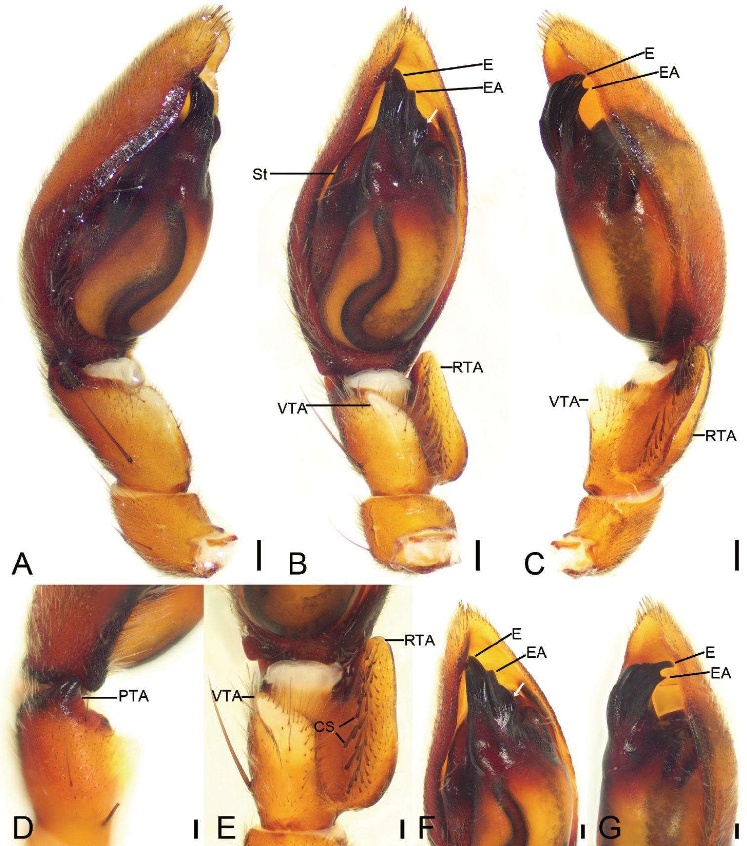
Spinirta sanxiandian Liu sp. nov., palp of male holotype A prolateral view B ventral view, white arrow shows the lateral apophysis located on anterolateral tegulum C retrolateral view D detail of PTA, dorso-prolateral view E detail of RTA, ventral view F detail of tegulum, white arrow shows the lateral apophysis located on anterolateral tegulum, ventral view G same, retrolateral view. Abbreviations: CS – cone-shaped spines, E – embolus, EA – embolic apophysis, PTA – prolateral tibial apophysis, RTA – retrolateral tibial apophysis, St – subtegulum, VTA – ventral tibial apophysis. Scale bars: 0.2 mm (A–C); 0.1 (D–G). |