|
||
|
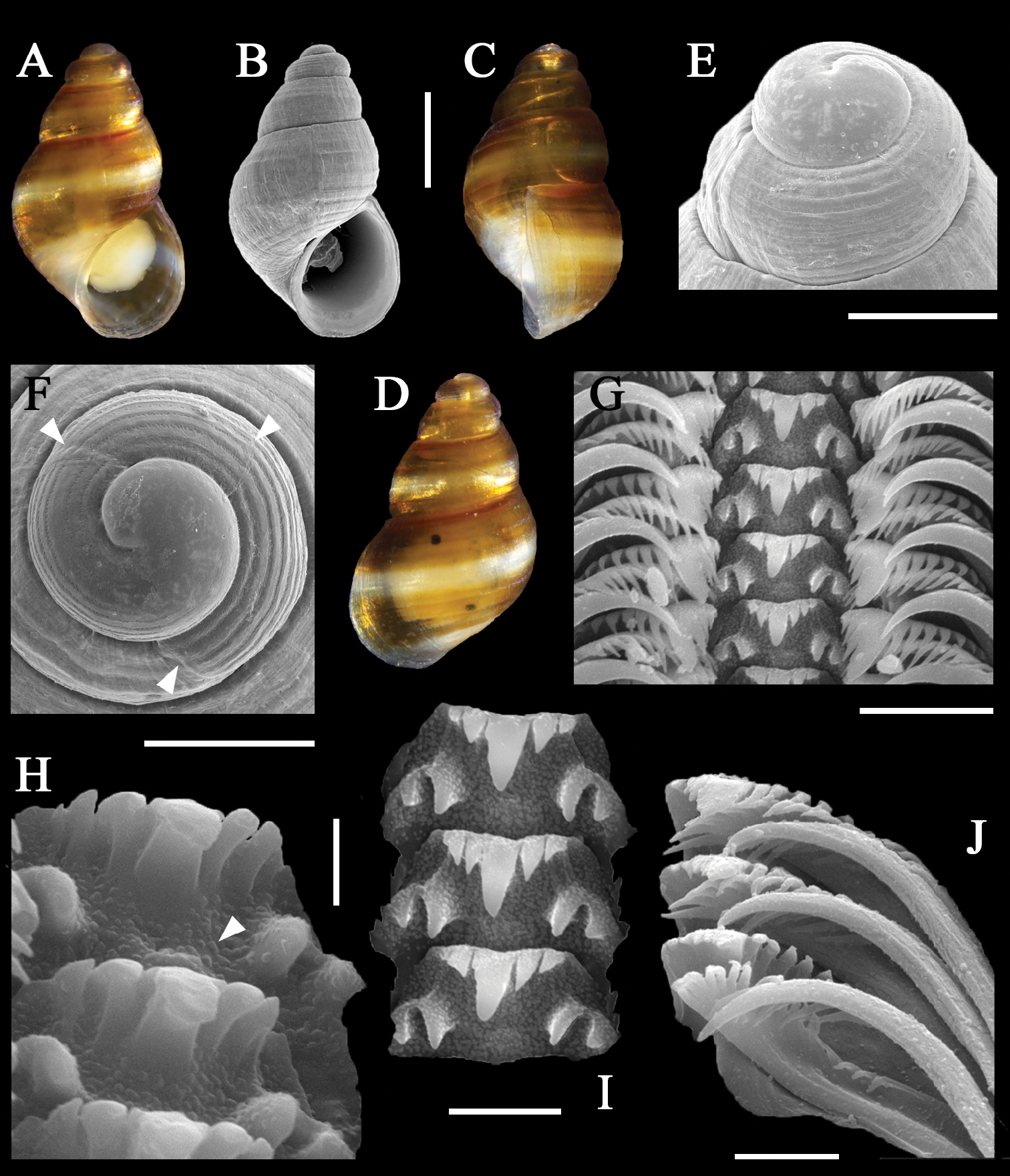
Paratype of Alvania wangi Xu, Qi & Kong, sp. nov. (A–D) shell A apertural view of shell B scanning electron micrographs of apertural view of shell C lateral view of shell D dorsal view of shell E protoconch F apical view of protoconch (top two arrowheads show the two growth lines of the protoconch; bottom arrowhead indicates demarcation between protoconch and teleoconch) G radula H oblique view of central teeth (arrowhead indicates pustules on base of central teeth) I central teeth J lateral and marginal teeth. Scale bars: 500 μm (A–D); 200 μm (E, F); 10 μm (G); 2 μm (H); 5 μm (I, J). |