|
||
|
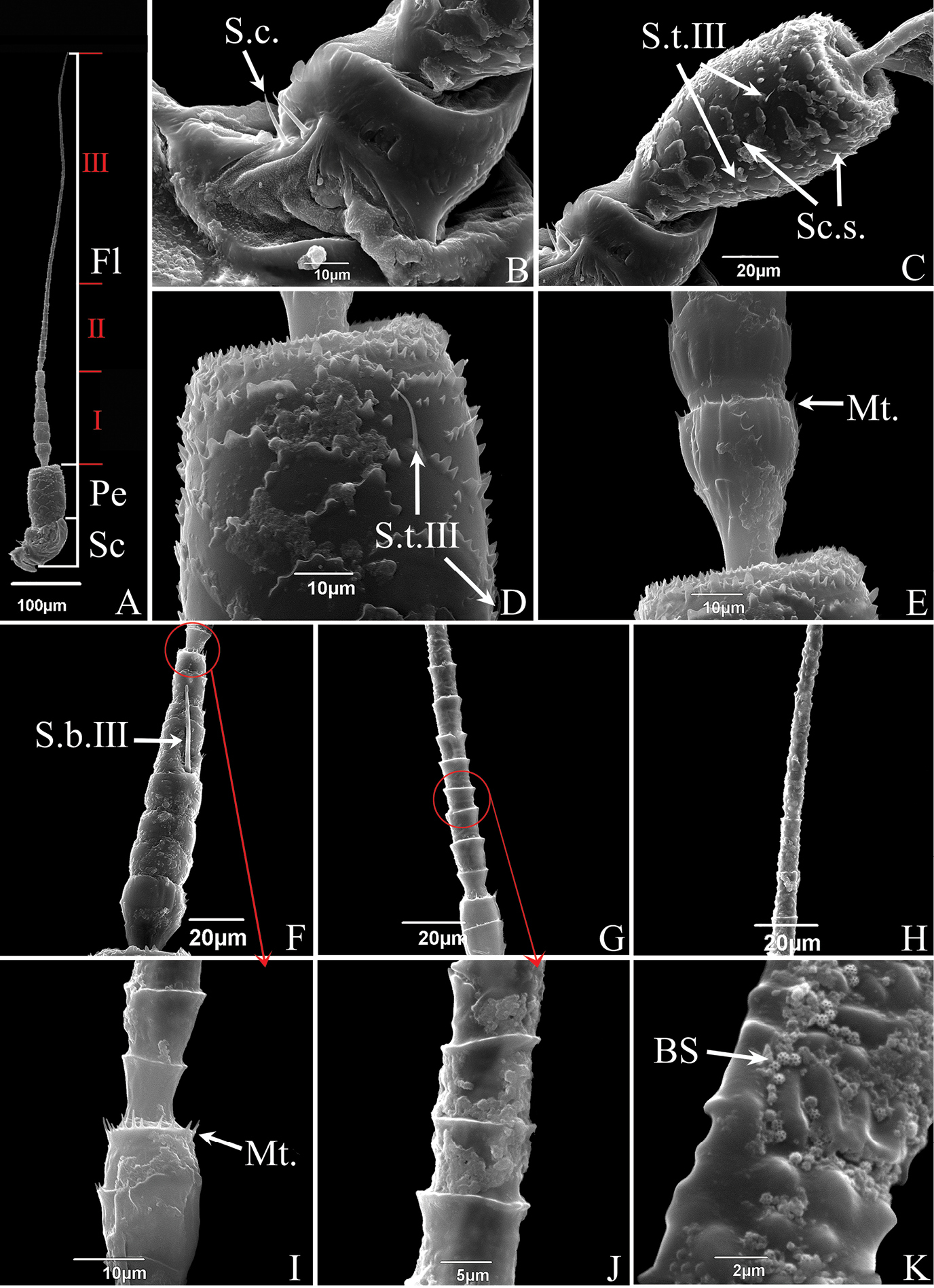
SEM of the antennae of S. shinshana A antenna, composed of three parts: scape (Sc), pedicel (Pe) and three regions of flagellum (Fl) B scape, showing smooth surface, with two sensilla chaetica (S.c.) C pedicel, showing scale-like structures (Sc.s.) and sensilla trichodea III (S.t.III) D enlarged view of pedicel, showing scaly structures and sensilla trichodea III (S.t.III) E junction between pedicel and flagellum, showing microtrichia (Mt.) F first region of flagellum, showing sensilla basiconica III (S.b.III) G second region of the flagellum H junction between second and third regions of flagellum, showing change in surface protrusions I junction between first part and second regions of flagellum, showing microtrichia (Mt.) J enlarged view of second part of flagellum, showing cylindrical subsegments K enlarged view of third part of flagellum, showing brochosomes (BS). |