|
||
|
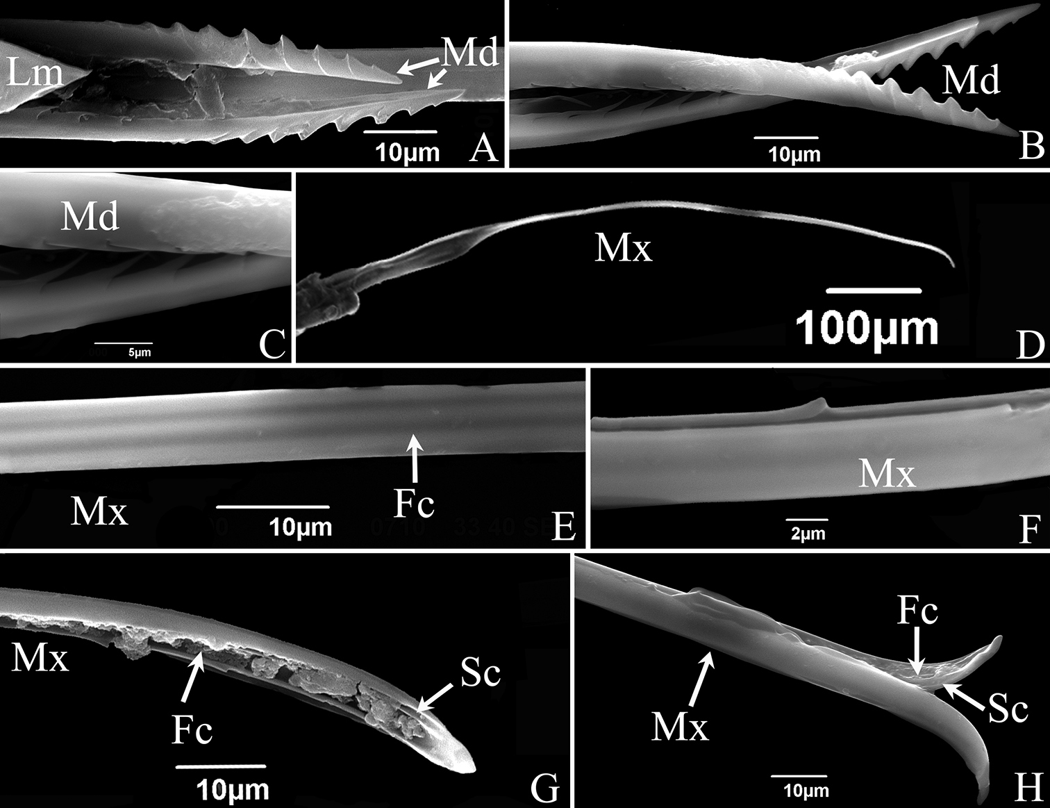
SEM of the stylet fascicle of S. shinshana A mandibular stylets (Md), showing relative position of mandibular stylets and labrum (Lm) B mandibular stylet (Md), showing serrate ridge on the convex external surface and zigzag structure on inner edge C enlarged middle of mandibular stylet (Md), showing zigzag structure on inner edge D maxillary stylets E dorsal view of middle section of maxillary stylets (Mx), showing lines indicating food canal (Fc) F lateral view of middle section of maxillary stylets (Mx), showing relatively blunt tooth-like protrusion G tip of maxillary stylet (Mx), showing salivary canal (Sc) and food canal (Fc) H tip of maxillary stylets (Mx), showing two stylets with different lengths. |