|
||
|
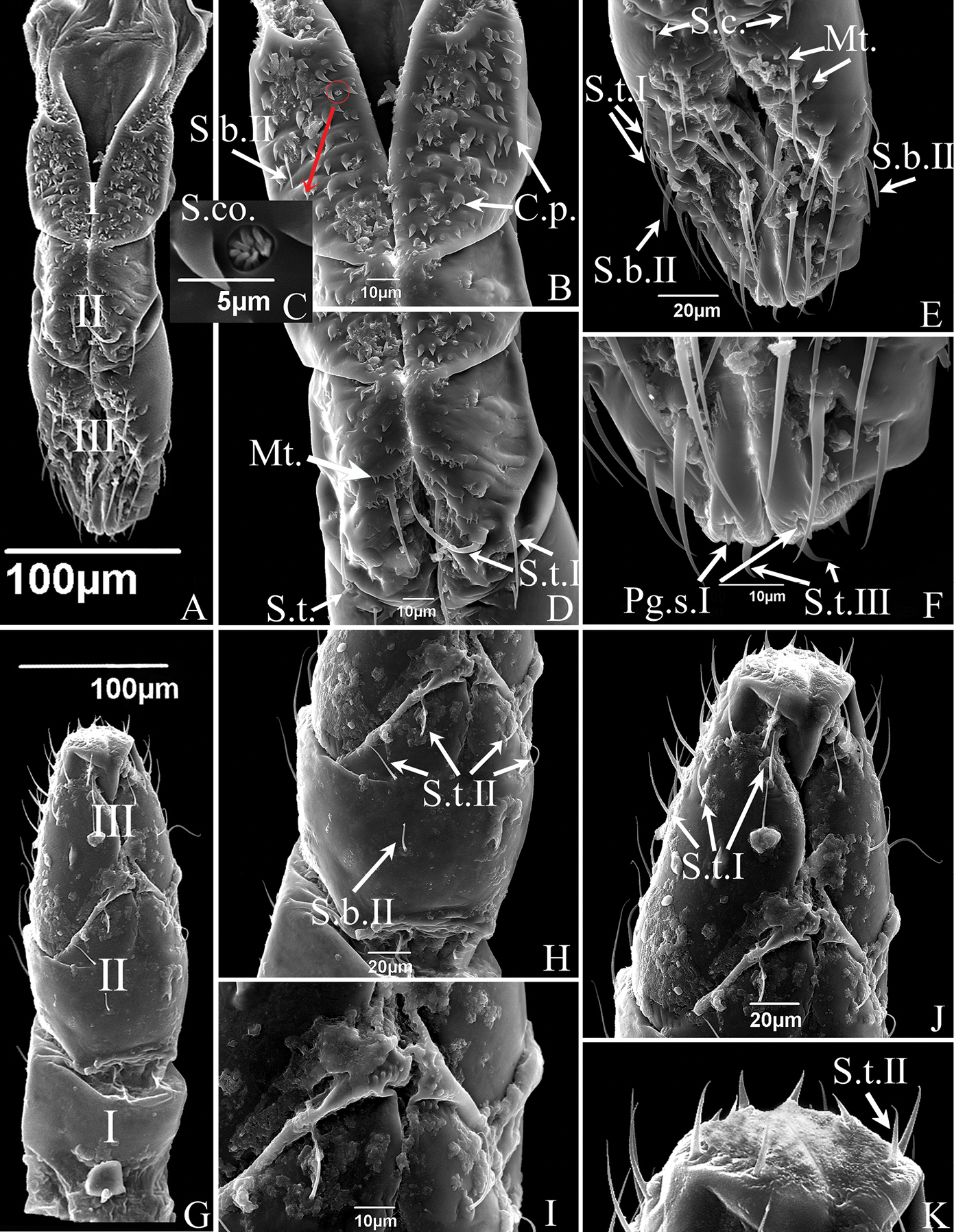
SEM of the labium of S. shinshana A anterior view of labium showing three-segmented labium (I-III), and sensilla symmetrically located on each side of the labial groove B anterior view of first segment of labium showing sensilla basiconica II (S.b.II) and cuticular processes (C.p) C sensilla coeloconica (S.co.) D the anterior view of second segment of labium showing sensilla trichodea I (S.t.I) and microtrichia (Mt.) E anterior view of third segment of labium showing sensilla trichodea I (S.t.I), sensilla basiconica II (S.b.II), sensilla chaetica (S.c.) and microtrichia (Mt.) F anterior view of labial tip showing peg sensilla I (Pg.s.I) and sensilla trichodea III (S.t.III) G dorsal view of mouthparts showing three-segmented labium (I-III) and some sensilla H dorsal view of second segment of labium showing sensilla trichodea II (S.t.II) and sensilla basiconica II (S.b.II) I junction of second and third labial segments showing spherical protrusions J dorsal view of third segment of labium showing sensilla trichodea I (S.t.I) K tip of labium, showing distribution of sensilla. |