|
||
|
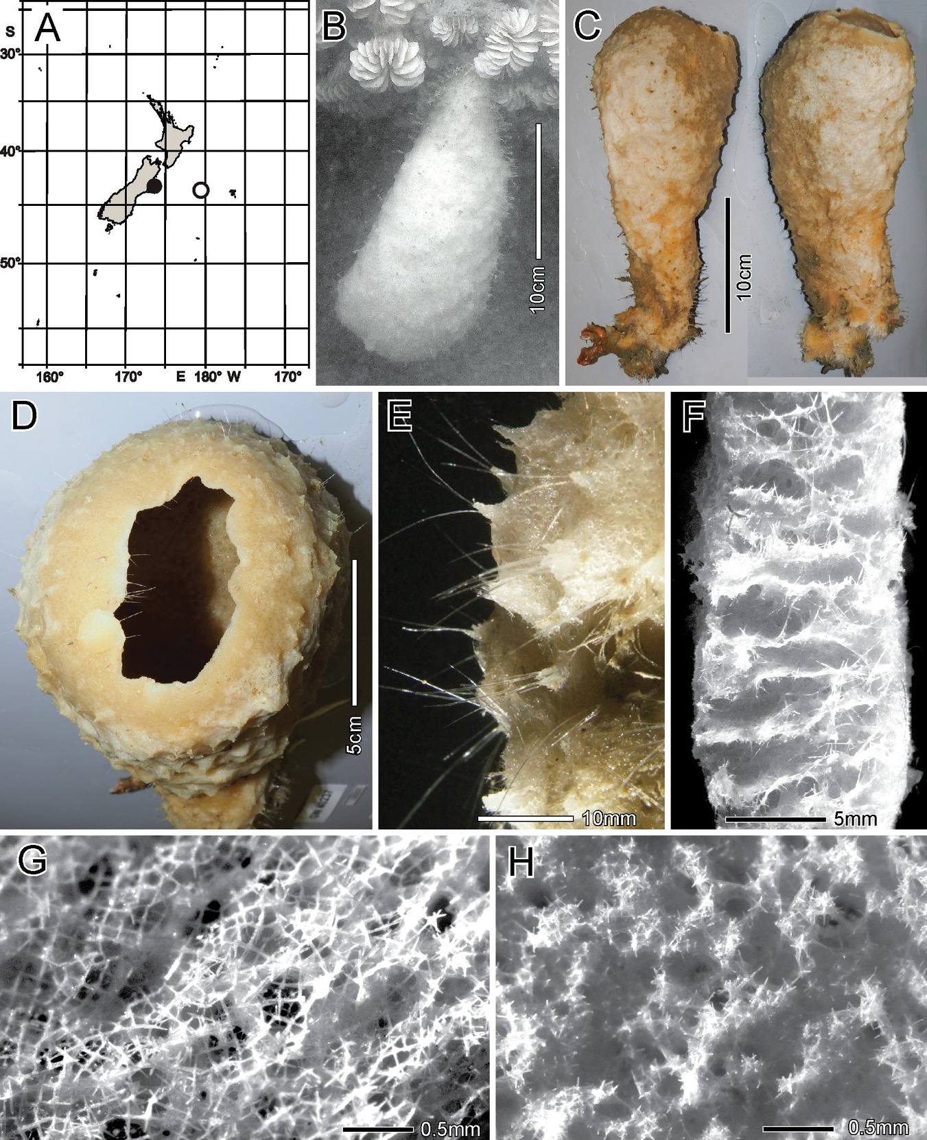
Scyphidium australiense Tabachnick, Janussen & Menschenina, 2008, NIWA 126237, distribution, skeleton and morphology A distribution in New Zealand waters, holotype as open circle, new specimen as filled circle B new specimen in situ (scale bar is approximate) C deck image (two sides, image by PJS) D osculum, deck image (by PJS) E preserved conulose outer surface of the lower body with prostal diactins F preserved wall section of the mid-body without conules G preserved dermal surface with intact pentactin lattice H preserved atrial surface with hexactins displaced from the atrial lattice. Image B captured by ROV Team GEOMAR, ROV Kiel 6000 onboard RV Sonne (voyage SO254), courtesy of Project PoribacNewZ, GEOMAR, and ICBM. |