|
||
|
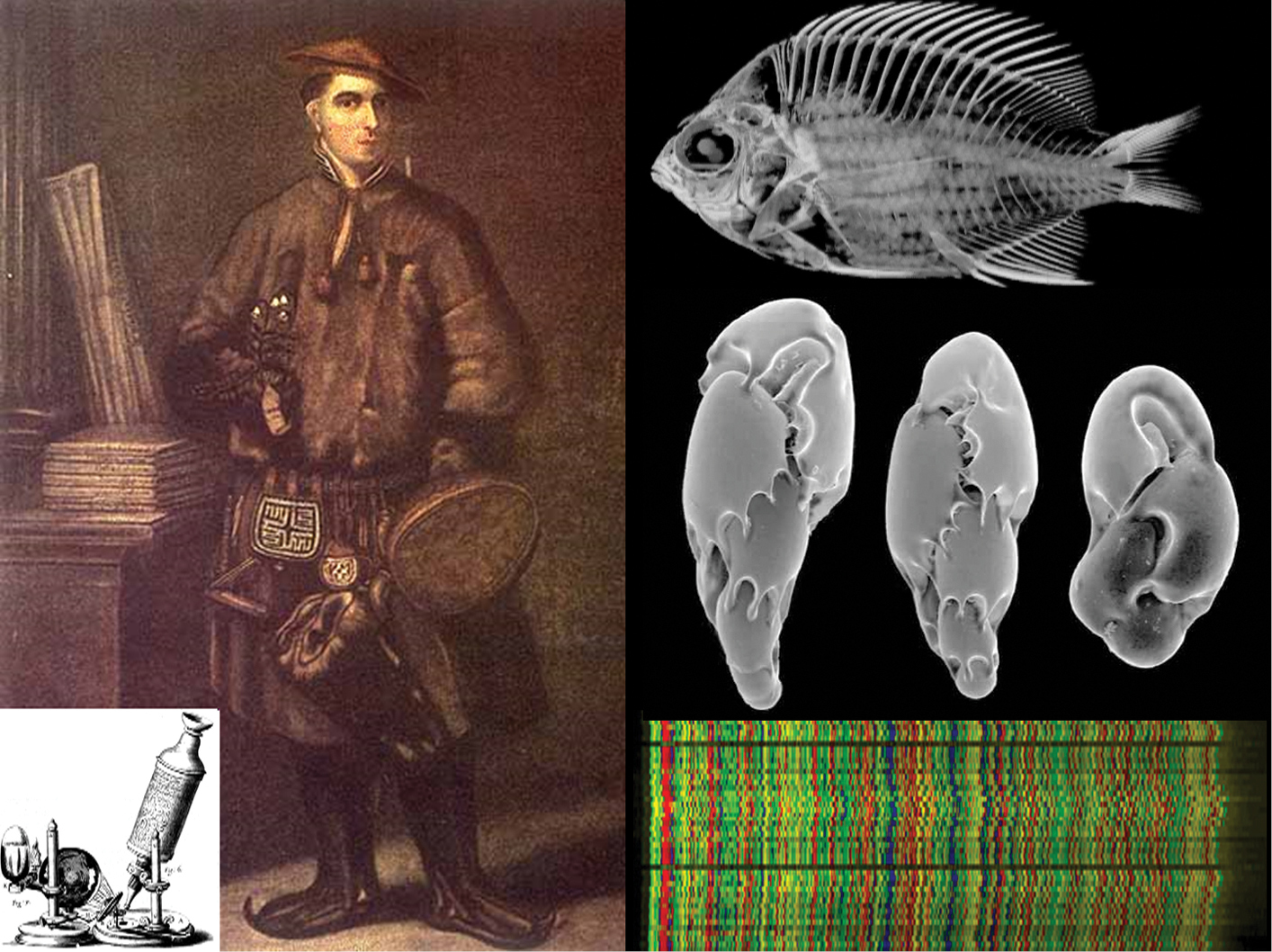
Carl Linnaeus used candle-lit microscopes with primitive optics to examine his specimens (left, H. Kingsbury). Modern technology allows us to generate high-resolution 3D CT scans of the internal structures of specimens without displacing a single scale (right top, Digimorph; Chromis abyssus), capture crisp images of tiny organisms through electron microscopy (right middle, NOAA; single-celled foraminifera), and read DNA sequences (right bottom, BOLD, unspecified taxon). |