|
||
|
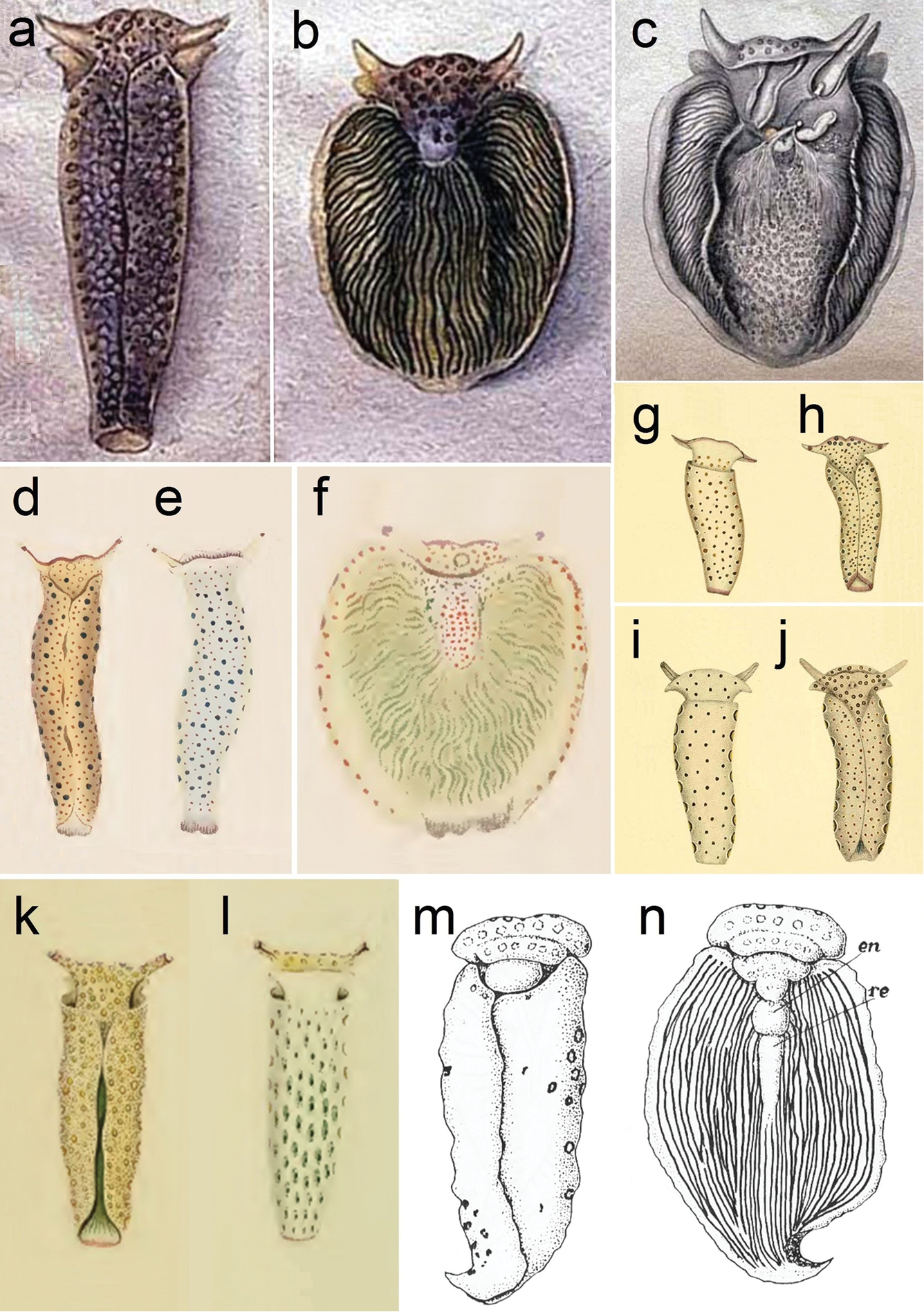
a–c Plakobranchus ocellatus, drawings by van Hasselt (1824): a dorsal view with parapodia folded up on the dorsal body surface b dorsal view with open parapodia, showing longitudinal lamellae c internal anatomy d–f P. ianthobaptus, drawings by Gould (1852): d dorsal view with parapodia folded up on the dorsal body surface e ventral view f dorsal view with open parapodia, showing longitudinal lamellae g–j two Plakobranchus species illustrated by Pease (1871)g–h P. gracilis: g ventral view h dorsal view i–j P. variegatus: i ventral view j dorsal view k–l drawings of P. chlorophacus by Bergh (1873): k dorsal view with parapodia folded up on the dorsal body surface l ventral view with ocellated spots m–n drawings of P. ocellatus by Marcus (1982): m dorsal view with parapodia folded up on the dorsal body surface n dorsal view with open parapodia, showing the longitudinal lamellae (en = pericardium; re = renal prominence). |