|
||
|
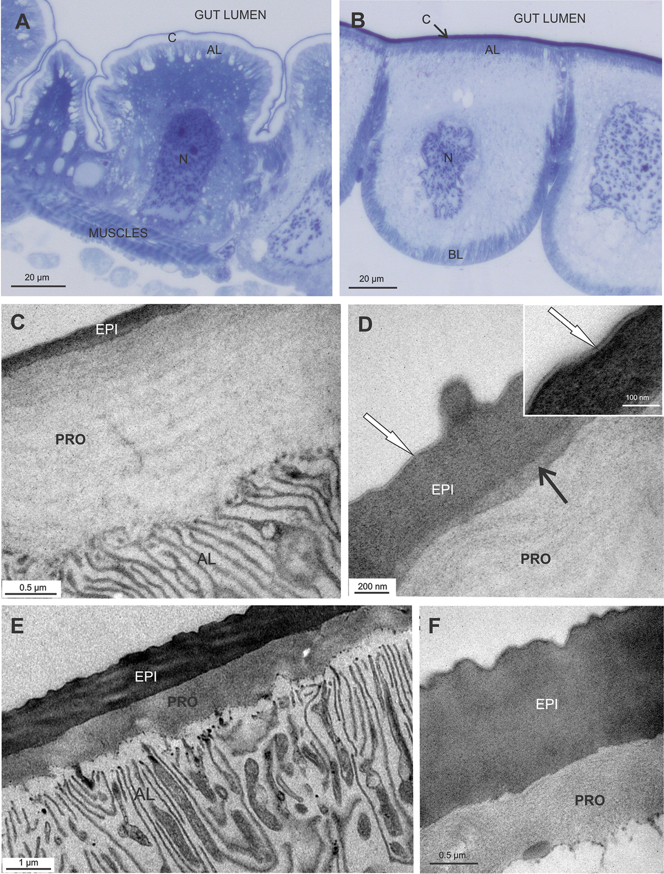
Hindgut epithelium and cuticle in P. scaber adults. A Semithin section of the hindgut anterior chamber. Gut cells protrude apically into the gut lumen. The apical membrane forms an apical labyrinth (AL), that is covered with the cuticle (C). N – nucleus of gut cell B Semithin section of the hindgut papillate region. Gut cells bulge basally into the hemocoel. Apical and basal labyrinths (AL, BL) are evident. Cuticle covers apical cell surface (C). N – nucleus of gut cell C, D Ultrastructure of the cuticle in anterior chamber. The cuticle is composed of thin electron dense epicuticle (EPI) and much thicker ''lamellated'' electron lucent procuticle (PRO). Several thin sublayers are discernible in the outermost part of the epicuticle (D inset - white →). A layer of medium electron density is visible between the epi- and procuticle (D - black →). A cuticular spine is present on the cuticle surface E, F Ultrastructure of the gut cuticle in papillate region. Epicuticle (EPI) and procuticle (PRO) are about the same thickness. Both are composed of morphologically homogenous matrix. Abundant mitochondria are observed closely to the membranes of the apical labyrinth (AL) F Several thin sublayers in the outermost region of the epicuticle are visible. |