|
||
|
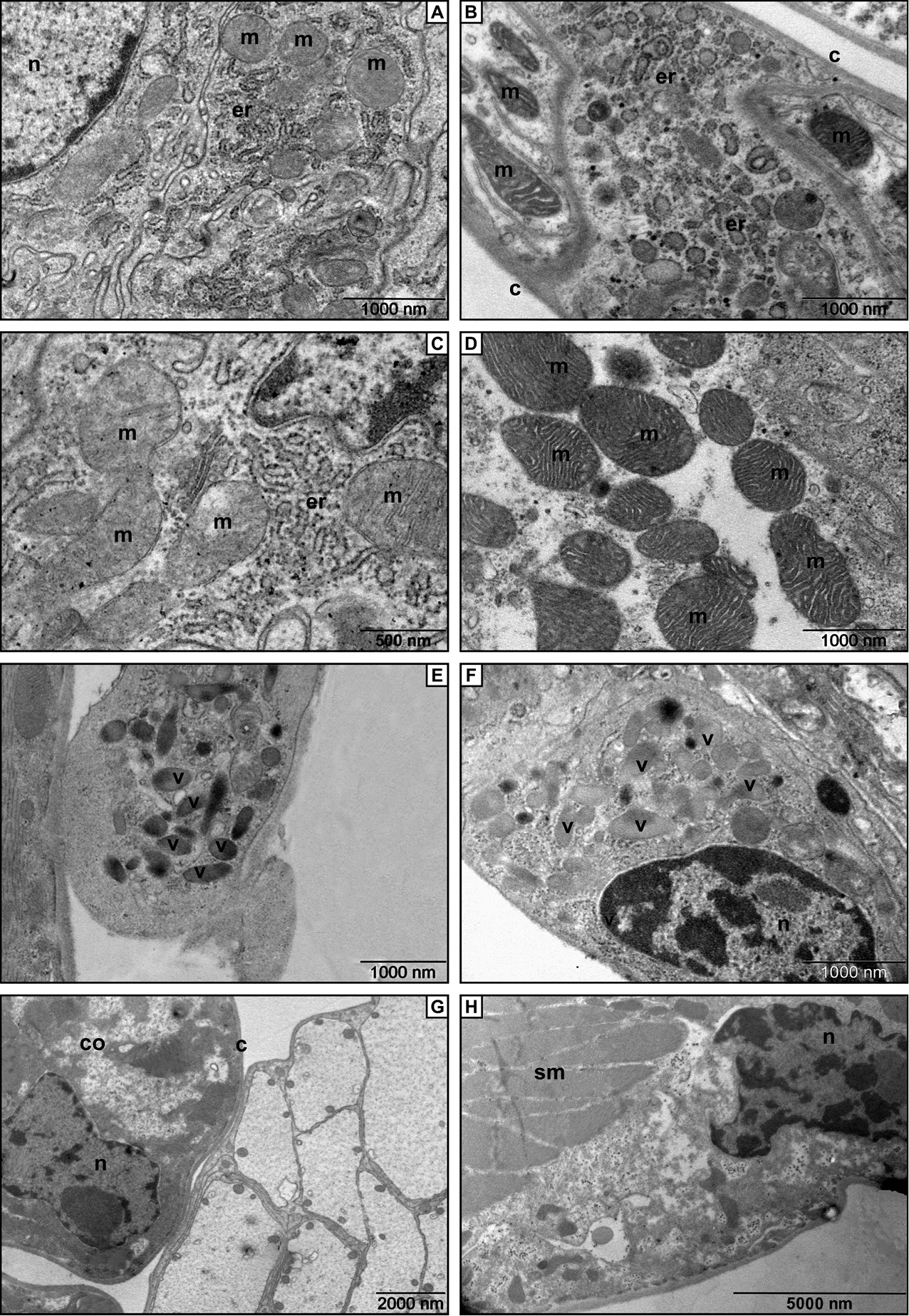
Electron micrographs of the cotyledon. A Fine structure of a cell from the medial portion of the cotyledon in Trachelipus rathkii. The most abundant cell organelles are mitochondria and rough endoplasmic reticulum B In the medial portion of the cotyledon the cells contain vesiculated rough endoplasmic reticulum and mitochondria (C. convexus) C Higher magnified detail of the cotyledon in T. rathkii. D High power micrograph of the cotyledon in Cylisticus convexus. Note the densely cristate mitochondria E Rounded ending of the cotyledon with electron dense vesicles (T. rathkii) F A cell with large vesicles containing moderately electron dense material (C. convexus) G Cotyledon ending of T. rathkii covered by a thin cuticle H Bundles of striated muscle fibers located at the base of cotyledon (C. convexus). Legends: c – cuticle, co – cotyledon, er – rough endoplasmic reticulum, m – mitochondria, n – nucleus, sm – striated muscle, v – vesicle. |