|
||
|
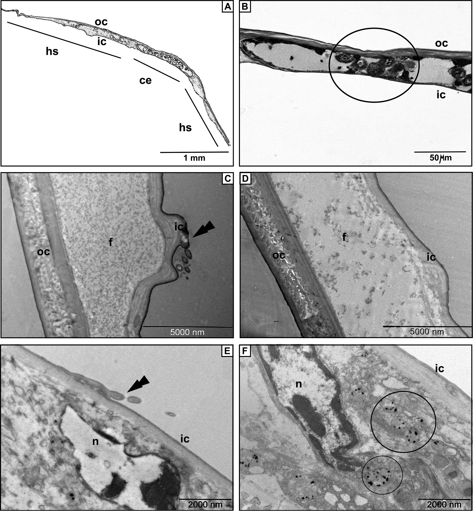
The structure of the oostegite. A Schematic drawing of the cross -sectioned oostegite B Semithin section after PAS staining with positive cytoplasm in the cells of the oostegite C An electron micrograph of the break between cells of Trachelipus rathkii oostegite. Note scale-like protrusion of the inner cuticle (arrow) D Identical detail in Cylisticus convexus. No protrusion was found E Cell in the oostegite of T. rathkii below a scale-like protrusion of the inner cuticle (arrow) F Cell in the oostegite of C. convexus. Note the membrane-bound electron dense inclusions. Legends: ce – cellular elements, f – fleecy precipitate, hs – hemolymph space, ic – inner cuticle, n – nucleus, oc – outer cuticle. |