|
||
|
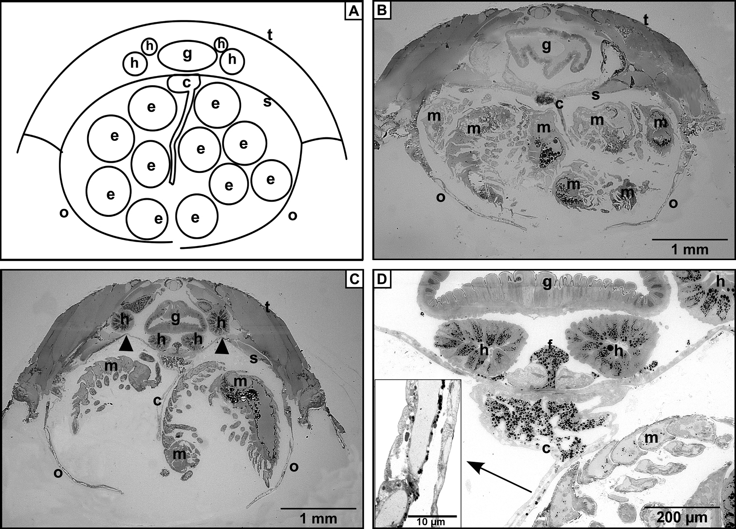
Cross sections of marsupium. A Schematic drawing of the brood pouch B Marsupium with developing mancas in the non-conglobating Trachelipus rathkii C Marsupium of conglobating Cylisticus convexus in the same stage. Note arching sternites (arrowheads) D Higher magnification image of the proximal part of the cotyledon in C. convexus. The cells are filled with darkly stained lipid droplets. Insert: Higher magnification reveals that along the longitudinal axis of cotyledon a beadlike array of lipid droplets lines up. Legends: c – cotyledon, e – egg, f – maternal fat body, g – gut, h – hepatopancreas, m – manca, o – oostegite, s – sternite, t – tergite. |