|
||
|
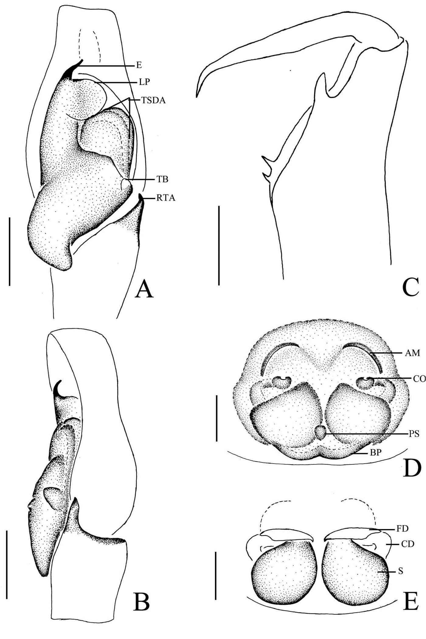
Phintella arcuata sp. n., A male palp, ventral view B male palp, retrolateral view C left chelicerae of male, posterior view D epigyne, ventral view E vulva, dorsal view. AM atrium margin BP basal plate CD copulatory duct E embolus FD fertilization duct LP lamellar process PL posterior lobe PS poriform structure RTA retrolateral tibial apophysis TB tegulum bump TSDA terminal sperm duct angle S spermathecae. Scale bars: 0.1 mm (A–E). |