|
||
|
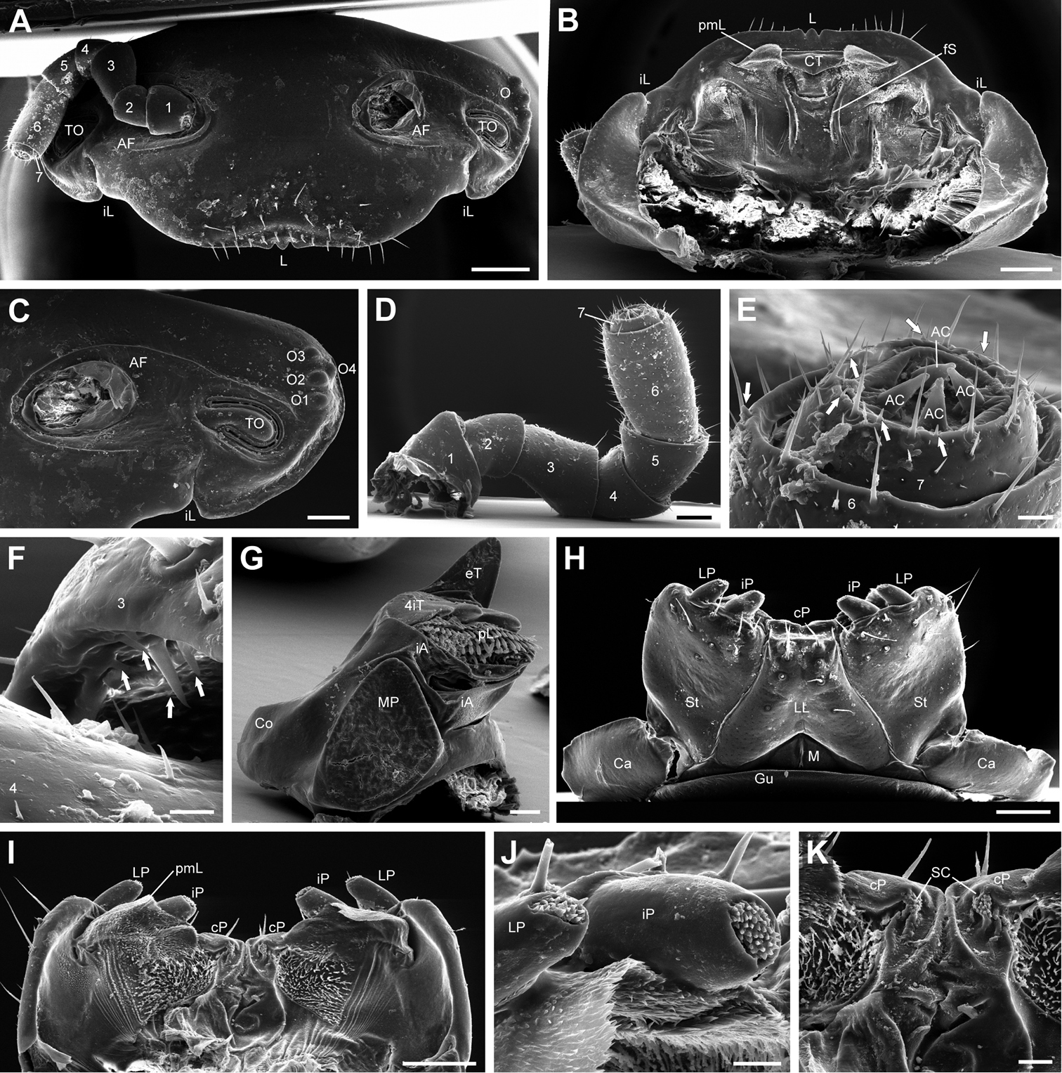
Eupeyerimhoffia archimedis (Strasser, 1965) male, SEM. A Head, frontal view B Head, ventral view C Head, detail of lateral area D Antenna, posterior viewE Antenna, antennomere 6 and 7 F Antenna, apical edge of antennomere 3 G Mandible, mesal view H Gnathochilarium, ventral view I Endochilarium, dorsal view J Gnathochilarium, lateral and inner palpi, dorsal view K Endochilarium, detail of median area, dorsal view. Abbreviations: 1-7 = Antennal segments number 1-7; 4iT = 4-combed inner tooth; AC = Apical cone; AF = Antennal fossa; Ca = Cardine; Co = Condylus; cP = Central pads; CT = Central tooth; eT = External tooth; fS = fringed seam; Gu = Gula; iA = Intermediate area; iL = Incisura lateralis; iP = Inner palpi; L = Labrum; LL = Lamella linguales; LP = Lateral palpi; M = Mentum; MP = Molar plate; O = Ocellaria; O1-O4 = Ocelli 1-4; pL = Pectinate lamellae; pmL = paramedian lobe; TO = Organ of Tömösváry; SC = Sensory clusters; St = Stipites. Arrows mark sensilla basiconica. Scale bar: 400 μm (A, B); 200 μm (C); 150 μm (D); 25 μm (E); 10 μm (F); 100 μm (G); 250 μm (H, I); 40 μm (J); 50 μm (K). |