|
||
|
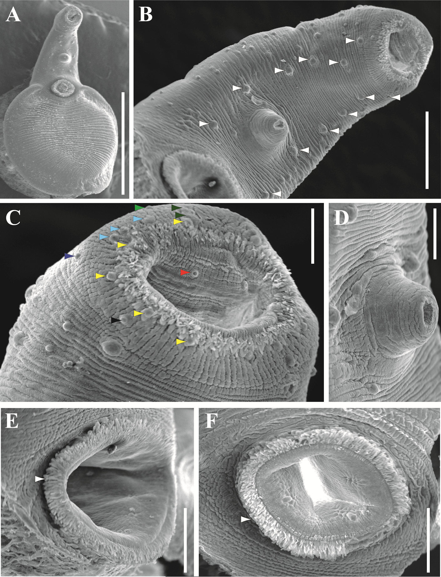
Scanning electron microscopy (SEM) images of Xystretrum solidum (from three specimens collected at Progreso Port, Yucatan, Mexico) A whole adult specimen (ventral view) with scattered rosette papillae on forebody B forebody, showing 6 pairs of robust papillae (white arrowhead) C oral sucker, showing 13 pairs of papillae: 5 on interior margin surrounding mouth (yellow arrowhead); one posterolateral to interior margin (dark blue arrowhead); three anterolateral to interior margin (light blue arrowhead); 2 on stylet scar (dark green arrowhead); one lateral to stylet scar (light green arrowhead); one on posterior external margin of oral sucker (black arrowhead); one inside of mouth (red arrowhead) (only right hand side papillae are indicated) D genital atrium detail E ventral sucker (side view), showing long papillae on inner margin (white arrowhead) F ventral sucker (ventral view), showing long papillae on inner margin (white arrowhead). Scale bars: 1000 µm (A); 200 µm (B); 500 µm (C, D); 100 µm (E, F). For more details of observed characters by SEM from other localities analyzed in this study, see Suppl. material 2: Figure S1. |