|
||
|
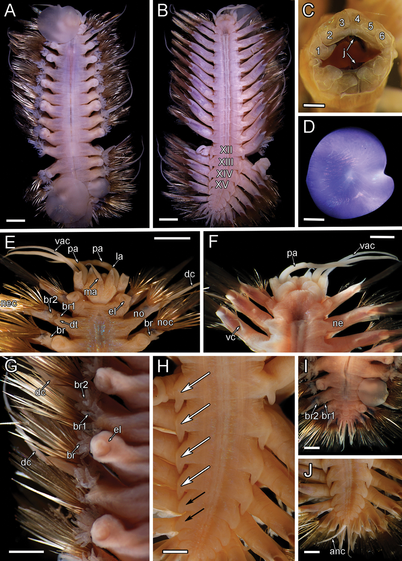
Macro photos and micrographs of P. elvisi sp. nov. holotype SIO-BIC A8488 A dorsal view B ventral view. Segments 12–15 are marked to indicate the presence of four pairs of papillae C frontal view of proboscis showing papillae and jaws. Numbers mark the papillae on the dorsal (six papillae) surface D left elytron from segment 2 E dorsal view of anterior F ventral view of anterior G left side branchiae on segments 7–11 H ventral papillae on segments 12–15 (four pairs) indicated by white arrows. Ventral lamellae on segments 16–17 (two pairs) indicated by black arrows I dorsal view of posterior J ventral view of posterior. Abbreviations: XII, segment 12; XIII, segment 13; XIV, segment 14; XV, segment 15; j, jaws; ma, median antenna; pa, palp; la, lateral antenna; vac, ventral anterior cirrus; el, elytrophore; br, single large group of branchiae on elytrigerous segment; noc, notochaetae; dc, dorsal cirrus; br1, branchiae small group 1 attached to dorsal tubercle on cirrigerous segment; br2, branchiae large group 2 attached near base of notopodium on cirrigerous segment; no, notopodium; nec, neurochaetae; vc, ventral cirrus; ne, neuropodium; dt, dorsal tubercle; anc, anal cirrus. Scale bars: 4 mm (A, B); 0.5 mm (C); 1 mm (D, H–J); 2 mm (E–G). |