|
||
|
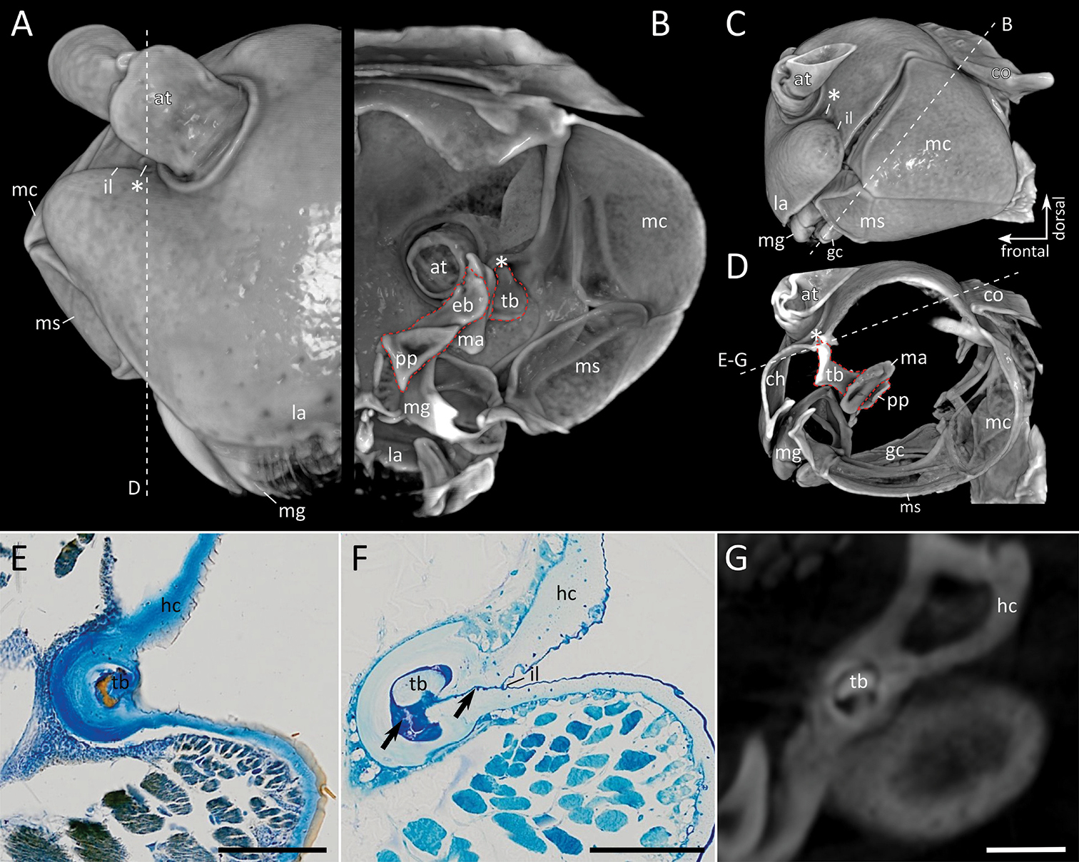
Polydesmus angustus, head A–D volume rendering based on micro-CT data: A Frontal view B cross-section, posterior view, plane indicated in C C lateral view D sagittal view, cutting plane indicated in A E–G details of connection of tentorial transverse bar to head capsule at incisura lateralis, plane as indicated in D: E histological section (Paraffin, Azan-staining) F histological section (Araldite, Toluidine blue) G optical section of micro-CT scan. Abbreviations: at = antenna, co = collum, eb = epipharyngeal bar of tentorium, gc = gnathochilarium, gls = gnathal lobe sclerite, hc = head capsule, il = incisura lateralis, la = labrum, mc = mandibular cardo, mg = mandibular gnathal lobe, ms = mandibular stipes, pp = posterior process of tentorium, tb = transverse bar of tentorium. Asterisk (*) indicates structure previously interpreted by Hennings (1906) as the Tömösváry organ in the Polydesmida. In the volume renderings the tentorium is marked with a red dotted line. Scale bar: 100 µm (E–G). |