|
||
|
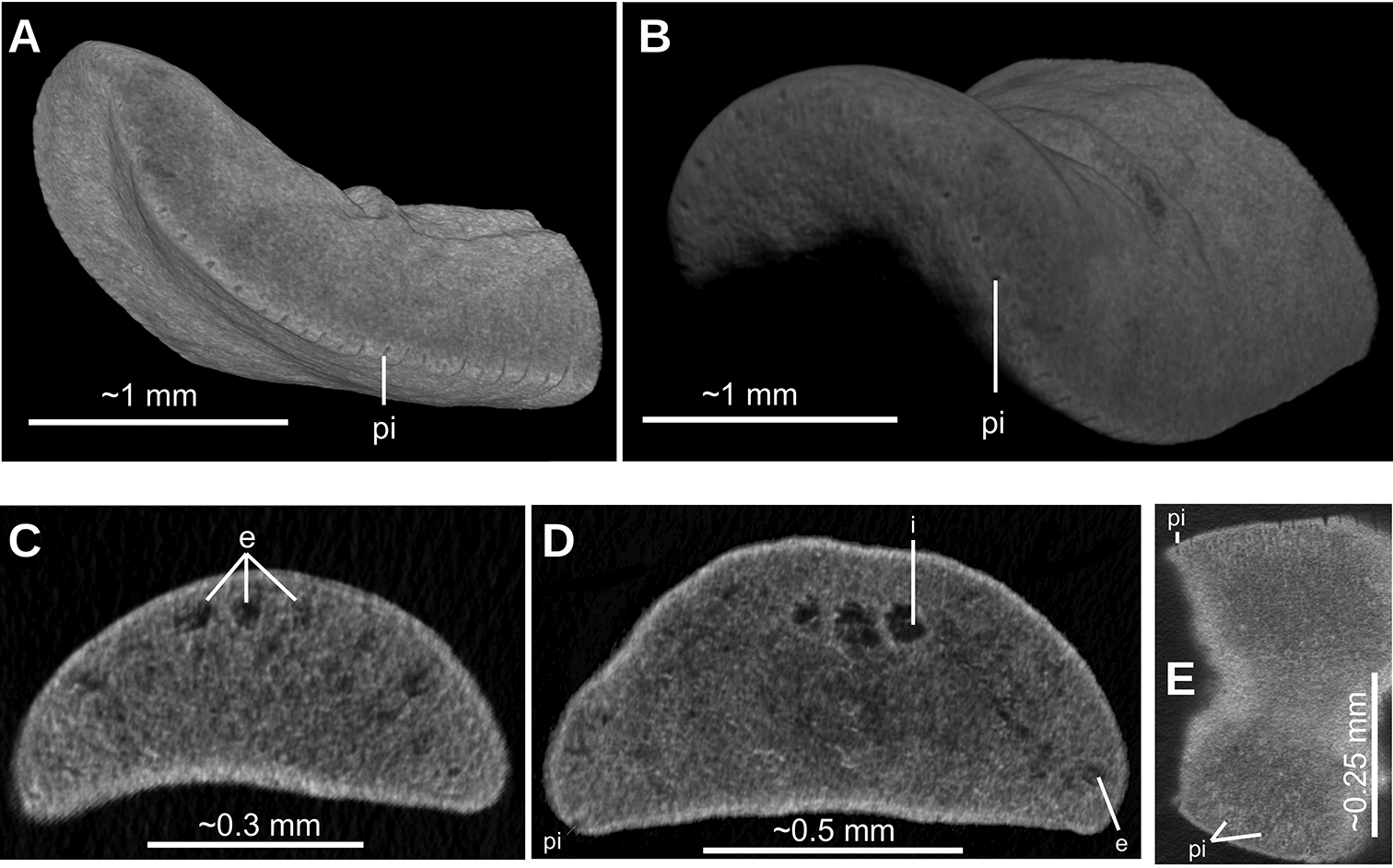
Paraba bresslaui (Schirch 1929), µCT-derived images of cephalic extremity of holotype A, B perspective view of 3D rendering C virtual transverse section showing anteriormost eyes, pixel size: 1.9 µm D virtual transverse section showing eye and sensory pit, pixel size: 1.9 µm E horizontal virtual section showing rows of sensory pits, pixel size: 1.9 µm. |